

Spine and Spinal Cord Trauma
Overview
Spine trauma refers to bony, ligamentous, or disc injury to the vertebral column, whereas Spinal Cord Injury (SCI) refers to acute neural damage resulting in sensory, motor, or autonomic dysfunction.
Classification:
- Primary Injury: Direct mechanical damage (contusion, laceration, compression, transaction) occurring at impact.
- Secondary Injury: Downstream cascade (ischemia, edema, inflammation, excitotoxicity) progressing over hours-to-days.
- Neurological Incompleteness: Defined by ASIA Impairment Scale (AIS); sacral sparing (S4–S5 sensory or voluntary anal contraction) confirms an incomplete SCI.
Sacral sparing (preservation of perianal sensation, deep anal pressure, or voluntary anal sphincter contraction) is the single most important physical exam marker defining an incomplete spinal cord injury, carrying a significantly better prognosis for recovery.
Approach
Step 1: Immobilization & Airway Stabilization
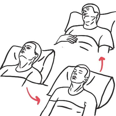
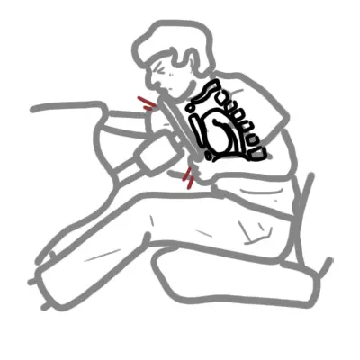
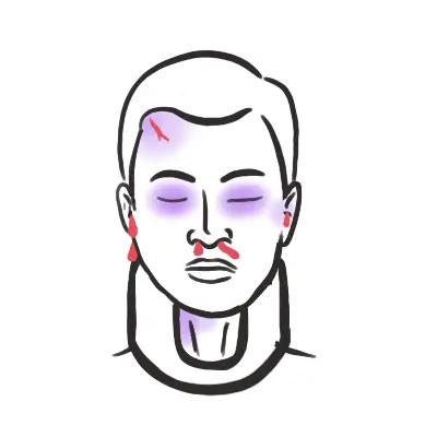
- Strict Spinal Motion Restriction: Rigid cervical collar + block/tape on hard backboard (log roll off board onto flat firm mattress as soon as possible to prevent pressure injuries).
- High Cervical Injury Check (C3–C5): “C3, C4, C5 keeps the diaphragm alive.” Lesions above C5 risk diaphragm paralysis; prepare for early intubation using video laryngoscopy with manual in-line stabilization (MILS).
Step 2: Hemodynamic & Neurogenic Shock Triage
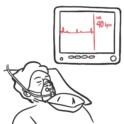
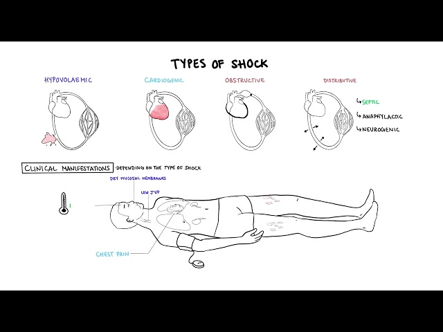
- Neurogenic Shock (T6 and above): Loss of sympathetic tone -> Triad of Hypotension + Bradycardia + Hypothermia/Warm Dry Extremities.
- Distinguish from Hypovolemic Shock: Hypovolemia causes Hypotension + Tachycardia + Cold/Clammy skin.
Step 3: Clinical Decision Rules for Imaging (Clearance)
- NEXUS Criteria or Canadian C-Spine Rule: Assess need for cervical imaging.
- Clear clinically ONLY if: Completely alert/oriented, no distracting injuries, no focal neurological deficits, no midline cervical tenderness, no intoxication. Otherwise -> CT Spine.
Neurogenic shock causes hypotension WITH bradycardia due to unopposed vagal tone (loss of sympathetic input above T6). Treating neurogenic shock with fluid overload alone causes pulmonary edema—early vasopressors/inotropes with alpha and beta activity (e.g., Norepinephrine) are required.
Differential Diagnosis
Incomplete Spinal Cord Syndromes:
- Central Cord Syndrome (Most Common):
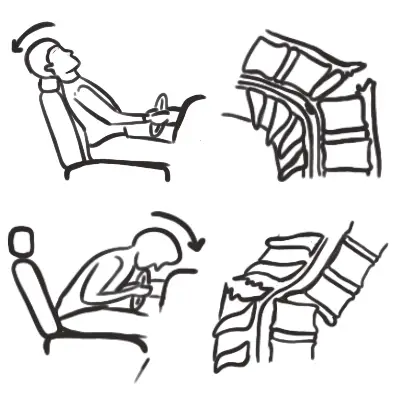
- Mechanism: Hyperextension injury in older adults with pre-existing cervical spondylosis.
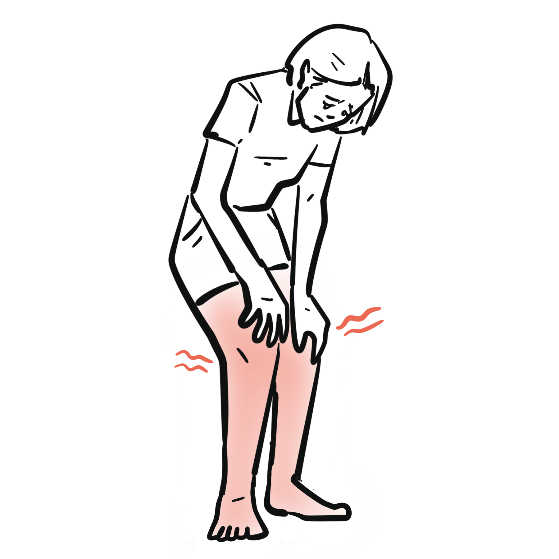
- Distinguishing features: Upper extremity weakness > Lower extremity weakness (“arms worse than legs”); variable sensory loss.
- Anterior Cord Syndrome:
- Mechanism: Hyperflexion injury or anterior spinal artery occlusion/ischemia.
- Distinguishing features: Loss of motor function + loss of pain/temperature sensation below lesion WITH preserved vibration and proprioception (dorsal columns intact). Worst prognosis.
- Brown-Séquard Syndrome (Hemicord Lesion):
- Mechanism: Penetrating trauma or lateral compression.
- Distinguishing features: Ipsilateral motor paralysis & loss of vibration/proprioception + Contralateral loss of pain/temperature 1–2 segments below the lesion.
- Posterior Cord Syndrome (Rare):
- Distinguishing features: Isolated loss of proprioception and vibration sensation (sensory ataxia); motor function and pain/temp preserved.
Conus Medullaris vs. Cauda Equina Syndrome:
- Conus Medullaris Syndrome (L1–L2 cord level):
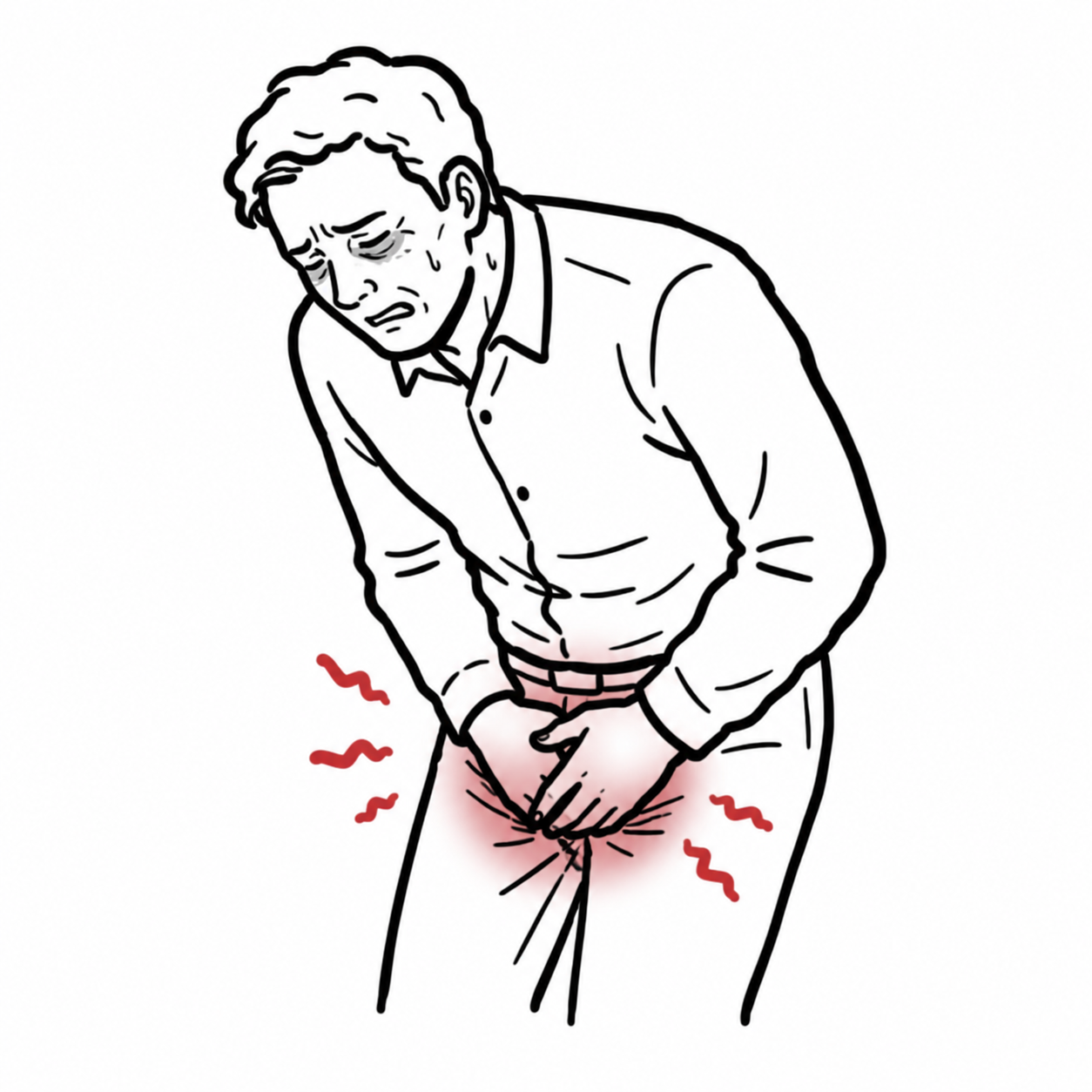
- Distinguishing features: Sudden onset, symmetric bilateral motor weakness, early bowel/bladder dysfunction, saddle anesthesia.
- Cauda Equina Syndrome (L2–S5 nerve roots):
- Distinguishing features: Gradual/subacute onset, asymmetric weakness, radicular pain, absent lower limb reflexes, urinary retention with overflow incontinence.
Anterior Cord Syndrome spares the dorsal columns (proprioception and light touch remain intact). Central Cord Syndrome affects upper extremities far more than lower extremities because the cervical corticospinal tracts are situated centrally within the spinal cord.
Investigations
First-Line / Bedside & Emergency Diagnostics:
- Bedside Vital Signs & ABG/VBG: Monitor for hypercapnia/hypoxia (cervical injuries) and hypotension/bradycardia (neurogenic shock).
- Focused ASIA Neurological Exam: Standardized motor (0–5 grade across 10 key muscle groups) and sensory (light touch and pinprick across 28 dermatomes) scoring.
- Urinary Catheter Placement: Quantify output and screen for urinary retention.
Targeted / Imaging Strategy:
- Non-Contrast CT Spine (Whole Spine if High-Energy Mechanism): Gold standard for identifying bony fractures, dislocations, retropulsed fragments, and subluxation.
- MRI Spine (Emergent):
- Indications: Neurological deficit unexplained by CT, soft tissue assessment (ligamentous disruption, intervertebral disc herniation), spinal epidural hematoma, cord contusion/edema, or suspected Cauda Equina/Conus Medullaris syndrome.
CT is for bones; MRI is for nerves, discs, and ligaments. If a patient has a focal neurological deficit after trauma but their CT spine is completely normal, obtain an immediate MRI to rule out cord edema, ligamentous injury, or disc herniation (SCIWORA – Spinal Cord Injury Without Radiologic Abnormality).
Critical Management
Neurogenic Shock & Hemodynamic Targets:
- Mean Arterial Pressure (MAP) Target: Maintain MAP 85–90 mmHg for the first 7 days post-injury to optimize spinal cord perfusion pressure and prevent secondary ischemic injury.
- First-Line Vasoactive Agent: Norepinephrine (provides alpha-1 vasoconstriction and beta-1 chronotropic/inotropic support).
Surgical Decompression:
- Timing: Early decompression (< 24 hours, ideally < 12 hours) in setting of acute cord compression or progressive neurological deterioration.
High-Dose Steroids (Methylprednisolone):
- Current Evidence: High-dose methylprednisolone (NASCIS protocol) is NOT universally recommended due to high rates of severe side effects (wound infection, sepsis, GI bleeding) with minimal motor recovery benefit.
Avoid high-dose methylprednisolone routine use in acute SCI—guidelines now consider it an optional treatment rather than a standard of care due to significant risks of systemic infection and adverse events.
References
- National Institute for Health and Care Excellence. Spinal injury: assessment and initial management (NG41) [Internet]. 2016 [cited 2026 Aug 20]. Available from: https://www.nice.org.uk/guidance/ng41
- British Orthopaedic Association. BOASt: assessment of the spine in the trauma patient [Internet]. 2025 [cited 2026 Aug 20]. Available from: https://www.boa.ac.uk/resource/boast-assessment-of-the-spine-in-the-trauma-patient.html

Members only discussions coming soon…