

Jaundice
Overview
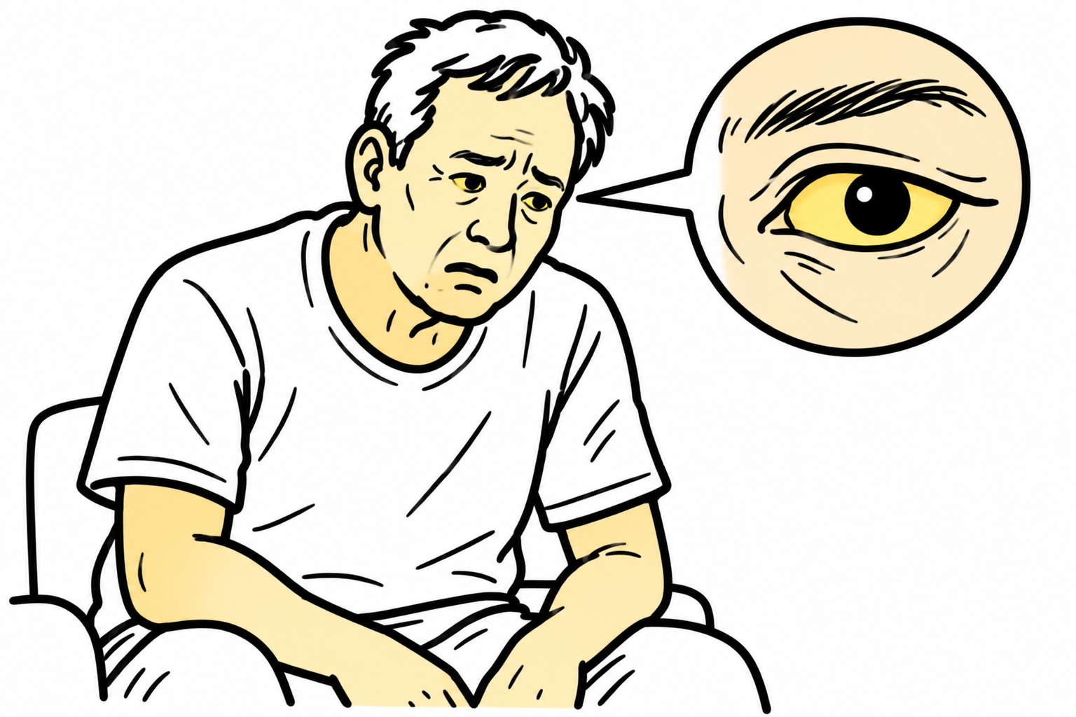
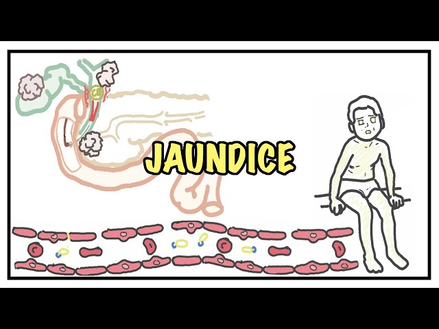
Jaundice (icterus) is the yellow pigmentation of the skin, sclerae, and mucous membranes caused by hyperbilirubinemia.
Normal total bilirubin is < 21 umol/L (< 1.2 mg/dL). Scleral icterus becomes clinically detectable when total bilirubin exceeds 35–50 umol/L (~2.0–3.0 mg/dL).
Pathophysiologic Categorization
- Classified mechanistically into Pre-hepatic (overproduction/hemolysis)
- Hepatic (parenchymal dysfunction/uptake/conjugation defect)
- Post-hepatic / Obstructive (biliary outflow tract obstruction).
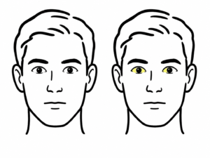
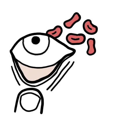
Bilirubin has a high affinity for elastin, which is why scleral icterus is the most sensitive early clinical sign of hyperbilirubinemia. Always examine the sclerae under natural light.
Approach
Step 1: Emergency Triage & Red Flag Screening
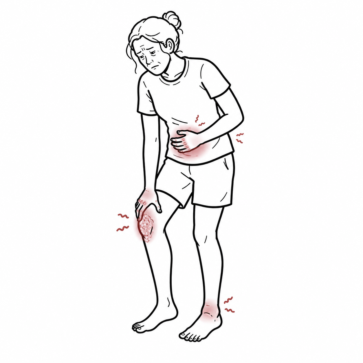
- Fever + RUQ Pain + Jaundice: -> Charcot’s Triad for Acute Cholangitis.
- Charcot’s Triad + Hypotension + Altered Mental Status: -> Reynold’s Pentad for Severe Suppurative Cholangitis (Emergency surgical/endoscopic decompression required).
- Rapidly Rising Bilirubin + Coagulopathy (INR >= 1.5) + Encephalopathy: -> Acute Liver Failure (ALF). Immediate liver transplant unit referral.
- Painless Jaundice + Palpable Gallbladder (Courvoisier’s Sign): -> Suspect Malignant Biliary Obstruction (e.g., Head of Pancreas Adenocarcinoma or Cholangiocarcinoma).
Step 2: Fractionate Bilirubin & Determine Pattern
- Predominantly Unconjugated (Indirect) Hyperbilirubinemia (> 80% Unconjugated):
- -> Pre-hepatic cause (Hemolysis, ineffective erythropoiesis) or impaired hepatic uptake/conjugation (Gilbert’s Syndrome).
- Predominantly Conjugated (Direct) Hyperbilirubinemia (> 50% Conjugated):
- Check Liver Function Tests (LFTs) to determine Hepatocellular vs. Cholestatic pattern.
- Hepatocellular Pattern: Markedly elevated ALT/AST (> 5x ULN) with modest ALP elevation -> Viral hepatitis, drugs/toxins, ischemia.
- Cholestatic Pattern: Markedly elevated ALP and GGT (> 3x ULN) with modest ALT/AST elevation -> Biliary tree obstruction or intrahepatic cholestasis.
Step 3: Clinical Decision Branching for Conjugated Cholestasis
- Urgent Abdominal Ultrasound:
- Dilated Intrahepatic / Extrahepatic Bile Ducts: -> Extrahepatic Obstructive Jaundice (Gallstones, Mass) -> Proceed to CT Abdomen / MRCP / ERCP.
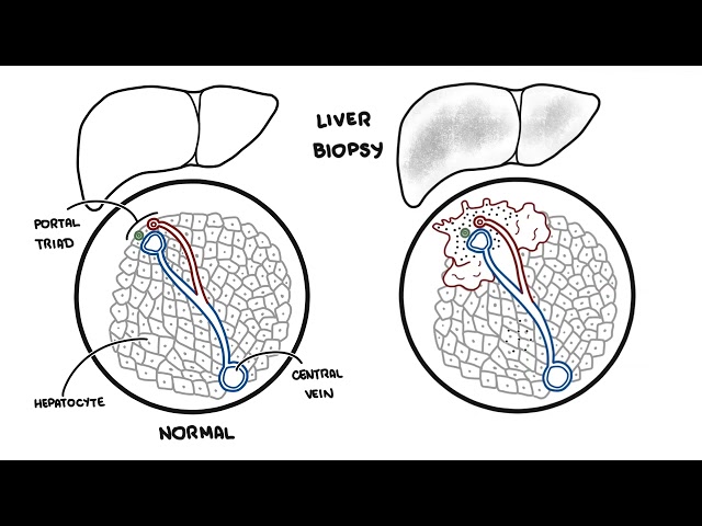
- Non-Dilated Bile Ducts: -> Intrahepatic Cholestasis / Parenchymal Disease (Primary Biliary Cholangitis, Primary Sclerosing Cholangitis, Drug-induced liver injury) -> Proceed to autoimmune panel, viral serologies, or liver biopsy.
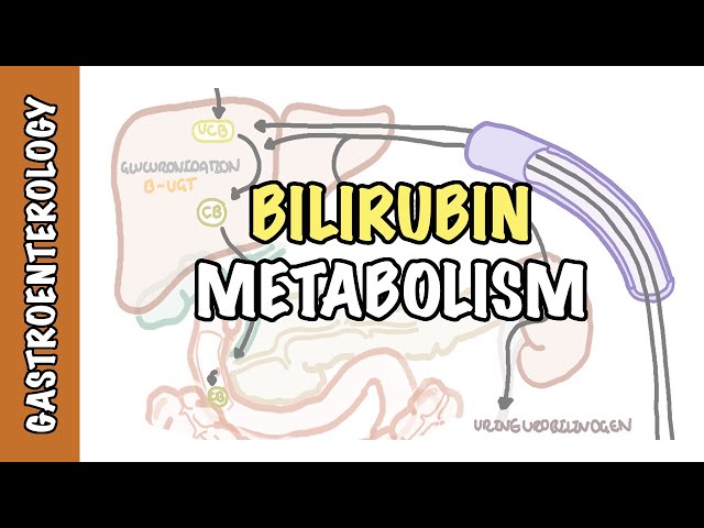
Unconjugated bilirubin is lipid-soluble and bound to albumin, so it cannot be excreted in urine. Dark urine (bilirubinuria) occurs ONLY in conjugated hyperbilirubinemia, serving as a rapid bedside clue for hepatic or post-hepatic disease.
Differential Diagnosis
Pre-Hepatic (Unconjugated Hyperbilirubinemia):
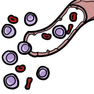
- Hemolytic Anemia (Intravascular or Extravascular):
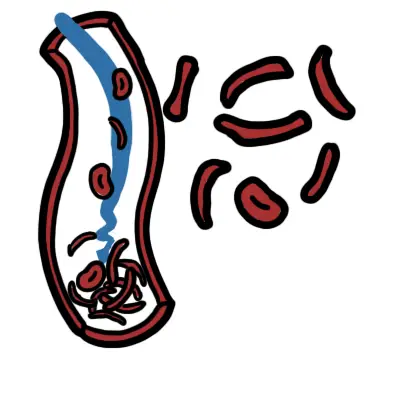
- Distinguishing features: Anemia, reticulocytosis, elevated LDH, decreased haptoglobin, bite/schistocyte cells on blood film; dark urine due to urobilinogen (not conjugated bilirubin).
- Gilbert’s Syndrome (Benign UGT1A1 Mutation):
- Distinguishing features: Mild asymptomatic unconjugated hyperbilirubinemia (typically < 50 umol/L) triggered by fasting, physical exertion, stress, or intercurrent illness; normal baseline LFTs, hemogram, and imaging.
Hepatic / Parenchymal (Mixed or Conjugated Hyperbilirubinemia):
- Acute Viral / Toxic / Ischemic Hepatitis:
- Distinguishing features: AST/ALT > 1000 U/L; history of paracetamol overdose, mushroom ingestion, viral exposures (Hep A, B, C, E), or prolonged shock/hypotension (“shock liver”).
- Cirrhosis / Alcohol-Related Liver Disease:
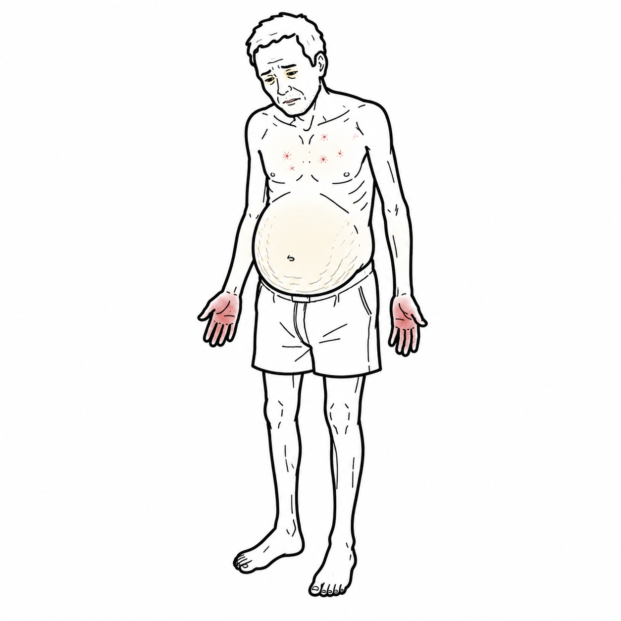
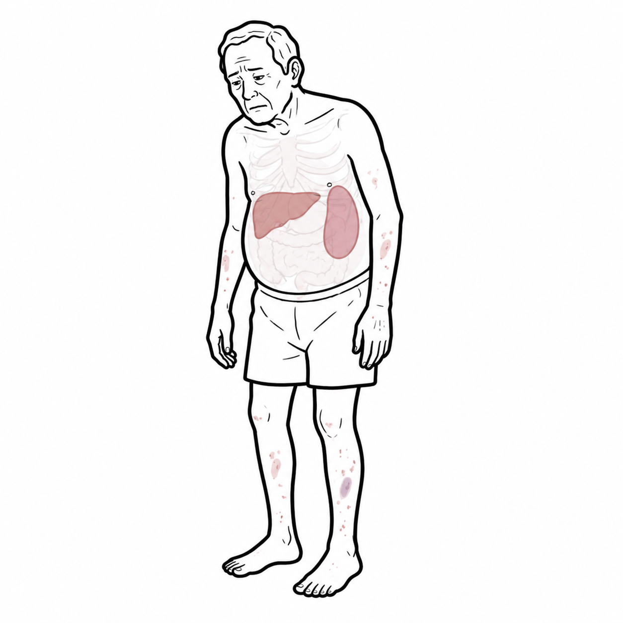
- Distinguishing features: Stigmata of chronic liver disease (spider naevi, palmar erythema, gynecomastia, caput medusae); AST:ALT ratio > 2:1 in alcohol-related disease.
- Primary Biliary Cholangitis (PBC) / Primary Sclerosing Cholangitis (PSC):
- Distinguishing features: PBC: Middle-aged females, severe pruritus, fatigue, positive Anti-Mitochondrial Antibodies (AMA). PSC: Strong association with Inflammatory Bowel Disease (Ulcerative Colitis); “beaded” appearance of intra/extrahepatic ducts on MRCP.
Post-Hepatic / Obstructive (Conjugated Hyperbilirubinemia):
- Choledocholithiasis (Gallstones in Common Bile Duct):
- Distinguishing features: Painful jaundice; colicky RUQ/epigastric pain, fluctuating jaundice, shadow-casting intraductal calculus on US or CT.
- Pancreatic Head Adenocarcinoma:
- Distinguishing features: Painless progressive jaundice, significant constitutional weight loss, Courvoisier’s sign (palpable non-tender gallbladder), double-duct sign on CT/MRCP.
- Cholangiocarcinoma (e.g., Klatskin Tumor at Biliary Confluence):
- Distinguishing features: Progressive obstructive jaundice, pruritus, elevated CA 19-9; intrahepatic duct dilation with normal/collapsed extrahepatic ducts.
Courvoisier’s Law states that in the presence of obstructive jaundice, a palpably enlarged, non-tender gallbladder is unlikely to be caused by gallstone disease (due to chronic fibrotic scarring of the gallbladder wall) and is more likely caused by a malignant obstruction (e.g., pancreatic head tumor).
Investigations
First-Line / Bedside & Laboratory Diagnostics:
- Total & Conjugated (Direct) Bilirubin: Confirms hyperbilirubinemia and determines whether conjugated or unconjugated.
- Liver Function Tests (LFTs): ALT, AST, ALP, GGT, Total Protein, Albumin.
- Coagulation Profile (INR / PT): Vital marker of hepatic synthetic function; elevated INR indicates significant parenchymal failure or Vitamin K malabsorption secondary to cholestasis.
- Full Blood Count & Hemolysis Panel: Reticulocyte count, LDH, Haptoglobin, Peripheral blood film, Direct Coombs test (if hemolysis suspected).
- Urinalysis: Check for bilirubin (dark tea-colored urine) and urobilinogen.
Targeted / Diagnostic Imaging Strategy:
- Transabdominal Ultrasound (First-Line Imaging Modality): Excellent initial test to evaluate for gallstones, gallbladder wall thickening, and to assess whether intra- or extra-hepatic bile ducts are dilated (CBD diameter > 6 mm is generally abnormal, or > 8 mm post-cholecystectomy).
- Contrast-Enhanced CT Abdomen/Pelvis: Evaluates pancreatic masses, focal hepatic lesions, abdominal lymphadenopathy, and defines the level of biliary obstruction.
- Magnetic Resonance Cholangiopancreatography (MRCP): Non-invasive gold standard diagnostic tool to visualize the entire biliary tree, strictures, choledocholithiasis, and anatomical variants without radiation or risk of pancreatitis.
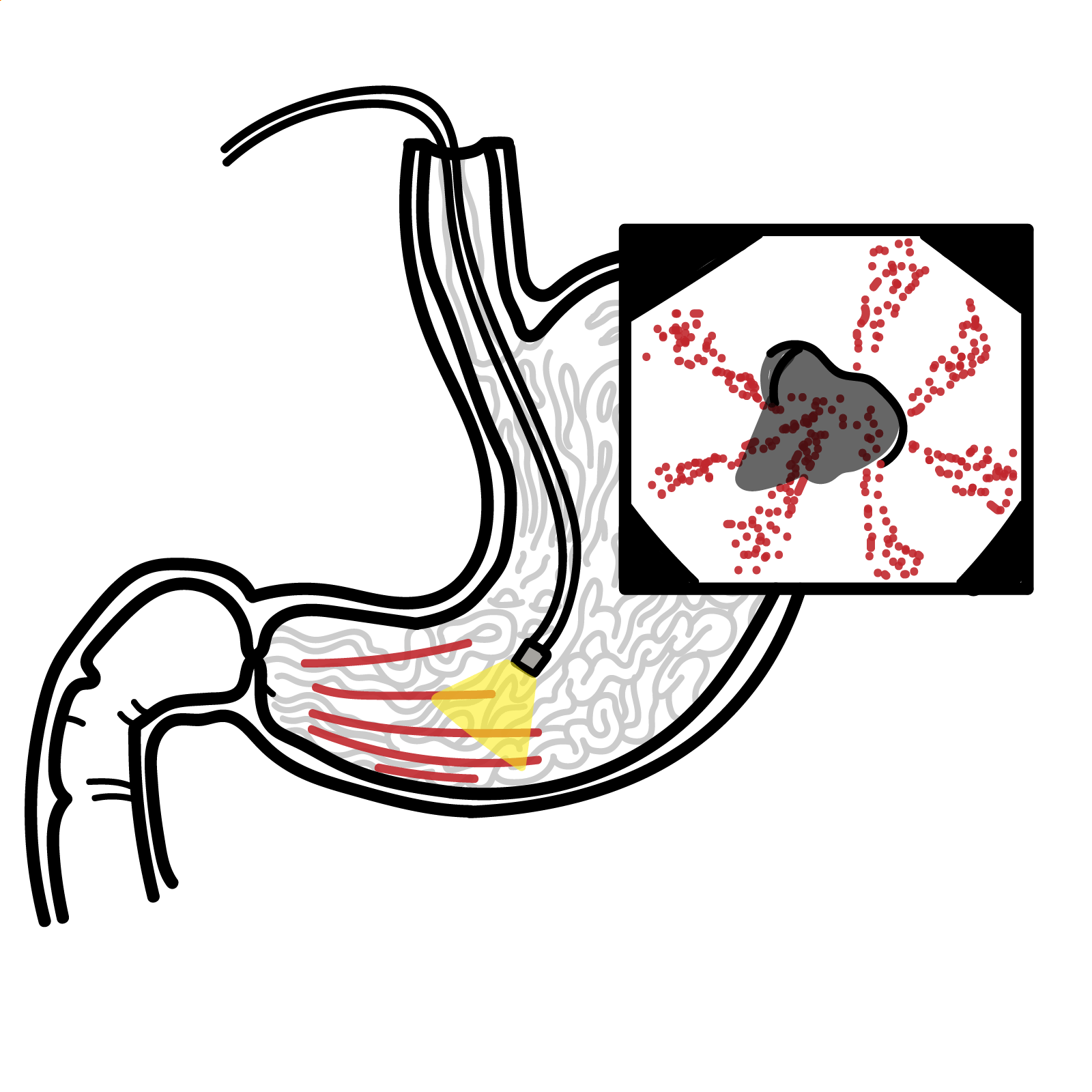
- Endoscopic Retrograde Cholangiopancreatography (ERCP): Invasive therapeutic modality reserved for relief of extrahepatic obstruction (stone extraction, sphincterotomy, or biliary stent placement for strictures/malignancy).
Transabdominal ultrasound is the mandatory first-line imaging test in any patient with conjugated hyperbilirubinemia to immediately separate extrahepatic surgical obstruction (dilated ducts) from intrahepatic medical jaundice (non-dilated ducts).
Critical Management
Charcot’s Triad / Acute Cholangitis (Emergency Intervention):
- Resuscitation: IV fluid resuscitation, keep NPO, blood cultures, and prompt administration of broad-spectrum IV antibiotics (e.g., Piperacillin/Tazobactam or Ceftriaxone + Metronidazole).
- Biliary Decompression: Urgent ERCP within 24 hours (or immediate if Reynold’s Pentad / septic shock is present) for biliary sphincterotomy and stenting/drainage.
Acute Liver Failure:
- Supportive & Specific Measures: Transfer to an ICU at a liver transplant center; administer N-acetylcysteine (NAC) early (beneficial in both paracetamol and non-paracetamol acute liver failure). Avoid sedatives.
Pruritus Management in Chronic Cholestasis:
- Medical Therapy: Cholestyramine (bile acid sequestrant; first-line), Rifampicin, Naltrexone, or Sertraline.
In acute cholangitis, antibiotic therapy alone is insufficient—definitive management requires prompt biliary decompression via ERCP to relieve the infected, obstructed system.
References
- Kwo PY, Cohen SM, Lim JK. ACG clinical guideline: evaluation of abnormal liver chemistries. Am J Gastroenterol. 2017;112(1):18-35. doi:10.1038/ajg.2016.517
- National Institute for Health and Care Excellence. Suspected cancer: recognition and referral (NG12) [Internet]. 2015 [updated 2026; cited 2026 Aug 20]. Available from: https://www.nice.org.uk/guidance/ng12

Members only discussions coming soon…