

Anaemia

Overview
Definition
Anaemia is defined as a reduction in circulating red blood cell (RBC) mass or total haemoglobin concentration below physiological reference limits, impairing oxygen delivery to peripheral tissues.
WHO / Australian Diagnostic Thresholds:
- Adult Males: Haemoglobin < 130 g/L
- Non-Pregnant Adult Females: Haemoglobin < 120 g/L
- Pregnant Females (1st/3rd trimester): Haemoglobin < 110 g/L (or < 105 g/L in 2nd trimester)
Morphological Classification (by Mean Corpuscular Volume [MCV]):
- Microcytic: MCV < 80 fL
- Normocytic: MCV 80–100 fL
- Macrocytic: MCV > 100 fL
Anaemia is a sign of an underlying disease process, never a complete diagnosis in itself. In adult males and post-menopausal females, Iron Deficiency Anaemia is gastrointestinal blood loss from malignancy (colorectal/gastric carcinoma) until proven otherwise.
Approach
Step 1: Emergency Triage & Hemodynamic Red Flags
- Active Major Hemorrhage: Massive GI bleed, trauma, ruptured ectopic -> Activate Massive Transfusion Protocol (MTP), maintain permissive hypotension (target SBP 80-90 mmHg), surgical/endoscopic source control.
- Symptomatic Tissue Hypoperfusion / Ischemia: Ischemic chest pain (Type 2 NSTEMI), syncope, severe resting dyspnea, acute heart failure, or altered mental status -> Urgent transfusion of packed red blood cells (PRBCs).
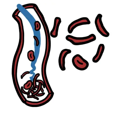
- Hemolytic Crisis / Thrombotic Microangiopathy (TMA): Thrombocytopenia + Microangiopathic Hemolytic Anaemia (MAHA) + schistocytes on blood film -> Suspect TTP / HUS (Emergency plasma exchange).
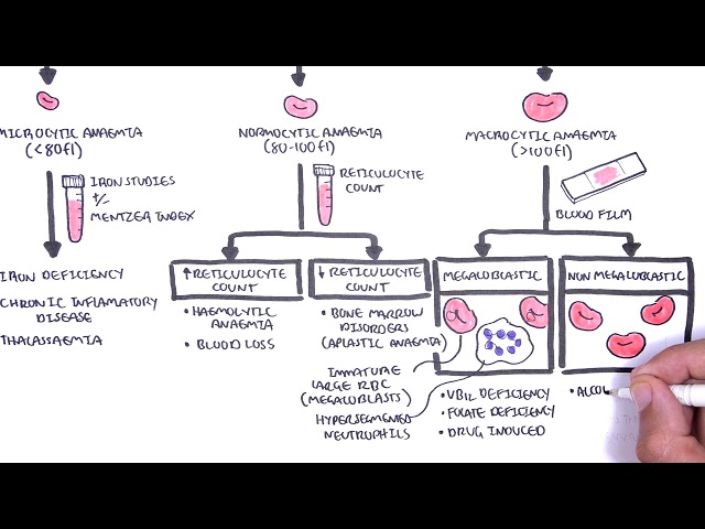
Step 2: Morphological Branching Logic (MCV-Guided)
- Microcytic (MCV < 80 fL):
- Check Serum Ferritin & Iron Studies -> Iron Deficiency Anaemia (Ferritin < 30 ug/L) vs. Anaemia of Chronic Disease (ACD) vs. Thalassaemia Minor (Mentzer Index < 13) vs. Sideroblastic Anaemia.
- Normocytic (MCV 80–100 fL):
- Check Reticulocyte Count:
- Elevated Reticulocytes (> 100 x 10^9/L or Reticulocyte Production Index > 2): Blood Loss or Active Hemolysis (Check LDH, Haptoglobin, Bilirubin, Coombs test).
- Low/Normal Reticulocytes: Chronic Kidney Disease (low EPO), Bone Marrow Failure (Aplastic Anaemia, Myelodysplasia), Early Iron/Mixed Deficiency, ACD.
- Check Reticulocyte Count:
- Macrocytic (MCV > 100 fL):
- Check Blood Film for Hypersegmented Neutrophils:
- Megaloblastic (Hypersegmented PMNs present): Vitamin B12 / Folate deficiency, Antimetabolite medications (Methotrexate, Hydroxyurea).
- Non-Megaloblastic (Round macrocytes): Alcohol excess, Liver disease, Hypothyroidism, Reticulocytosis, Myelodysplastic Syndrome (MDS).
- Check Blood Film for Hypersegmented Neutrophils:
Ferritin is an acute-phase reactant and can be falsely normal or elevated in infection, inflammation, or liver disease. In the presence of systemic inflammation (CRP > 5 mg/L), a ferritin < 100 ug/L still indicates co-existing iron deficiency.
Differential Diagnosis
Microcytic Anaemias (MCV < 80 fL – “TAILS”):
- Iron Deficiency Anaemia (IDA – Most Common):
- Distinguishing features: Low ferritin (< 30 ug/L), high Total Iron Binding Capacity (TIBC / Transferrin), low transferrin saturation (< 16-20%), high Red Cell Distribution Width (RDW); microcytic hypochromic film with pencil/cigar cells.
- Thalassaemia Trait / Minor (Alpha or Beta):
- Distinguishing features: Normal or high RBC count with profound microcytosis (MCV < 70 fL) disproportionate to mild anaemia; normal ferritin/iron studies; low RDW; Mentzer Index (MCV / RBC count) < 13; elevated HbA2 on Hb electrophoresis in Beta Thalassaemia.
- Anaemia of Chronic Disease / Inflammation (ACD):
- Distinguishing features: Hepcidin-mediated iron trapping; normal or elevated ferritin (> 100 ug/L), low serum iron, low/normal TIBC, low transferrin saturation.
Macrocytic Anaemias (MCV > 100 fL):
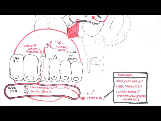
- Vitamin B12 Deficiency (Pernicious Anaemia / Malabsorption):
- Distinguishing features: Megaloblastic film, hypersegmented neutrophils (>= 5 lobes), elevated methylmalonic acid (MMA) and homocysteine; neurological symptoms (subacute combined degeneration of the spinal cord: loss of vibration/proprioception, ataxia).
- Folate Deficiency:
- Distinguishing features: Dietary deficit/alcoholism/hemolysis; hypersegmented neutrophils; normal MMA with elevated homocysteine; no neurological deficits.
- Non-Megaloblastic Causes:
- Distinguishing features: Alcohol abuse (MCV 100-110 without severe anaemia), Myelodysplastic Syndrome (cytopenias + dysplastic cells), Hypothyroidism.
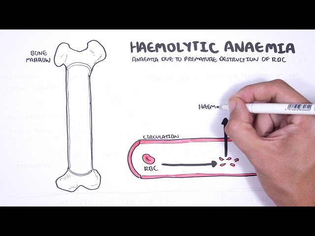
Hemolytic Anaemias (Normocytic / Macrocytic + High Reticulocytes):
- Autoimmune Hemolytic Anaemia (AIHA – Warm vs Cold):
- Distinguishing features: Spherocytes on film, positive Direct Antiglobulin Test (DAT / Coombs test), elevated LDH, undetectable haptoglobin, indirect hyperbilirubinemia.
- Microangiopathic Hemolytic Anaemia (TTP / HUS / DIC):
- Distinguishing features: Severe thrombocytopenia, prominent schistocytes / helmet cells on blood film, elevated LDH, normal coagulation in TTP/HUS vs abnormal coagulopathy in DIC.
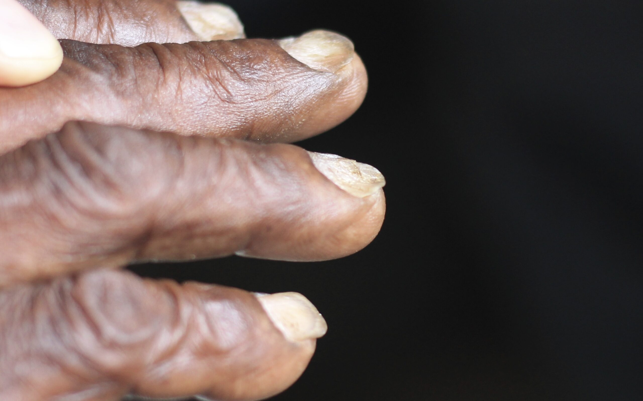
Koilonychia: spoon-shaped nails associated with iron deficiency. Image: CHeitz, Wikimedia Commons, CC BY 2.0.
What to notice
Detailed image description
Source: https://commons.wikimedia.org/wiki/File:Koilonychia_iron_deficiency_anemia.jpg — Creative Commons Attribution 2.0.
The Mentzer Index (MCV / RBC count in 10¹²/L) rapidly differentiates iron deficiency anaemia from thalassaemia trait: a value <13 strongly suggests Thalassaemia Trait, whereas a value >13 suggests Iron Deficiency Anaemia.
Investigations
First-Line / Basic Diagnostic Laboratory Panel:
- Full Blood Count (FBC) & Red Cell Indices: Hb, MCV, MCH, RDW, Platelets, White Cell Count.
- Peripheral Blood Film (MANDATORY): Identifies diagnostic morphologies:
- Pencil cells / Microcytes: Iron deficiency.
- Target cells / Basophilic stippling: Thalassaemia / Hemoglobinopathies.
- Hypersegmented neutrophils / Oval macrocytes: Megaloblastic anaemia (B12/Folate).
- Schistocytes / Helmet cells: MAHA (TTP/HUS/DIC).
- Spherocytes: AIHA or Hereditary Spherocytosis.
- Bite cells / Heinz bodies: G6PD deficiency.
- Reticulocyte Count & Reticulocyte Production Index (RPI): Assesses bone marrow regenerative response.
- Iron Studies: Serum Iron, Transferrin/TIBC, Transferrin Saturation (Tsat), Serum Ferritin.
Targeted / Specialized Diagnostic Strategy:
- GI Investigation (Mandatory in Unexplained IDA): Gastroscopy + Colonoscopy (rule out bleeding malignancy, celiac disease via duodenal biopsy).
- Hemolysis Workup: Direct Antiglobulin Test (DAT / Coombs), Serum Haptoglobin (undetectable in intravascular hemolysis), LDH, Unconjugated Bilirubin, Urine hemosiderin.
- Hemoglobinopathy Screen: Hemoglobin Electrophoresis / HPLC (detects HbA2, HbF, HbS).
- Active B12 (Holotranscobalamin) & Serum / RBC Folate: Secondary testing with Serum Methylmalonic Acid (MMA) and Homocysteine if B12 is borderline (150–250 pmol/L).
- Bone Marrow Aspirate & Trephine Biopsy: Indicated for unexplained cytopenias, suspected Aplastic Anaemia, Myelodysplastic Syndrome, or acute leukemia.
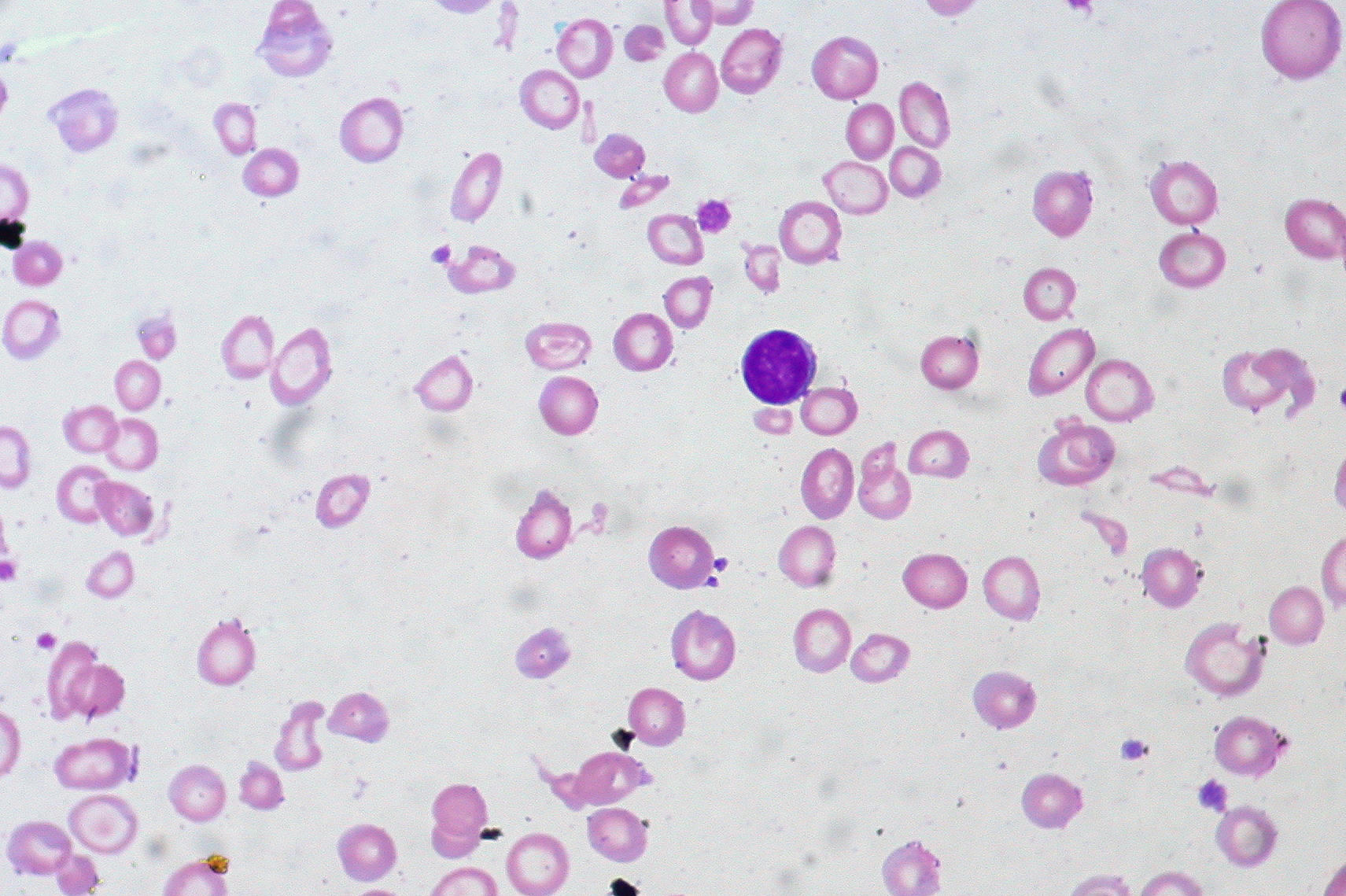
Peripheral blood film showing severe microcytic, hypochromic iron-deficiency anaemia. Image: Ed Uthman, Wikimedia Commons, CC BY 2.0.
What to notice
Detailed image description
Source: https://commons.wikimedia.org/wiki/File:Iron-deficiency_Anemia,_Peripheral_Blood_Smear_(4422704616).jpg — Creative Commons Attribution 2.0.
In severe hereditary hemolytic conditions like Thalassaemia Major, marked extramedullary hematopoiesis and bone marrow expansion lead to classical skeletal changes such as the “hair-on-end” appearance on skull X-rays and facial bone prominence.
Critical Management
Acute Transfusion Guidelines (Restrictive Transfusion Strategy):
- Hemodynamically Stable Inpatients: Transfusion threshold is Hb < 70 g/L (Target Hb 70–90 g/L).
- Acute Coronary Syndrome (ACS) / Active Myocardial Ischemia: Transfusion threshold is Hb < 80 g/L (Target Hb 80–100 g/L).
- Active Massive Hemorrhage: Transfuse based on hemodynamic response and blood loss rather than static lab values; activate MTP.
Specific Etiology-Driven Interventions:
- Iron Deficiency Anaemia:
- Oral Iron: Ferrous sulfate/fumarate (100–200 mg elemental iron every second day optimizes absorption and reduces GI intolerance).
- IV Iron (Ferric Carboxymaltose / Ferric Derisomaltose): Indicated for severe anaemia (Hb < 80 g/L), 2nd/3rd trimester pregnancy, IBD, chronic kidney disease, or oral intolerance/malabsorption.
- Vitamin B12 Deficiency with Neurological Symptoms:
- Hydroxocobalamin 1000 ug IM on alternate days until neurological symptoms stabilize, then maintenance every 2–3 months. (Always replace B12 before or concurrently with Folate to prevent precipitating subacute combined degeneration).
- Thrombotic Thrombocytopenic Purpura (TTP):
- Emergency Plasma Exchange (PLEX) + High-dose IV Methylprednisolone + Caplacizumab. Do NOT transfuse platelets (precipitates microvascular thrombosis).
Never administer oral or IV folic acid alone in severe megaloblastic anaemia without first confirming normal Vitamin B12 levels. Folate corrects the hematological indices while masking the progression of irreversible neurological damage from Vitamin B12 deficiency.
References
- World Health Organization. Guideline on haemoglobin cutoffs to define anaemia in individuals and populations [Internet]. Geneva: World Health Organization; 2024 [cited 2026 Aug 14]. Available from: https://www.who.int/publications/i/item/9789240088542
- Gerber GF. Red blood cell production [Internet]. Rahway (NJ): Merck & Co., Inc.; 2024 [cited 2026 Aug 14]. Available from: https://www.merckmanuals.com/professional/hematology-and-oncology/approach-to-the-patient-with-anemia/red-blood-cell-production
- ARUP Laboratories. Anemia: choose the right test [Internet]. Salt Lake City (UT): ARUP Laboratories; [cited 2026 Aug 14]. Available from: https://arupconsult.com/content/anemia
- Australian Red Cross Lifeblood. Diagnosis and investigation of iron deficiency anaemia [Internet]. Melbourne (AU): Australian Red Cross Lifeblood; [cited 2026 Aug 14]. Available from: https://www.lifeblood.com.au/health-professionals/clinical-practice/clinical-indications/iron-deficiency-anaemia/diagnosis-investigation
- Snook J, Bhala N, Beales ILP, Cannings D, Kightley C, Logan RPH, et al. British Society of Gastroenterology guidelines for the management of iron deficiency anaemia in adults. Gut. 2021;70(11):2030–2051. doi:10.1136/gutjnl-2021-325210
- National Institute for Health and Care Excellence. Vitamin B12 deficiency in over 16s: diagnosis and management [Internet]. London: NICE; 2024 [cited 2026 Aug 14]. Available from: https://www.nice.org.uk/guidance/ng239
- Carson JL, Stanworth SJ, Guyatt G, Valentine S, Dennis J, Bakhtary S, et al. Red blood cell transfusion: 2023 AABB international guidelines. JAMA. 2023;330(19):1892–1902. doi:10.1001/jama.2023.12914



Members only discussions coming soon…