

Bradycardia
Overview
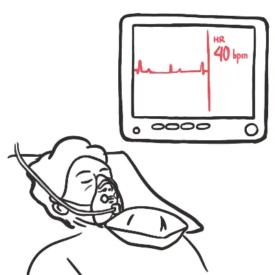
Bradycardia is defined as a heart rate (HR) < 60 beats per minute (bpm). Clinical significance depends on physiological context and the presence of symptomatic tissue hypoperfusion (typically clinically significant when HR < 50 bpm).
Core Pathophysiology: Caused by either impaired impulse generation at the Sinoatrial (SA) node (sinus node dysfunction) or impaired impulse conduction through the Atrioventricular (AV) node / His-Purkinje system (AV block).
A heart rate < 60 bpm in a trained athlete or during sleep can be physiological. Focus treatment on whether the patient is symptomatic with signs of inadequate end-organ perfusion (hypotension, altered mental status, chest pain, acute heart failure).
Approach
Step 1: Primary Assessment & Hemodynamic Instability Screening
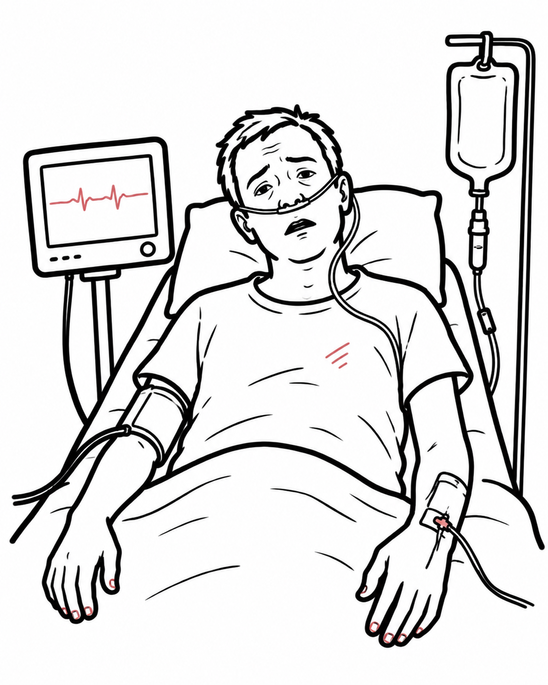
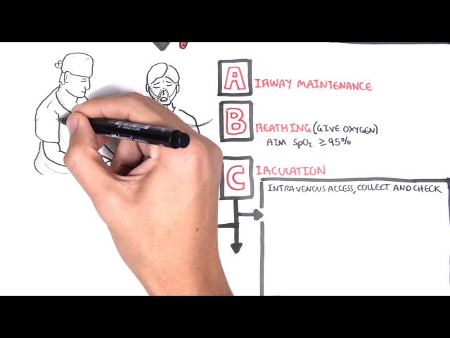
- Confirm HR < 50-60 bpm; attach cardiac monitor, pulse oximeter, and BP cuff. Secure IV access and 12-lead ECG.
- Screen for Unstable Features (Signs of Hypoperfusion):
- Hypotension / Shock (SBP < 90 mmHg).
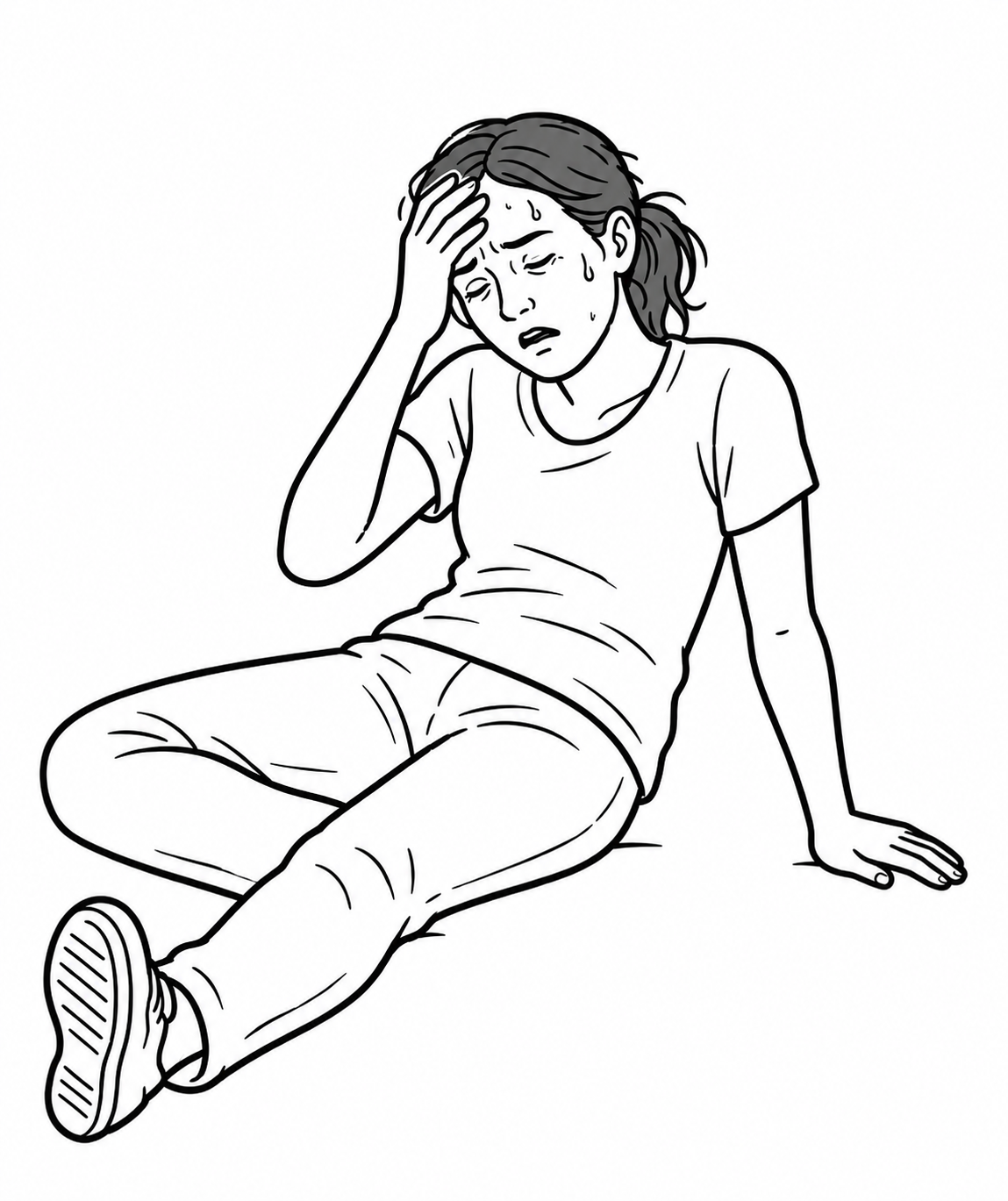
- Altered mental status (acute confusion, lethargy, syncope).
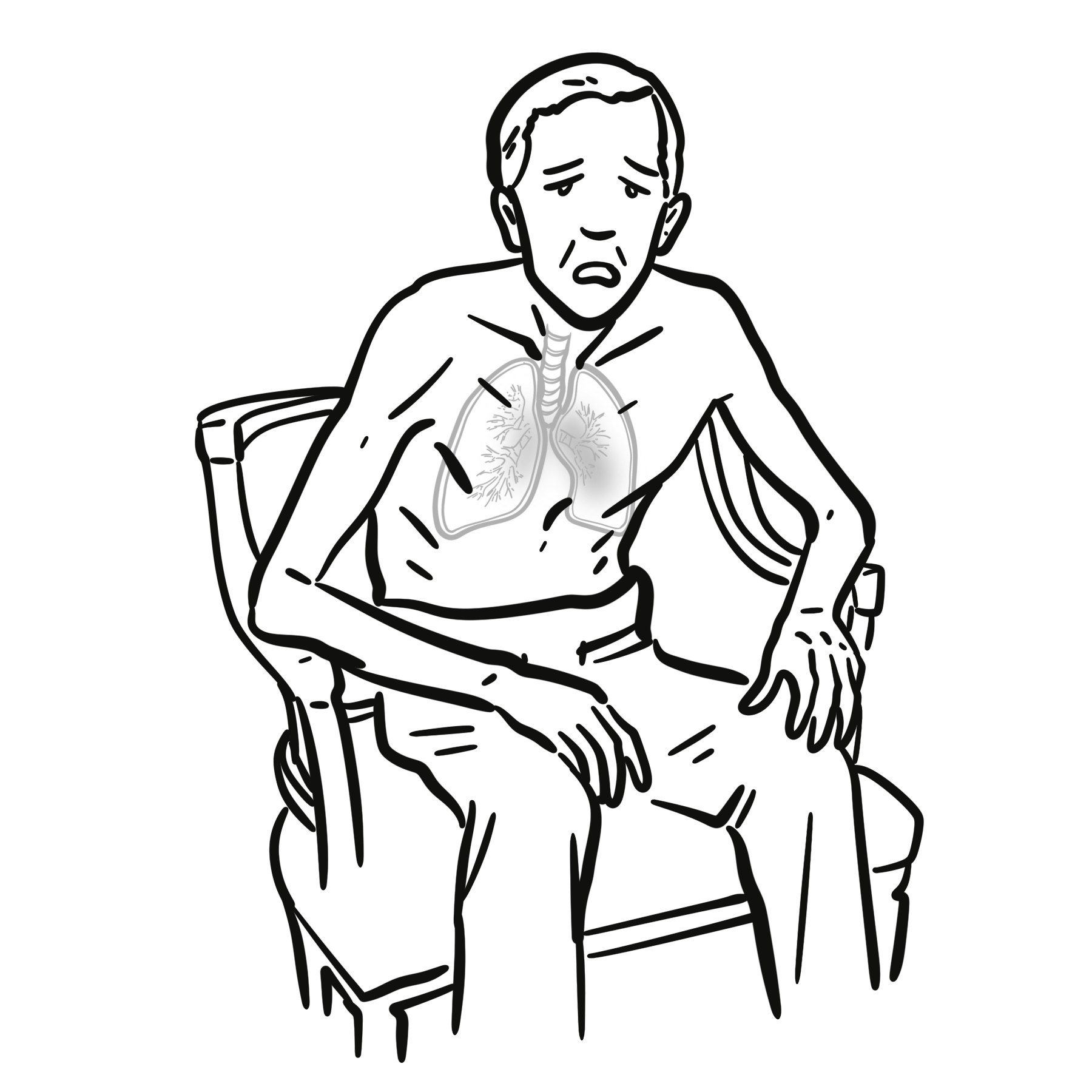
- Signs of acute heart failure (pulmonary edema, elevated JVP).
- Chest discomfort (ischemic pain).
Step 2: Branching Logic (Symptomatic vs. Asymptomatic)
- UNSTABLE (Presence of ANY unstable feature):
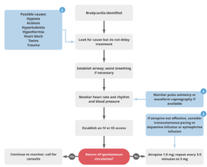
- First-line: Atropine 1 mg IV bolus.
- If refractory to Atropine: Transcutaneous Pacing (TCP) OR Inotrope/Chronotrope Infusion (Epinephrine or Dopamine) OR Transvenous Pacing.
- STABLE (No unstable features present):
- Obtain 12-lead ECG to determine rhythm (Sinus bradycardia vs. Mobitz I vs. Mobitz II / 3rd-degree block).
- Monitor, identify underlying cause, hold AV-nodal blocking drugs, and obtain cardiology consult.
Atropine works by blocking vagal tone at the AV node. It is generally INEFFECTIVE in Mobitz Type II or 3rd-Degree AV block with a wide QRS escape rhythm (infranodal block). Do not waste time with repeat Atropine doses in high-degree infranodal blocks—move straight to transcutaneous pacing or epinephrine.
Differential Diagnosis
Intrinsic Cardiac Causes (Structural / Conduction Disease):
- Sinus Node Dysfunction (Sick Sinus Syndrome): Tachy-brady syndrome, sinus arrest/exit block.
- Acute Myocardial Infarction:
- Degenerative Conduction Disease: Lev’s disease (calcification of cardiac skeleton), Lenègre’s disease.
Extrinsic / Systemic Causes (Potentially Reversible):
- Drug-Induced (Most Common Overall): Beta-blockers, Non-dihydropyridine Calcium Channel Blockers (Verapamil, Diltiazem), Digoxin, Antiarrhythmics (Amiodarone), Cholinesterase inhibitors, Clonidine.
- Electrolyte Imbalance: Hyperkalemia (causes “peaked T waves”, loss of P waves, and severe bradycardia/sine wave), Severe Hypokalemia, Hypermagnesemia.
- Metabolic / Endocrine: Severe Hypothermia (“Osborn / J waves” on ECG), Hypothyroidism / Myxedema coma.
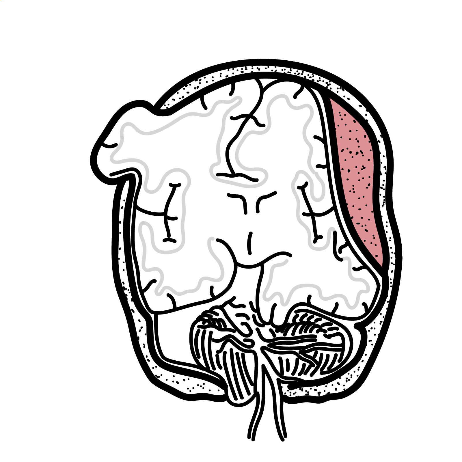
- Increased Intracranial Pressure (Cushing’s Triad): Bradycardia + Severe Hypertension + Irregular Respirations.
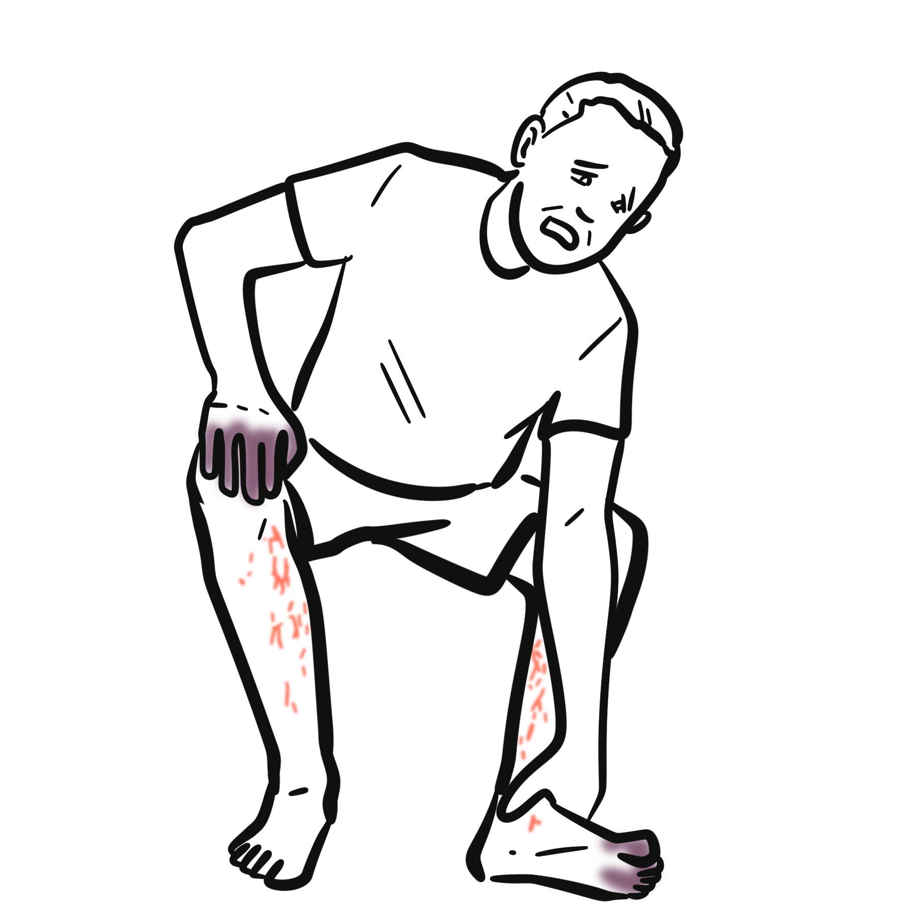
- Infectious / Infiltrative: Lyme disease (Lyme carditis with AV block), Amyloidosis, Sarcoidosis, Myocarditis, Endocarditis (perivalvular abscess).
Hyperkalemia is the great mimicker on ECG. Any slow, wide-complex rhythm without clear P waves should be treated as life-threatening Hyperkalemia until proven otherwise—administer IV Calcium Gluconate/Chloride immediately for cardiac membrane stabilization.
Investigations
First-Line / Bedside Diagnostics:
- 12-Lead ECG: Essential to define the specific rhythm (Sinus Bradycardia, Junctional Escape, 1st Degree, Mobitz I / Wenckebach, Mobitz II, or 3rd-Degree Complete Heart Block).
- Continuous Cardiac Telemetry: Monitor for intermittent pauses or high-grade conduction failure.
- Point-of-Care Glucose & Electrolytes (Serum Potassium): Rapid rule-out of severe hyperkalemia.
- Troponin: Evaluate for acute coronary syndrome.
Targeted / Secondary Workup:
- Serum Drug Levels: Digoxin level, acetaminophen/salicylate screens (if toxic ingestion suspected).
- Thyroid Function Tests (TSH, Free T4): Rule out hypothyroidism.
- Echocardiogram: Evaluate structural heart disease, ejection fraction, wall motion abnormalities, or valvular pathology.
- Lyme Serology / Inflammatory Markers (ESR/CRP): Indicated in young patients with unexplained high-degree AV block.
Differentiate Mobitz I (Wenckebach) from Mobitz II on ECG: Mobitz I shows progressive PR lengthening until a drop occurs (benign, AV nodal). Mobitz II shows constant PR intervals with unpredictable dropped QRS complexes (high risk of progression to complete heart block; requires pacemaker).
Critical Management
Unstable Symptomatic Bradycardia (ACLS Protocol):
- Atropine:1 mg IV bolus every 3-5 minutes (Maximum cumulative dose: 3 mg).
- Dosing note: Doses < 0.5 mg can cause paradoxical reflex bradycardia.
- Transcutaneous Pacing (TCP): Initiate immediately if Atropine fails or if high-degree block (Mobitz II / 3rd degree) is present. Set demand rate to 60-80 bpm; increase current (mA) until electrical AND mechanical capture (palpable pulse) is confirmed.
- Inotrope Infusions (Alternative to Pacing):
- Epinephrine Infusion: 2-10 mcg/min IV infusion.
- Dopamine Infusion: 5-20 mcg/kg/min IV infusion.
Specific Antidotes / Reversal Agents:
- Beta-Blocker Toxicity: IV Glucagon (3-10 mg slow IV push) + High-Dose Insulin Euglycemic Therapy (HIET).
- Calcium Channel Blocker Toxicity: IV Calcium Chloride/Gluconate + High-Dose Insulin Euglycemic Therapy (HIET).
- Digoxin Toxicity: Digoxin-specific antibody fragments (DigiFab).
Always confirm mechanical capture during transcutaneous pacing by checking a femoral or radial pulse. Do not rely solely on the electrical spikes on the monitor screen.
References
- Heart Rhythm Society. 2018 ACC/AHA/HRS guideline on the evaluation and management of patients with bradycardia and cardiac conduction delay [Internet]. 2018 [cited 2026 Aug 20]. Available from: https://www.hrsonline.org/resource/2018-accahahrs-guideline-evaluation-and-management-patients-bradycardia-and-cardiac-conduction/
- European Society of Cardiology. 2021 ESC guidelines on cardiac pacing and cardiac resynchronization therapy [Internet]. 2021 [cited 2026 Aug 20]. Available from: https://www.escardio.org/guidelines/clinical-practice-guidelines/all-esc-practice-guidelines/cardiac-pacing-and-cardiac-resynchronization-therapy/


Members only discussions coming soon…