

Peripheral Neuropathy
Approach
Step 1: Identify Acute / Subacute “Red Flags” (Immediate Triage)
- Rapidly Progressive / Ascending Weakness: Suspect Guillain-Barré Syndrome (GBS) / Acute Inflammatory Demyelinating Polyneuropathy (AIDP) $\rightarrow$ Assess forced vital capacity (FVC) / negative inspiratory force (NIF) and autonomic stability immediately.
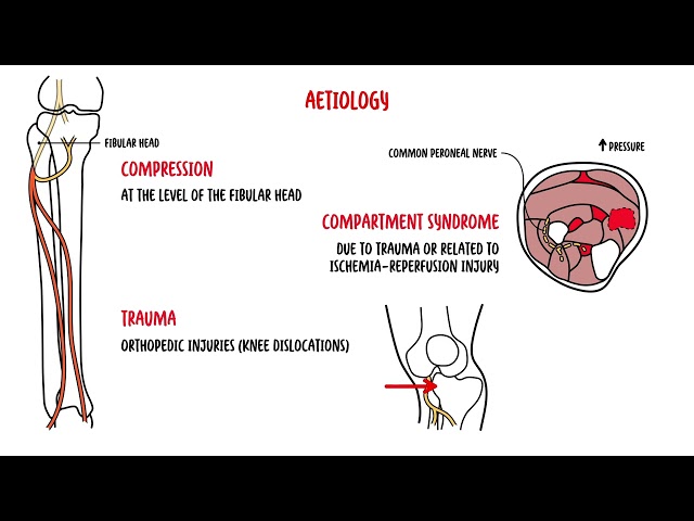
- Asymmetric Motor-Dominant Deficits / Multfocal Drops (e.g., wrist/foot drop): Suspect Mononeuritis Multiplex (Vasculitis) (High-risk for rapid progression), systemic tissue ischemia.
- Autonomic Collapse: Severe orthostasis, neurogenic bladder, cardiac dysrhythmias (e.g., Amyloidosis, Diabetic Autonomic Neuropathy).
Step 2: Define Phenotype & Pattern
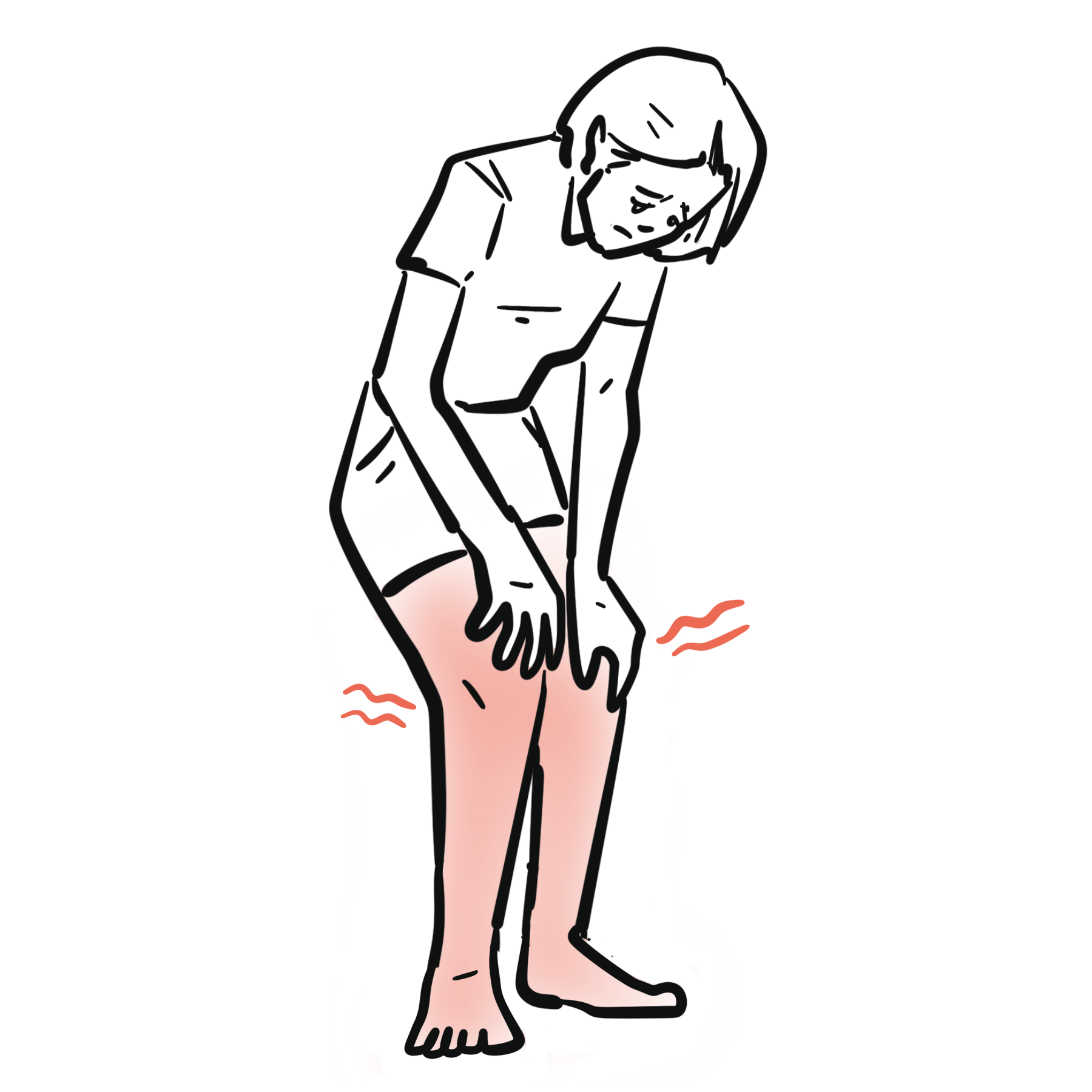
- Symmetrical Distal Length-Dependent (“Stocking-Glove”): Classic sensory-predominant metabolic/toxic presentation.
- Asymmetrical / Multifocal: Vasculitis, radiculopathy, mononeuropathy multiplex, compressive syndromes.
- Pure Motor vs. Pure Sensory vs. Mixed / Autonomic: Motor-predominant suggests GBS, CIDP, lead, porphyria, or ALS; pure sensory suggests ganglionopathy (Sjögren’s, paraneoplastic, B6 toxicity).
Ascending weakness with reduced/absent reflexes is GBS until proven otherwise. Check respiratory mechanics (FVC/NIF) early—do not wait for arterial blood gas changes or hypoxia.
Differential Diagnosis
Distal Symmetrical Polyneuropathy (DSPN) – Most Common (>80%):
- Diabetic / Metabolic
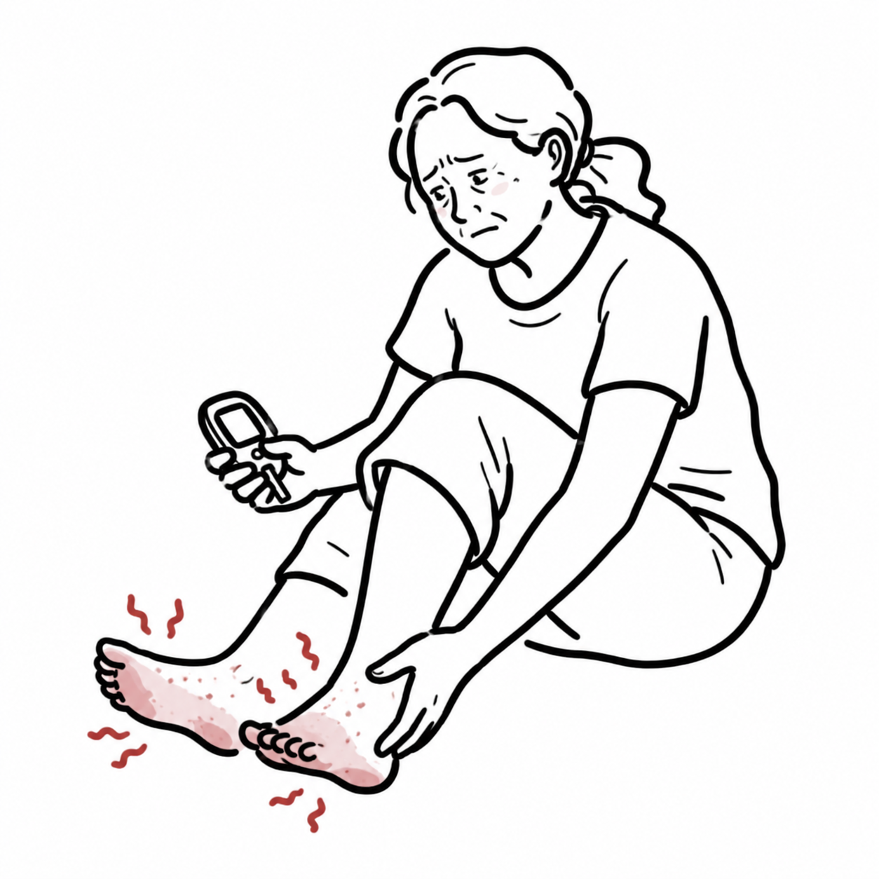
- Distinguishing features: Pain, burning feet, insidious onset, loss of vibration/monofilament sensation.
- Toxic / Medication-Induced: Alcohol use disorder, chemotherapy (platinum agents, paclitaxel, vincristine), isoniazid, metronidazole, vitamin B6 toxicity.
- Distinguishing features: Temporal relationship with drug onset/dose escalation; sensory ataxia.
- Nutritional / Deficiency: Vitamin B12 deficiency, copper deficiency, thiamine (B1), folate.
- Distinguishing features: Subacute combined degeneration (B12) with concurrent posterior column signs (loss of proprioception) and upper motor neuron features (hyperreflexia/Babinski).
Immune-Mediated / Inflammatory:
- GBS / AIDP: Acute (<4 weeks), ascending symmetric weakness, hyporeflexia/areflexia, facial diplegia.
- CIDP (Chronic Inflammatory Demyelinating Polyneuropathy): Progressive or relapsing symmetric proximal AND distal weakness/sensory loss >8 weeks; diminished reflexes.
- Vasculitic Neuropathy (Mononeuritis Multiplex): Acute/subacute pain, asymmetric multifocal mononeuropathies (e.g., right radial nerve + left peroneal nerve), systemic symptoms (fever, weight loss, rash).
Monoclonal Gammopathies & Paraneoplastic:
- MGUS / Multiple Myeloma / AL Amyloidosis / POEMS:
- Distinguishing features: Painful sensory-motor neuropathy, autonomic involvement (amyloidosis), organomegaly, endocrinopathy, skin changes (POEMS).
Normal HbA1c does NOT rule out diabetic/metabolic neuropathy. Impaired Glucose Tolerance (2-hour Oral Glucose Tolerance Test) is the single most common cause of idiopathic small-fiber neuropathy.
Investigations
First-Line / Initial Diagnostic Workup (All Patients with DSPN):
- Fasting Blood Glucose & HbA1c: (or 2-hour Oral Glucose Tolerance Test if HbA1c is normal).
- Serum Vitamin B12 & Methylmalonic Acid (MMA): MMA is essential if B12 is borderline (150–300 pg/mL) to confirm functional deficiency.
- Serum Protein Electrophoresis (SPEP) + Immunofixation (IFE) + Serum Free Light Chains: Screen for monoclonal gammopathy/paraneoplastic processes.
- Serum Creatinine / eGFR & LFTs: Rule out uremic neuropathy and hepatic disease.
- TSH: Screen for hypothyroidism.
Targeted / Diagnostic Workup (Guided by Phenotype):
- Nerve Conduction Studies (NCS) & Electromyography (EMG): Essential to differentiate Axonal vs. Demyelinating patterns and localize focal vs. diffuse disease.
- Lumbar Puncture (CSF Analysis): Look for albuminocytological dissociation (elevated protein with normal WBC count) in suspected GBS or CIDP.
- Autoimmune / Vasculitis Panel: ANA, ANCA, ENA (SSA/SSB for Sjögren’s), ESR/CRP, Cryoglobulins, HIV, Hepatitis B/C.
- Skin Punch Biopsy: Assesses intraepidermal nerve fiber density (IENFD) for pure small-fiber neuropathy when EMG/NCS is normal.
NCS/EMG evaluates LARGE myelinated fibers only. If a patient presents with burning foot pain, intact reflexes, and normal vibration/proprioception, NCS will be normal—suspect Small-Fiber Neuropathy (confirm with skin biopsy or thermal testing).
Critical Management
Acute Inflammatory Demyelinating Polyneuropathy (GBS):
- Airway / Respiration: Transfer to ICU/HDU if FVC drops <20 mL/kg or NIF < -30 cmH2O.
- Targeted Therapy:IVIG (0.4 g/kg/day for 5 days) OR Plasma Exchange (PLEX). Both are equally effective.
- Contraindication: Do NOT give systemic corticosteroids in GBS (ineffective and slows recovery).
Vasculitic Neuropathy:
- Immediate Immunosuppression: High-dose IV pulse methylprednisolone followed by oral steroids + Cyclophosphamide or Rituximab to halt ischemic nerve infarction.
Steroids are contraindicated in acute GBS, but are a main stay of therapy for CIDP and Vasculitic Neuropathy. Never delay IVIG/PLEX in suspected GBS while waiting for CSF results.
References
- England JD, Gronseth GS, Franklin G, Carter GT, Kinsella LJ, Cohen JA, et al. Practice parameter: evaluation of distal symmetric polyneuropathy—role of laboratory and genetic testing. Neurology. 2009;72(2):185-192. doi:10.1212/01.wnl.0000336370.51010.a1
- National Institute for Health and Care Excellence. Suspected neurological conditions: recognition and referral (NG127) [Internet]. 2019 [updated 2023; cited 2026 Aug 20]. Available from: https://www.nice.org.uk/guidance/ng127







Members only discussions coming soon…