

Altered level of Conciousness
Overview
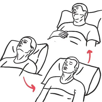
Altered Level of Consciousness (ALOC) refers to an acute or subacute impairment in arousal (wakefulness, mediated by the ascending reticular activating system [ARAS]) and/or awareness (cognition, mediated by the cerebral cortex). It spans a spectrum from confusion/delirium to lethargy, obtundation, stupor, and coma.
Core Pathophysiology: ALOC results from either:
- Diffuse bihemispheric cortical dysfunction (metabolic, toxic, infectious, or hypoxic states).
- Focal brainstem lesion directly disrupting the ARAS.
Hypoglycemia and hypoxia are the two most common, immediately reversible, life-threatening causes of ALOC. Always perform a point-of-care fingerstick blood glucose test and check oxygen saturation before initiating lengthy diagnostic workups.
APPROACH
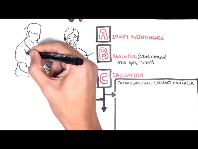
Step 1: Primary Survey & Immediate Resuscitation (ABCDE)
- Airway: Assess patency and protective reflexes. Glasgow Coma Scale (GCS) <= 8 -> Prepare for endotracheal intubation.
- Breathing: Oxygenation/ventilation check (SpO2, ABG/VBG for pCO2 retention).
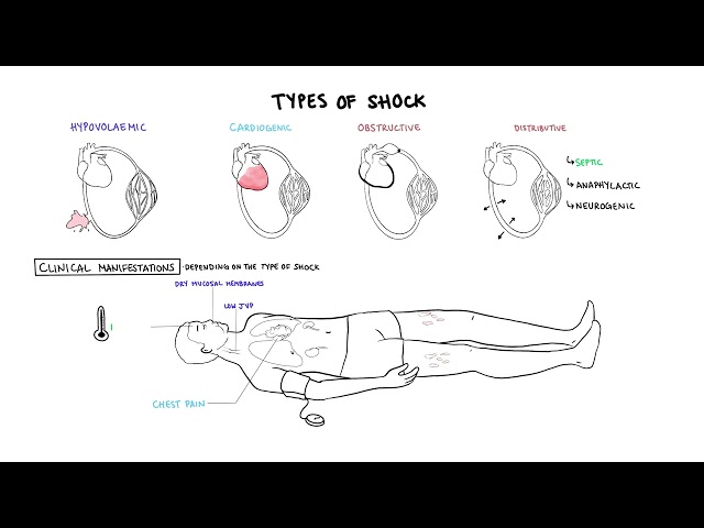
- Circulation: IV access, monitor BP and HR (hypotension/shock vs. Cushing’s triad [bradycardia, severe hypertension, irregular respirations] indicating elevated intracranial pressure).
- Disability: GCS score, pupil size/reactivity, bedside glucose check (DON’T EVER FORGET GLUCOSE – “DEFG”).
- Exposure: Temperature (hypothermia vs. hyperthermia/sepsis), toxidrome clues, trauma marks.
Step 2: Rapid Empiric Reversals (If Indicated)
- Hypoglycemia: 50 mL D50W IV (or 100 mL D10W).
- Opioid Overdose (Pinpoint pupils + respiratory depression): Naloxone 0.4–2 mg IV/IM/IN.
- Wernicke Encephalopathy / Alcohol Use: Thiamine 100–500 mg IV (give before or alongside glucose).
Step 3: Clinical Decision Branching (AEIOU TIPS Mnemonic)
- Focal Neurological Deficits / Asymmetry: -> Immediate Non-Contrast Head CT +/- CTA (Rule out ischemic stroke, ICH, mass effect, herniation).
- Fever / Meningismus / Rash: -> Lumbar Puncture (after CT if focal signs/papilledema present) + Empiric IV Antibiotics/Acyclovir.
- Diffuse Symmetrical Non-Focal Exam: -> Metabolic/Toxicology/Sepsis workup.
“GCS 8, Intubate!” Loss of airway protection reflexes increases the risk of aspiration pneumonitis and acute hypercapnic respiratory failure.
DIFFERENTIAL DIAGNOSIS
Use the “AEIOU TIPS” Mnemonic for Etiologies:
- A – Alcohol / Acidosis: Acute intoxication, withdrawal, DKA, AKA, uremic acidosis.
- E – Epilepsy / Encephalopathy / Endocrine: Post-ictal state, non-convulsive status epilepticus (NCSE), hepatic encephalopathy, thyroid storm / myxedema coma, adrenal crisis.
- I – Infection: Meningitis, encephalitis, sepsis, UTI/pneumonia in elderly.
- O – Oxygenation / Opiates: Hypoxia, hypercapnia (CO2 narcosis), opioid/sedative toxicity.
- U – Uremia: Severe acute kidney injury or end-stage renal disease.
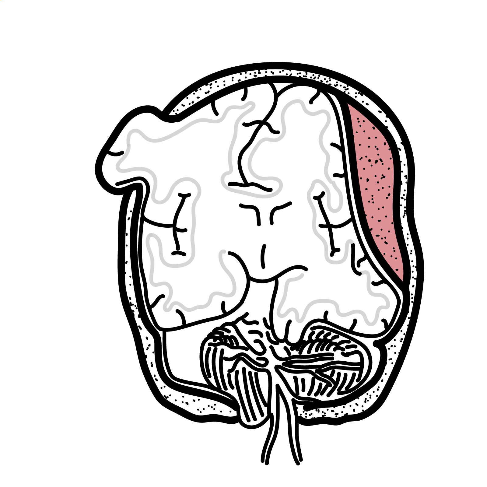
- T – Trauma / Temperature / Tumor: TBI (epidural/subdural hematoma, contusion), heat stroke, hypothermia, intracranial mass.
- I – Insulin (Hypoglycemia): Absolute emergency; mimics stroke/focal deficits.
- P – Poisoning / Psychogenic: Toxidromes (anticholinergics, sympathomimetics, TCAs), catatonia, conversion disorder.
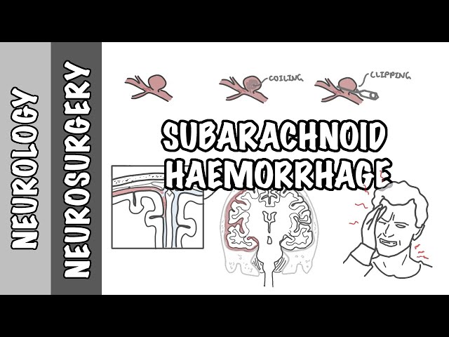
- S – Stroke / Space-Occupying Lesion: Ischemic stroke (especially basilar artery occlusion), ICH, SAH, venous sinus thrombosis.
Non-Convulsive Status Epilepticus (NCSE) is a frequently missed cause of unexplained coma or prolonged post-ictal state. Consider urgent continuous EEG if the patient fails to improve after seizure cessation.
INVESTIGATIONS
First-Line / Bedside & Emergency Diagnostics:
- Point-of-Care Blood Glucose: Immediate bedside test.
- Venous / Arterial Blood Gas (VBG/ABG): Evaluate pH, pCO2 (hypercapnia), pO2, lactate, carbon monoxide (HbCO).
- Basic Bloods: FBC, Electrolytes, Urea, Creatinine, LFTs, Calcium, Magnesium, Phosphate.
- Toxicology Screen: Serum paracetamol, salicylate levels, ethanol; urine drug screen.
- 12-Lead ECG: Check for conduction delays (TCA overdose), ischemia, or arrhythmias.
- Chest X-ray / Urinalysis: Screen for hidden infectious sources.
Targeted / Secondary Neuro-Diagnostic Workup:
- Non-Contrast Head CT: Indicated in trauma, focal exam, papilledema, acute sudden onset, fever with altered mental status, or unexplained ALOC.
- CT Angiography (CTA) Head/Neck: Rule out basilar artery thrombosis or arterial dissection.
- Lumbar Puncture (CSF Analysis): Cell count, protein, glucose, Gram stain, bacterial/viral PCR (HSV, VZV, enterovirus) for suspected CNS infection.
- Electroencephalogram (EEG): Rule out NCSE or subclinical seizures.
- MRI Brain (Diffusion-Weighted Imaging): Superior detection for early ischemic stroke, brainstem lesions, encephalitis, or posterior reversible encephalopathy syndrome (PRES).
Do NOT delay empiric antibiotics and antiviral therapy (Acyclovir) for suspected bacterial meningitis or HSV encephalitis while waiting to perform a CT scan or Lumbar Puncture.
CRITICAL MANAGEMENT
Immediate Stabilization Targets:
- Airway: Secure airway early if GCS <= 8, or if significant pooling of secretions/hypoventilation exists.
- Hemodynamics: Maintain Mean Arterial Pressure (MAP) >= 65 mmHg (or higher if elevated ICP is suspected to ensure adequate Cerebral Perfusion Pressure [CPP = MAP – ICP]).
- Glycemic Control: Treat hypoglycemia immediately with IV dextrose; avoid rapid overcorrection of severe hyperosmolar states.
Suspected Elevated Intracranial Pressure (ICP):
- Positioning: Elevate head of bed 30 degrees; maintain head in neutral midline position.
- Hyperosmolar Therapy: Administer IV Hypertonic Saline (3%) or IV Mannitol (0.5–1 g/kg) if signs of brain herniation (unilateral dilated pupil, decerebrate posturing) are present.
- Hyperventilation: Temporary bridging measure (target pCO2 30–35 mmHg) only during active herniation crises.
In suspected Wernicke Encephalopathy, always administer IV Thiamine before or simultaneously with IV Glucose to prevent precipitating or worsening irreversible brainstem/thalamic damage.


Members only discussions coming soon…