

Acute Kidney Injury
Overview
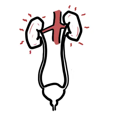
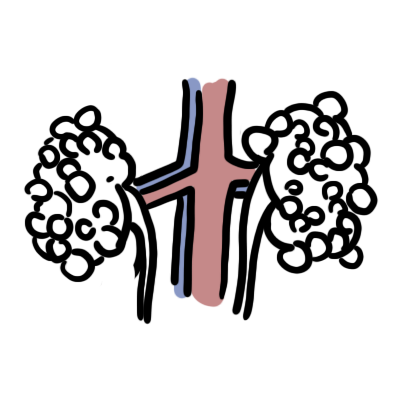
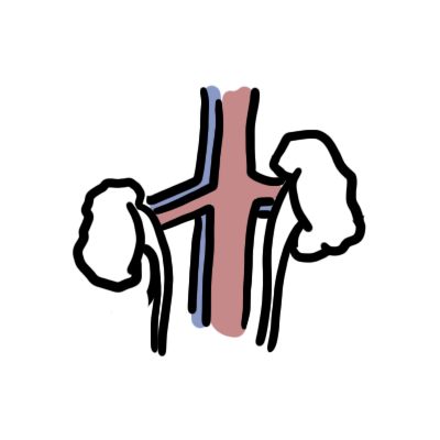
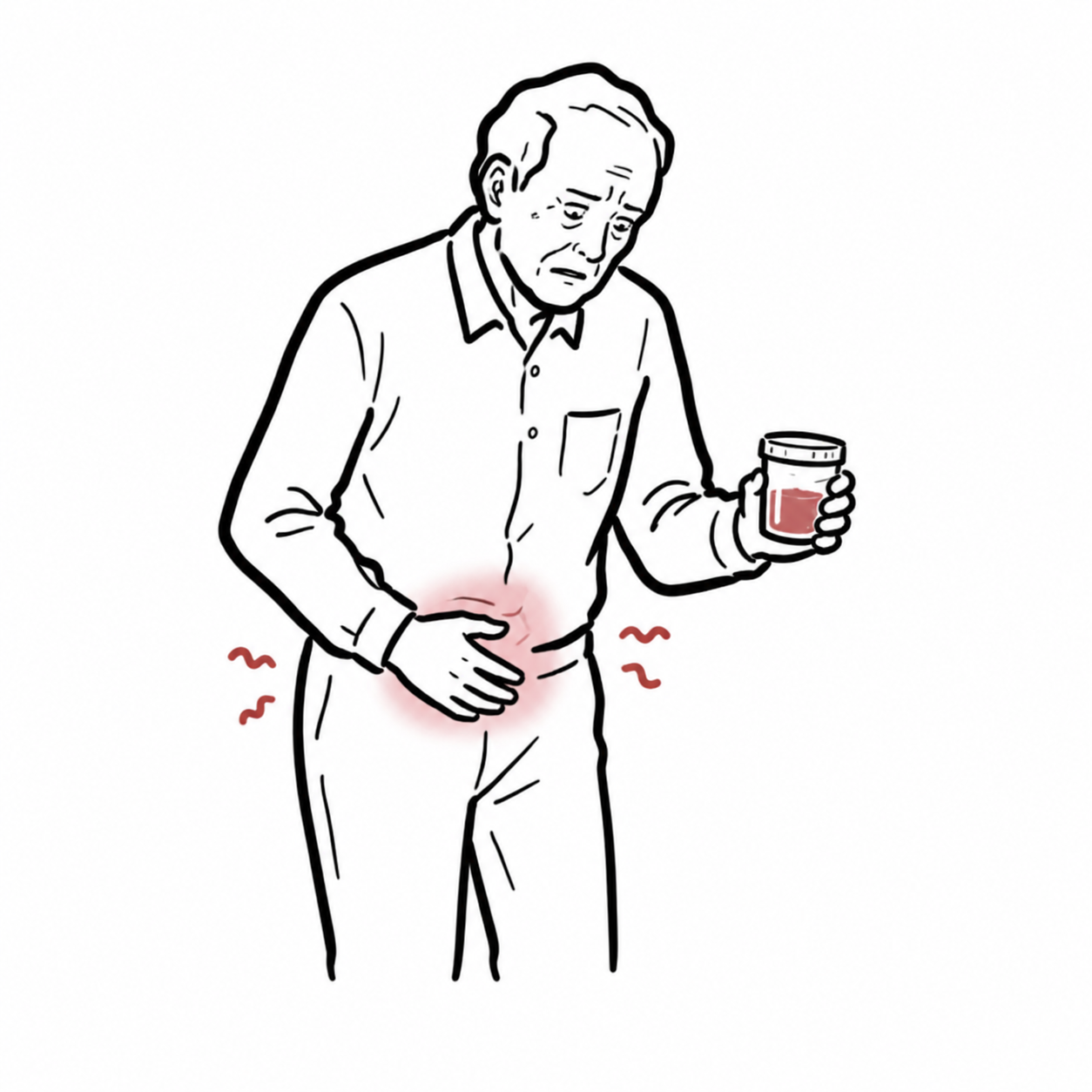
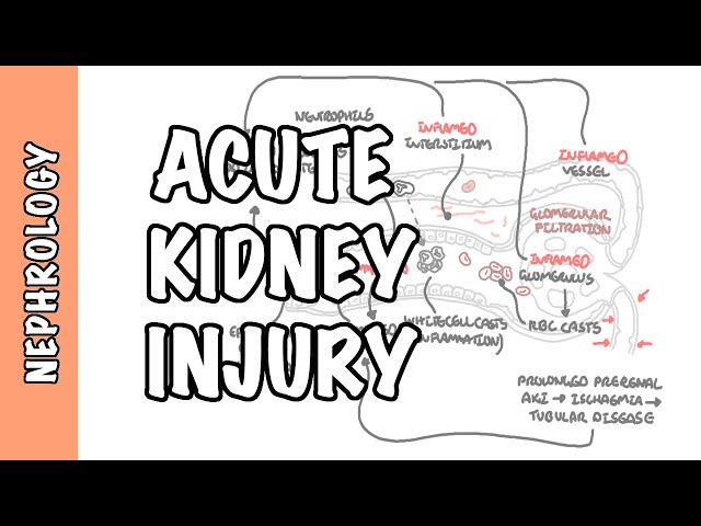
Acute Kidney Injury (AKI) is an acute, rapidly progressive decline in renal function occurring over hours to days, leading to nitrogenous waste retention (urea, creatinine) and disruption of fluid, electrolyte, and acid-base homeostasis.
KDIGO Diagnostic Criteria (Any ONE of the following):
- Increase in Serum Creatinine by >= 26.5 umol/L within 48 hours.
- Increase in Serum Creatinine to >= 1.5 times baseline within the prior 7 days.
- Urine Output < 0.5 mL/kg/h for >= 6 consecutive hours.
Pathophysiologic Categorization
- Prerenal (renal hypoperfusion)
- Intrinsic (structural parenchymal damage)
- Postrenal (urinary tract obstruction)
Serum creatinine is a delayed marker of kidney injury—it may take 24–48 hours to rise after an acute decline in GFR. Oliguria (< 0.5 mL/kg/h for 6 hours) is often the earliest sensitive sign of evolving AKI.
Approach
Step 1: Emergency Triage & Red Flag Screening (Life-Threatening Complications)
- Severe Hyperkalemia (K+ >= 6.5 mmol/L or ECG changes): Risk of fatal cardiac arrhythmias.
- Severe Metabolic Acidosis (pH < 7.10, HCO3- < 10 mmol/L): Impaired myocardial contractility, vasoplegia.
- Uremic Emergency: Uremic pericarditis (friction rub), uremic encephalopathy (asterixis, confusion, seizures), uremic bleeding.
- Refractory Volume Overload: Acute pulmonary edema, hypoxia, severe hypertension unresponsive to diuretics.
Step 2: Rapid Diagnostic Branching (Prerenal vs. Intrinsic vs. Postrenal)
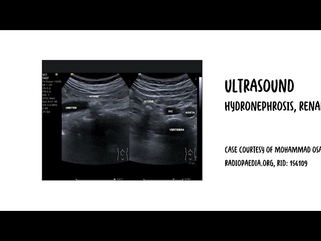
- Postrenal Exclude First: Bedside renal tract ultrasound or bladder scan to rule out urinary retention/obstruction.
- Prerenal vs. Intrinsic Differentiation:
- Prerenal: High urine osmolality (> 500 mOsm/kg), Low Fractional Excretion of Sodium (FENa < 1% or FEUrea < 35% if on diuretics), Urine Na+ < 20 mmol/L, Urea:Creatinine ratio > 100:1.
- Intrinsic (ATN): Low urine osmolality (< 350 mOsm/kg), High Fractional Excretion of Sodium (FENa > 2%), Urine Na+ > 40 mmol/L, Urea:Creatinine ratio < 40:1.
Step 3: Clinical Decision Branching
- Postrenal Obstruction Present: -> Immediate urinary decompression (Urinary catheter, suprapubic catheter, or nephrostomy).
- Volume Depleted / Prerenal: -> Fluid resuscitation with isotonic crystalloids (e.g., Plasmalyte or 0.9% Normal Saline).
- Volume Overloaded / Oliguric: -> Fluid restriction + IV Loop Diuretics (Furosemide challenge); evaluate for Renal Replacement Therapy (RRT) if non-responsive.
In patients taking loop diuretics, FENa can be falsely elevated (> 1%) even in prerenal states. Use Fractional Excretion of Urea (FEUrea < 35%) instead to confirm prerenal hypoperfusion.
Differential Diagnosis
Prerenal Azotemia (~50–60% of cases – Intact Parenchyma, Hypoperfusion):
- Volume Depletion: Hemorrhage, GI losses (vomiting/diarrhea), renal losses (diuretics), third-spacing (pancreatitis, sepsis, burns).
- Decreased Effective Arterial Blood Volume: Heart failure (Cardiorenal Syndrome), Cirrhosis (Hepatorenal Syndrome), Severe Sepsis.
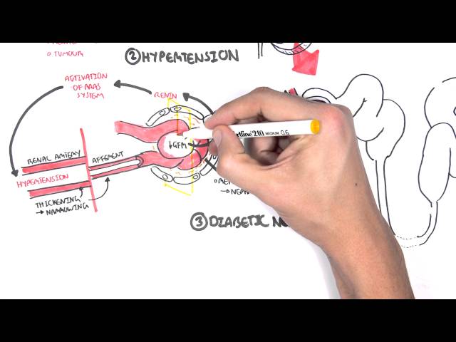
- Hemodynamic Autoregulatory Impairment: NSAIDs (inhibit afferent arteriolar vasodilation), ACEi / ARBs (inhibit efferent arteriolar vasoconstriction), Calcineurin inhibitors (Tacrolimus/Cyclosporine).
Intrinsic Renal Disease (~35–40% of cases – Parenchymal Damage):
- Acute Tubular Necrosis (ATN – Most Common Intrinsic Cause):
- Ischemic: Prolonged prerenal hypoperfusion, shock, cardiac arrest.
- Nephrotoxic: Exogenous (Aminoglycosides, Cisplatin, Amphotericin B, Iodinated Radiocontrast) or Endogenous (Rhabdomyolysis [myoglobinuria], Hemolysis [hemoglobinuria], Bence-Jones proteins in Multiple Myeloma).
- Distinguishing features: Muddy brown granular casts on urine microscopy.
- Acute Interstitial Nephritis (AIN):
- Triggers: Drug-induced (NSAIDs, Penicillins, Cephalosporins, PPIs, Sulfonamides) or Autoimmune (Sjögren’s, Sarcoidosis, Lupus).
- Distinguishing features: Classic triad (Classic in < 30%: Fever, Rash, Eosinophilia); WBC casts and eosinophiluria on urinalysis.
- Acute Glomerulonephritis (AGN):
- Etiologies: ANCA Vasculitis, Anti-GBM (Goodpasture), Lupus Nephritis, Post-Infectious GN, IgA Nephropathy.
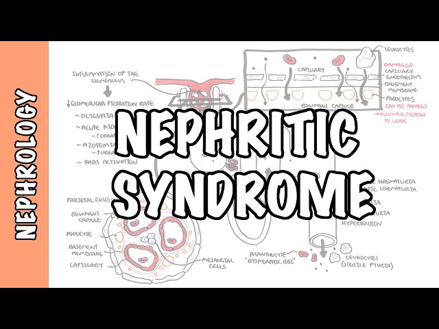
- Distinguishing features: Nephritic syndrome (Hypertension, Oliguria, Edema), Dysmorphic RBCs and RBC casts on urine microscopy, heavy proteinuria.
- Acute Vascular / Thrombotic Microangiopathy (TMA):
- Etiologies: Renal Artery Occlusion, Cholesterol Embolization (post-angiography with livedo reticularis/blue toes), HUS/TTP.
Postrenal Acute Kidney Injury (~5–10% of cases – Obstruction):
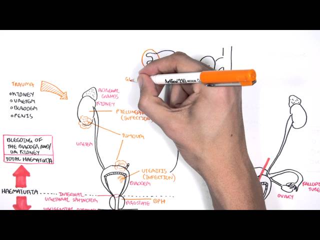
- Lower Urinary Tract Obstruction: Benign Prostatic Hyperplasia (BPH), Prostate Ca, Neurogenic Bladder, Urethral Stricture, Clot Retention.
- Upper Urinary Tract Obstruction (Bilateral or Unilateral in Single Kidney): Ureteral Calculi, Retroperitoneal Fibrosis, Pelvic Malignancy (Cervical/Colorectal Ca).
Key urinalysis microscopic clues: Muddy brown casts = Acute Tubular Necrosis; RBC casts / Dysmorphic RBCs = Glomerulonephritis; WBC casts / Eosinophiluria = Acute Interstitial Nephritis.
Investigations
First-Line / Bedside & Basic Diagnostic Tests:
- Bedside Bladder Scan & Urinary Catheterization: Assess post-void residual volume (PVR > 200 mL suggests lower tract obstruction).
- Urinalysis & Urine Microscopy (CRITICAL):
- Dipstick: Proteinuria, hematuria, leukocyte esterase, specific gravity.
- Microscopy: Look for casts (RBC, WBC, Muddy Brown), crystals, dysmorphic RBCs.
- Urine Electrolytes & Biochemistry: Urine Na+, Urine Creatinine, Urine Osmolality, Urine Urea -> Calculate FENa or FEUrea.
- Serum Biochemistry: Electrolytes (K+, Na+, HCO3-, Ca2+, PO4-), Urea, Creatinine, Lactate, Creatine Kinase (CK – rule out rhabdomyolysis).
- Full Blood Count & Blood Film: Check for leukocytosis, eosinophilia (AIN), or schistocytes/thrombocytopenia (TMA/HUS/TTP).
Targeted / Specialized Workup:
- Renal Tract Ultrasound: Gold standard imaging to rule out hydronephrosis/postrenal obstruction and evaluate kidney size/echogenicity (small shrunken kidneys suggest Chronic Kidney Disease).
- Serological Autoimmune / Glomerular Panel (If AGN Suspected): ANA, ANCA (p-ANCA/mpo, c-ANCA/pr3), Anti-GBM antibodies, Complement levels (C3, C4), Serum Protein Electrophoresis (SPEP) & Free Light Chains.
- Renal Biopsy: Indicated when intrinsic etiology is unclear, or when rapidly progressive glomerulonephritis (RPGN), AIN, or vasculitis is suspected to guide targeted immunosuppressive therapy.
A normal renal ultrasound does NOT completely exclude postrenal obstruction in early hyperacute obstruction, severe dehydration, or retroperitoneal fibrosis where collecting system dilation has not yet developed.
Critical Management
Emergency Management of Life-Threatening Complications:
- Hyperkalemia (K+ > 6.5 mmol/L or ECG changes):
- Acute Pulmonary Edema / Volume Overload: IV Furosemide (high-dose e.g., 80–200 mg IV) + Supplemental O2/NIV -> Emergency Hemodialysis if refractory.
Indications for Emergency Renal Replacement Therapy (RRT) – Mnemonic “AEIOU”:
- A – Acidosis: Refractory metabolic acidosis (pH < 7.15).
- E – Electrolytes: Refractory severe hyperkalemia (K+ > 6.5 mmol/L).
- I – Ingestion / Intoxication: Toxic alcohol, Salicylate, Lithium, Theophylline overdoses.
- O – Overload: Refractory volume overload / Pulmonary edema.
- U – Uremia: Symptomatic uremia (Uremic pericarditis, encephalopathy, neuropathy, bleeding).
General Supportive & Nephroprotective Measures:
- Stop Nephrotoxic Drugs: Hold NSAIDs, ACEi/ARBs, Aminoglycosides, Vancomycin, Amphotericin, Contrast agents.
- Dose Adjustments: Adjust all renally cleared medications according to estimated GFR.
- Fluid Management: Match fluid intake to losses; avoid fluid overload once volume euvolemia is achieved.
Use the “AEIOU” mnemonic for emergency dialysis indications. Do not delay Nephrology consultation or vascular access for acute RRT if a patient develops any refractory feature of “AEIOU”.
References
- National Institute for Health and Care Excellence. Acute kidney injury: prevention, detection and management (NG148) [Internet]. 2019 [updated 2024; cited 2026 Aug 20]. Available from: https://www.nice.org.uk/guidance/ng148
- Kidney Disease: Improving Global Outcomes. Acute kidney injury and acute kidney disease guideline [Internet]. [cited 2026 Aug 20]. Available from: https://kdigo.org/guidelines/acute-kidney-injury/





Members only discussions coming soon…