

Haematuria

Overview
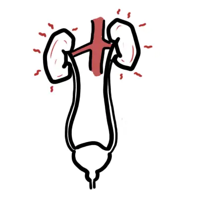
Haematuria is defined as the presence of red blood cells (RBCs) in the urine.
Classification
- Macroscopic (Gross) Haematuria: Urine visible as pink, red, or cola-coloured to the naked eye. Carries a high risk of underlying urological malignancy (~20–25%).
- Microscopic (Non-Visible) Haematuria (NVH): >= 3 RBCs per high-power field (HPF) on formal automated or manual urine microscopy. Dipstick screening requires microscopy confirmation.
Pathophysiologic Categorization
- Glomerular (Renal Parenchymal): Bleeding originating from damaged glomerular capillaries (Nephritic syndromes).
- Non-Glomerular (Extra-Glomerular): Bleeding originating anywhere along the urinary tract architecture (kidneys, ureters, bladder, prostate, urethra).
A positive urine dipstick for blood indicates hemoglobin or myoglobin but is NOT diagnostic of haematuria. True microscopic haematuria requires confirmation by formal urine microscopy showing >= 3 RBCs/HPF.
Approach
Step 1: Emergency Triage & Red Flag Screening
- Gross Haematuria + Clot Retention / Anuria: -> Acute Urinary Retention secondary to clot obstruction. Emergency urethral catheterisation (large-bore 20–24 Fr 3-way catheter) + Continuous Bladder Irrigation (CBI).
- Macroscopic Haematuria + Hemodynamic Instability / Shock: -> Severe ongoing blood loss. Resuscitate with IV crystalloid/blood products, urgent Urology consult for cystoscopic clot evacuation and hemostasis.
- Haematuria + Systemic Vasculitis / Rapidly Declining Renal Function: -> Suspect Rapidly Progressive Glomerulonephritis (RPGN) / ANCA Vasculitis / Anti-GBM disease. Emergency Nephrology referral.
Step 2: Differentiate Glomerular vs Non-Glomerular Origin
- Glomerular Origin:
- Urinary Clues: Dysmorphic RBCs (> 80%), RBC casts, heavy proteinuria (> 1 g/24h or PCR > 100 mg/mmol), “cola/tea-coloured” urine without blood clots.
- Clinical Clues: Nephritic syndrome triad (Hypertension, Oliguria, Edema), elevated serum creatinine.
- Non-Glomerular / Urological Origin:
- Urinary Clues: Morphic / Uniform RBCs, absence of RBC casts, minimal/no proteinuria, bright red or pink urine, presence of blood clots (pathognomonic for non-glomerular origin).
- Clinical Clues: Dysuria, flank pain, palpable abdominal mass, lower urinary tract symptoms (LUTS).
Step 3: Clinical Decision Branching for Investigation
- Confirmed Glomerular Origin: -> Nephrology pathway (Autoimmune serologies, renal ultrasound, consideration of Renal Biopsy).
- Non-Glomerular / Visible or Unexplained Non-Visible Haematuria (Age >= 35 or Risk Factors): -> Urology pathway (CT Urogram + Flexible Cystoscopy).
Blood clots in urine NEVER originate from a glomerular source—clotting factors are excluded by the intact glomerular basement membrane. The presence of clots definitively confirms a non-glomerular (urological) source of bleeding.
Differential Diagnosis
Urological / Non-Glomerular (Malignant & Benign):
- Urothelial Carcinoma (Bladder, Ureter, Renal Pelvis) & Renal Cell Carcinoma (RCC):
- Distinguishing features: Painless macroscopic haematuria in patients > 45 years or smokers; constitutional weight loss, palpable flank mass (RCC triad: flank pain, mass, haematuria seen in < 10%).
- Urolithiasis (Renal / Ureteral Calculi):
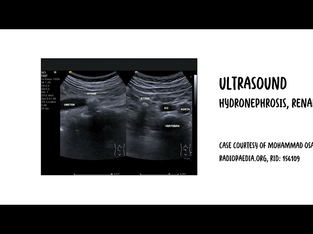
- Distinguishing features: Sudden onset severe, writhing colicky flank pain radiating to groin/scrotum, dysuria, costovertebral angle tenderness, microscopic or gross haematuria.
- Benign Prostatic Hyperplasia (BPH) / Prostatic Urethral Bleeding:
- Distinguishing features: Older males; LUTS (hesitancy, weak stream, nocturia), enlarged non-tender prostate on DRE; initial or terminal haematuria.
- Urinary Tract Infection (Hemorrhagic Cystitis / Pyelonephritis):
- Distinguishing features: Dysuria, urinary frequency, urgency, suprapubic pain, fever; positive urine dipstick for nitrites and leukocyte esterase, WBCs on microscopy.
Renal Parenchymal / Glomerular:
- IgA Nephropathy (Berger’s Disease – Most Common Glomerular Cause):
- Distinguishing features: Synpharyngitic haematuria (gross haematuria occurring concurrently or 1–2 days during an upper respiratory tract infection); dysmorphic RBCs, mild proteinuria.
- Post-Infectious Glomerulonephritis (PIGN):
- Distinguishing features: Occurs 1–3 weeks after a Group A Streptococcal pharyngitis or impetigo infection; low serum C3 complement, elevated ASOT/Anti-DNase B, edema, hypertension.
- ANCA-Associated Vasculitis / Anti-GBM Disease (Goodpasture):
- Distinguishing features: Rapidly progressive AKI, pulmonary hemorrhage (hemoptysis), constitutional symptoms; positive p-ANCA/c-ANCA or Anti-GBM antibodies.
Pigmenturia / Pseudo-Haematuria (Dipstick / Visual Mimics):
- Myoglobinuria (Rhabdomyolysis) / Hemoglobinuria (Intravascular Hemolysis): Positive dipstick for blood, but zero RBCs on microscopy.
- Dietary / Drug Causes: Beetroot, blackberries, Rifampicin, Nitrofurantoin, Phenazopyridine (negative dipstick for blood).
Differentiate IgA Nephropathy from Post-Streptococcal Glomerulonephritis by timing: IgA Nephropathy presents during or within 24–48 hours of an infection (“synpharyngitic”), whereas PSGN presents 1–3 weeks after the infection.
Transient haematuria: Exercise (‘joggers’ nephritis’), Menstruation, Sexual activity, UTI, Viral illnesses and Trauma.
Investigations
First-Line / Bedside & Laboratory Diagnostics:
- Mid-Stream Urine (MSU) Formal Microscopy & Culture: Confirm >= 3 RBCs/HPF, evaluate RBC morphology (dysmorphic vs uniform), casts, and rule out concurrent infection.
- Urine Protein-to-Creatinine Ratio (PCR) or Albumin-to-Creatinine Ratio (ACR): Quantify proteinuria (PCR > 50 mg/mmol suggests renal parenchymal disease).
- Serum Biochemistry & Hematology: Creatinine, eGFR, Urea, Electrolytes, Full Blood Count (anemia, thrombocytopenia), Coagulation profile (INR/APTT).
- Urine Cytology: Three morning urine samples for malignant urothelial cells (high specificity, low sensitivity; useful for carcinoma in situ).
Targeted Urology Workup (Upper & Lower Tract Assessment):
- CT Urogram (Triple-Phase CT Abdomen/Pelvis – Non-contrast, Nephrographic, Excretory phases): Gold standard imaging modality to evaluate the upper urinary tract for renal masses, urothelial tumors, and urolithiasis.
- Flexible Cystoscopy: Gold standard lower tract evaluation to visually inspect the bladder, ureteric orifices, and urethra. Mandatory in all visible haematuria and unexplained non-visible haematuria in adults >= 35 years.
- Renal Tract Ultrasound: Preferred upper tract imaging modality in young patients (< 35 years), pregnant women, or those with severe renal failure (eGFR < 30 mL/min) to avoid iodinated contrast/radiation.
The diagnostic evaluation for unexplained macroscopic haematuria or high-risk non-visible haematuria requires BOTH upper tract imaging (CT Urogram) AND lower tract visualization (Flexible Cystoscopy). Neither test alone is sufficient.
Critical Management
Acute Severe Gross Haematuria & Clot Retention:
- Catheterisation: Insert a 20–24 Fr 3-way Foley catheter.
- Manual Clot Evacuation: Perform vigorous manual flushing with a 60 mL Toomey syringe and sterile normal saline until all organized blood clots are cleared and urine runs clear.
- Continuous Bladder Irrigation (CBI): Connect 3-way catheter to normal saline irrigation; titrate infusion rate to keep outflow clear/light pink and prevent recurrent clot formation.
Glomerular / Vasculitic Emergencies:
- Immunosuppression: High-dose IV Methylprednisolone pulse therapy + Cyclophosphamide or Rituximab for ANCA vasculitis / Anti-GBM disease; consideration of Plasma Exchange (PLEX) if severe renal failure or pulmonary hemorrhage is present.
Never start Continuous Bladder Irrigation (CBI) through a 3-way catheter before manually evacuating all pre-existing intravesical blood clots. Initiating irrigation over an obstructed clot will cause severe bladder over-distension, bladder rupture, or urosepsis.
References
- American Urological Association; Society of Urodynamics, Female Pelvic Medicine & Urogenital Reconstruction. Microhematuria: AUA/SUFU guideline (2025) [Internet]. 2025 [cited 2026 Aug 20]. Available from: https://www.auanet.org/guidelines-and-quality/guidelines/microhematuria
- National Institute for Health and Care Excellence. Suspected cancer: recognition and referral (NG12) [Internet]. 2015 [updated 2026; cited 2026 Aug 20]. Available from: https://www.nice.org.uk/guidance/ng12

















Members only discussions coming soon…