

Hepatocellular Carcinoma

Overview
Hepatocellular carcinoma (HCC) is a malignant tumour arising from hepatocytes and the most common primary liver malignancy. It usually develops in chronic liver disease, particularly cirrhosis, although chronic hepatitis B can cause HCC without cirrhosis. It is more common in men and older adults; the burden is greatest in regions with endemic viral hepatitis. Early disease is often asymptomatic. Later features include weight loss, right upper-quadrant discomfort, hepatomegaly or unexplained hepatic decompensation. Early detection enables potentially curative treatment, whereas advanced disease and impaired liver reserve substantially limit treatment options.
Definition
- Hepatocellular carcinoma
- a primary malignant epithelial tumour showing hepatocellular differentiation.
Anatomy & Physiology
- The liver receives blood from the portal vein and hepatic artery; venous drainage is through hepatic veins into the inferior vena cava.
- Hepatocytes perform metabolic, detoxification and synthetic functions. Albumin, bilirubin and coagulation measures help assess hepatic reserve.
- HCC develops increasing arterial blood supply, explaining arterial-phase enhancement on dynamic imaging. Portal venous invasion can worsen portal hypertension and facilitate intrahepatic spread.
Aetiology & Risk Factors
Aetiology
- Chronic hepatitis B or C infection.
- Alcohol-associated liver disease and metabolic dysfunction-associated steatotic liver disease (MASLD), especially with steatohepatitis and cirrhosis.
- Other chronic liver diseases, including hereditary haemochromatosis and alpha-1 antitrypsin deficiency.
- Aflatoxin B1 exposure from contaminated food, particularly in conjunction with hepatitis B.
Risk Factors
- Cirrhosis of any aetiology is the principal clinical risk state.
- Increasing age, male sex, family history of HCC and persistent HBV replication.
- Obesity, diabetes, smoking and heavy alcohol consumption contribute to risk.
- Risk persists after hepatitis C cure in patients with cirrhosis; antiviral treatment reduces but does not eliminate risk. [1,3]
HCC can occur without cirrhosis, especially in chronic hepatitis B. Normal liver enzymes do not exclude HCC.
Pathophysiology
Repeated hepatocyte injury and regeneration promote oxidative stress, genomic damage and selection of malignant clones. Progressive fibrosis creates a pro-carcinogenic environment. HBV can additionally promote carcinogenesis through viral DNA integration; aflatoxin can cause characteristic TP53 mutations. Dysregulated growth signalling, angiogenesis and immune evasion permit tumour progression.
| Pathway | Consequence |
|---|---|
| Chronic injury → inflammation → fibrosis/cirrhosis | Regenerative and dysplastic nodules develop in a susceptible liver. |
| Genetic/epigenetic alterations → malignant hepatocytes | HCC develops; cirrhosis is not obligatory in every pathway. |
| Angiogenesis and vascular invasion | Arterialisation, portal-vein tumour thrombus and intrahepatic dissemination. |
| Progressive tumour burden ± extrahepatic spread | Liver dysfunction, constitutional symptoms and metastatic disease. |
Gross and Microscopic Pathology
- Gross: solitary mass, multiple nodules or a diffuse infiltrative tumour; haemorrhage, necrosis and vascular invasion may occur.
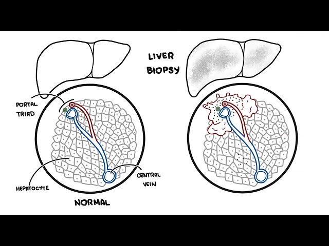
- Microscopy: atypical hepatocytes arranged in thickened trabeculae, pseudoglands or solid sheets; bile production can support hepatocellular differentiation.
- Fibrolamellar carcinoma is a distinct clinicopathological entity, typically arising in younger people without cirrhosis. [5]
Why can a small HCC in a severely cirrhotic liver be harder to treat than a larger tumour in a liver with preserved function?
Clinical manifestation
- Early HCC: often asymptomatic and detected during surveillance.
- Constitutional symptoms: weight loss, anorexia, fatigue and early satiety.
- Local features: right upper-quadrant pain or fullness; firm or irregular hepatomegaly.
- New or worsening decompensation: ascites, jaundice, encephalopathy or variceal bleeding.
- Examination: muscle wasting, hepatomegaly and signs of chronic liver disease or portal hypertension.
- Less common: hypoglycaemia, hypercalcaemia or erythrocytosis from paraneoplastic effects. [3,5]
Sudden severe abdominal pain with hypotension in a patient with HCC may indicate tumour rupture and haemoperitoneum: urgent resuscitation and interventional/surgical assessment are required.
Diagnosis
Diagnostic Criteria/Scoring Systems
In patients with cirrhosis or at-risk chronic HBV, validated CT/MRI LI-RADS criteria can establish HCC without biopsy; LR-5 denotes definite HCC. Criteria depend on lesion size and combinations of major imaging features. Indeterminate lesions require multidisciplinary review, further imaging, interval assessment or biopsy. Outside the validated at-risk population, histological confirmation is generally required.
Biopsy is useful when imaging is atypical or another malignancy is suspected. A negative biopsy does not exclude HCC, particularly in a small lesion. Bleeding and rare needle-track seeding are relevant risks.
Investigations
| Investigation | Purpose / important findings |
|---|---|
| Multiphasic liver CT or MRI | Assess tumour number, size, enhancement and vascular invasion. Non-rim arterial enhancement with non-peripheral washout is characteristic in the appropriate risk population. |
| AFP | May be elevated; a normal result does not exclude HCC and an elevated result is not diagnostic. Useful for trends and follow-up when informative. |
| FBC, liver biochemistry, albumin, INR, renal function | Assess cytopenias, cholestasis, synthetic function and treatment fitness. |
| HBV/HCV testing and aetiological work-up | Identify and manage underlying liver disease. |
| Staging imaging | Chest CT and liver imaging assess spread; additional investigations follow symptoms or risk. Routine PET is not generally used. |
Surveillance and recall
- Offer six-monthly liver ultrasound with AFP to appropriate at-risk patients, including most patients with cirrhosis and selected people with chronic HBV. Surveillance is appropriate when detection would change management.
- A subcentimetre ultrasound lesion usually prompts repeat ultrasound and AFP in 3–6 months. A lesion ≥1 cm, AFP ≥20 ng/mL or a rising AFP warrants diagnostic multiphasic CT or contrast-enhanced MRI.
- Consider MRI or CT surveillance when ultrasound visualisation is inadequate, following specialist assessment.
Classification
The Barcelona Clinic Liver Cancer (BCLC) system integrates tumour burden, liver function and cancer-related performance status. The table summarises typical categories; treatment allocation requires individual multidisciplinary assessment. Child–Pugh, MELD and/or ALBI provide complementary measures of hepatic reserve.
| BCLC stage | Typical features | Usual treatment direction |
|---|---|---|
| 0: Very early | Single tumour ≤2 cm; preserved liver function; ECOG 0 | Ablation or resection; transplant assessment when indicated. |
| A: Early | Single tumour, or ≤3 tumours each ≤3 cm; preserved function; ECOG 0 | Resection, transplantation or ablation. |
| B: Intermediate | Multifocal beyond stage A; no vascular invasion/extrahepatic spread; ECOG 0 | Selected transplantation, TACE or systemic therapy according to burden and suitability. |
| C: Advanced | Vascular invasion and/or extrahepatic spread; generally ECOG 0–2 with adequate liver function | Systemic therapy. |
| D: End stage | Poor performance status and/or severe liver dysfunction without a transplant option | Symptom-focused and palliative care. |
Treatment
Management should be coordinated by a hepatology, hepatobiliary surgery, transplant, radiology and oncology multidisciplinary team. Treat underlying liver disease, optimise nutrition and manage portal hypertension alongside cancer therapy.
Potentially curative treatment
- Resection: selected localised tumours with adequate future liver remnant; in cirrhosis, usually well-compensated disease without clinically significant portal hypertension.
- Liver transplantation: treats both HCC and the diseased liver. Classical Milan criteria are a single tumour ≤5 cm or up to three tumours each ≤3 cm, without macrovascular invasion or extrahepatic spread. Selected expanded criteria/downstaging pathways are centre-specific.
- Thermal ablation: radiofrequency or microwave treatment, particularly for small tumours when surgery is unsuitable. Location and proximity to vessels or bile ducts affect suitability.
Locoregional treatment
- Transarterial chemoembolisation (TACE): commonly used for suitable intermediate-stage disease with sufficient liver reserve.
- Radioembolisation (TARE/SIRT) or external-beam radiotherapy: options in selected patients, including when ablation or embolisation is unsuitable.
- Locoregional therapy can bridge to transplantation or downstage selected tumours. Avoid repeated ineffective treatment that compromises liver function; reassess for systemic therapy.
Systemic treatment
- For unresectable/advanced disease or progression beyond suitable locoregional therapy, established first-line options include atezolizumab–bevacizumab or durvalumab–tremelimumab.
- Nivolumab–ipilimumab is another first-line option supported by CheckMate-9DW and FDA approval in 2025. Local approval and funding determine access.
- Lenvatinib or sorafenib may be appropriate when immunotherapy combinations are unsuitable. Later-line choices depend on prior treatment, liver function and availability.
Before atezolizumab–bevacizumab, assess and manage high-risk varices and bleeding risk. Immune checkpoint inhibitors require careful assessment in transplant recipients and patients with significant autoimmune disease.
Supportive care and follow-up
- Address pain, fatigue, malnutrition and psychological needs; integrate palliative care according to symptom burden.
- Treat HBV/HCV when indicated; encourage alcohol cessation and metabolic risk management.
- Continue imaging-based follow-up after curative treatment because recurrence risk remains high; after resection, cross-sectional surveillance is generally every 3–6 months.
Complications and Prognosis
Complications
- Liver failure and portal hypertension: manage ascites, encephalopathy and variceal bleeding with specialist input.
- Tumour rupture: resuscitation and haemostatic intervention, commonly arterial embolisation.
- Vascular invasion, biliary obstruction and distant metastases.
- Treatment toxicity: hepatic decompensation after surgery/locoregional therapy; bleeding, hypertension or proteinuria with VEGF inhibition; immune hepatitis, colitis and endocrinopathies with checkpoint inhibitors.
Prognosis
- Outcome depends on tumour burden, vascular invasion, hepatic reserve, performance status and treatment response.
- Early-stage disease may be cured; selected transplant recipients have five-year survival exceeding 70%, but this is not representative of all HCC.
- Recurrence after resection is common, approaching 50–70% at five years.
- Modern systemic therapy improves advanced-disease survival: IMbrave150 reported median overall survival of 19.2 months with atezolizumab–bevacizumab versus 13.4 months with sorafenib in its selected trial population. Individual outcomes vary substantially.
References
1. Singal AG, Llovet JM, Yarchoan M, Mehta N, Heimbach JK, Dawson LA, et al. AASLD Practice Guidance on prevention, diagnosis, and treatment of hepatocellular carcinoma. Hepatology. 2023;78(6):1922–1965. doi:10.1097/HEP.0000000000000466.
2. European Association for the Study of the Liver. EASL Clinical Practice Guidelines on the management of hepatocellular carcinoma. J Hepatol. 2025;82(2):315–374. doi:10.1016/j.jhep.2024.08.028.
3. PDQ Adult Treatment Editorial Board. Primary liver cancer treatment (PDQ): health professional version [Internet]. Bethesda (MD): National Cancer Institute; [cited 2026 Oct 6]. Available from: https://www.cancer.gov/types/liver/hp/adult-liver-treatment-pdq
4. Liu Y, Wu F. Global burden of aflatoxin-induced hepatocellular carcinoma: a risk assessment. Environ Health Perspect. 2010;118(6):818–824. doi:10.1289/ehp.0901388.
5. Llovet JM, Kelley RK, Villanueva A, Singal AG, Pikarsky E, Roayaie S, et al. Hepatocellular carcinoma. Nat Rev Dis Primers. 2021;7(1):6. doi:10.1038/s41572-020-00240-3.
6. Reig M, Forner A, Rimola J, Ferrer-Fàbrega J, Burrel M, Garcia-Criado Á, et al. BCLC strategy for prognosis prediction and treatment recommendation: the 2022 update. J Hepatol. 2022;76(3):681–693. doi:10.1016/j.jhep.2021.11.018.
7. Cheng AL, Qin S, Ikeda M, Galle PR, Ducreux M, Kim TY, et al. Updated efficacy and safety data from IMbrave150: atezolizumab plus bevacizumab vs. sorafenib for unresectable hepatocellular carcinoma. J Hepatol. 2022;76(4):862–873. doi:10.1016/j.jhep.2021.11.030.
8. Abou-Alfa GK, Lau G, Kudo M, Chan SL, Kelley RK, Furuse J, et al. Tremelimumab plus durvalumab in unresectable hepatocellular carcinoma. NEJM Evid. 2022;1(8):EVIDoa2100070. doi:10.1056/EVIDoa2100070.
9. US Food and Drug Administration. FDA approves nivolumab with ipilimumab for unresectable or metastatic hepatocellular carcinoma [Internet]. 2025 Apr 11 [cited 2026 Oct 6]. Available from: https://www.fda.gov/drugs/resources-information-approved-drugs/fda-approves-nivolumab-ipilimumab-unresectable-or-metastatic-hepatocellular-carcinoma
10. Taddei TH, Brown DB, Yarchoan M, Mendiratta-Lala M, Llovet JM. Critical Update: AASLD Practice Guidance on prevention, diagnosis, and treatment of hepatocellular carcinoma. Hepatology. 2025;82(1):272–274. doi:10.1097/HEP.0000000000001269.













Members only discussions coming soon…