Anal Fissure

Notes

Overview

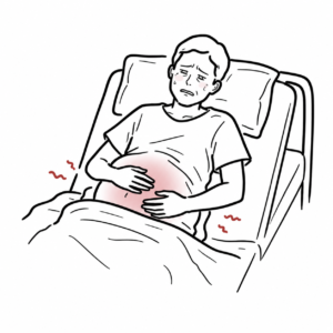
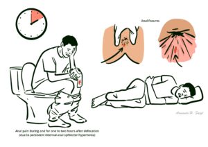
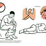
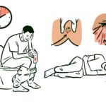
Anal fissure is a split in the skin of the distal anal canal. It presents with severe pain on defecation, like passing “glass shards”, and rectal bleeding. It is more frequently diagnosed in younger and middle-aged individuals. Anal fissures are mostly caused by passage of hard stool but can also be a result of other conditions, like Crohn’s disease.1,2

Definition

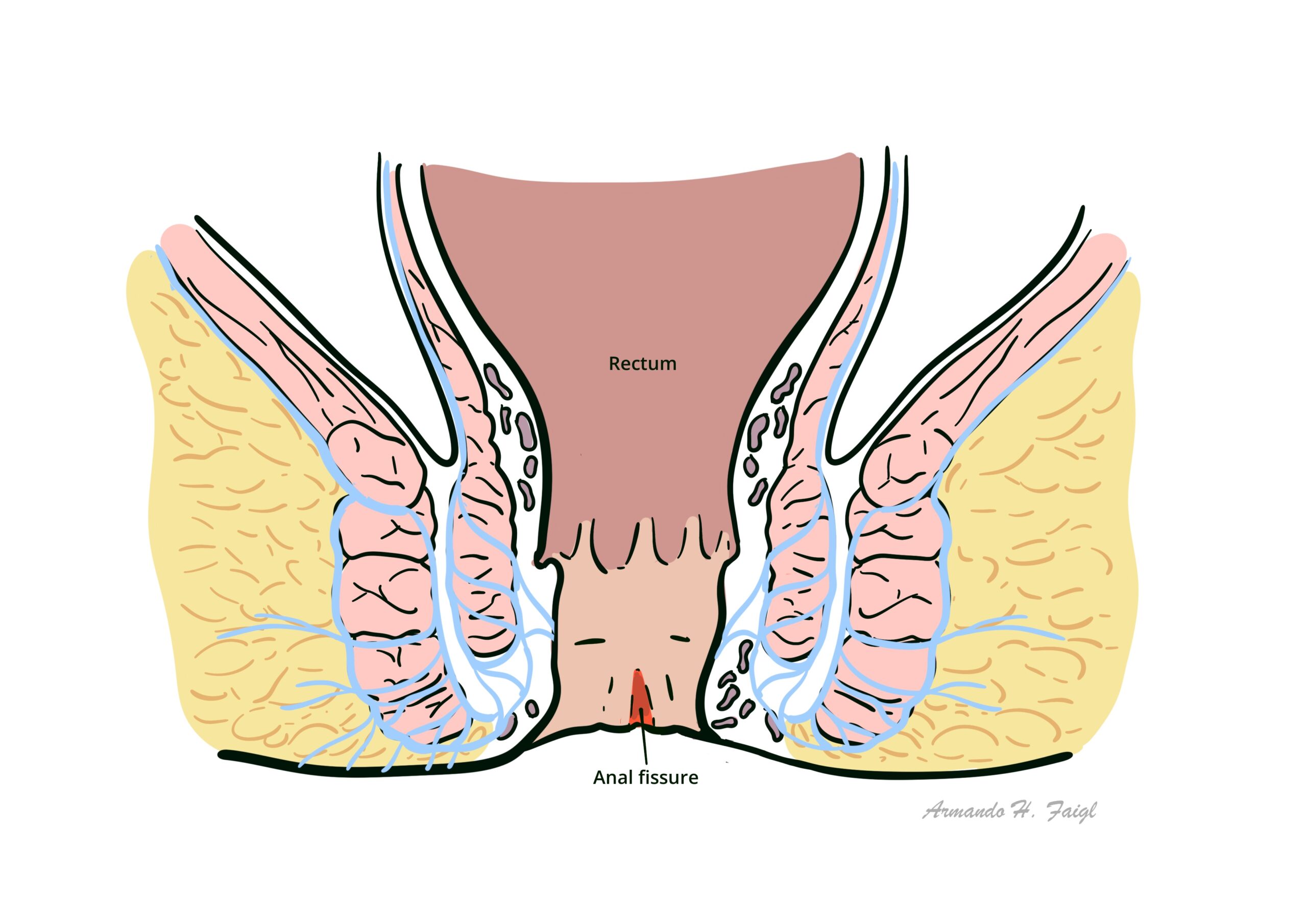
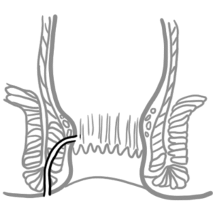
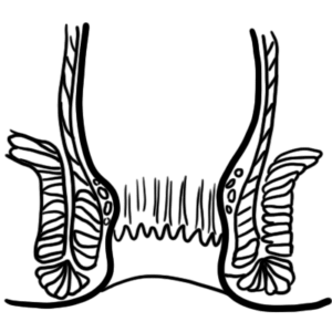
Anal fissure: linear tear within the anal canal that usually extends from the dentate line toward the anal verge.
Fistula in ano: abnormal communication between anorectal lining and perineal or vaginal epithelium; nearly always associated with anal abscess.
Perianal abscess: abscess within soft tissue surrounding the anal canal.
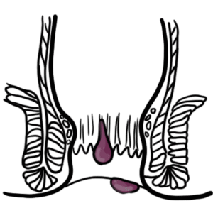
Anal haemorrhoids: dilation of either the internal or external haemorrhoidal plexi.

Anorectal Anatomy

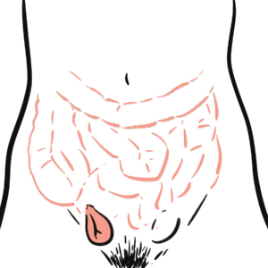
Anal canal – channel connecting the rectum to the anus, located within the anal triangle of the perineum, between the two ischioanal fossae.

Anal Sphincters

  • Internal sphincter
    • Involuntary control – continuation of circular fibers of the colon so it receives autonomic supply
    • Surrounds upper 3/4 of anal canal
  • External sphincter
    • Voluntary control
    • Surrounds entire length of anal canal
    • Consists of three parts – subcutaneous, superficial and deep

Intersphincteric plane

  • Between external sphincter muscle laterally and the longitudinal muscle (inner sphincteric muscle) medially
  • Contains anal glands
  • Can be opened up surgically to provide access for operations on the sphincter muscles

Anal glands

  • Apocrine glands
  • Occasionally infected and act as a source of anal fistula

Dentate (pectinate) line

  • Junction of the proximal and distal segments of the anal canal
  • Divides the anus into proximal 2/3 and distal 1/3
  • Situated at the middle of internal sphincter
Above Dentate lineBelow Dentate line
EmbryologyHindgut (endoderm)Proctodeum (ectoderm)
EpitheliumColumnarStratified squamous
Arterial supplySuperior rectal a.Middle rectal a.Inferior rectal a.
Venous drainageSuperior rectal v.Middle rectal v.Inferior rectal v.
Nerve supplyVisceral – Inferior hypogastric plexus(insensitive to pain)Somatic – Inferior rectal nerves: branches of Pudendal nerve(sensitive to pain)

Hilton’s Line also called while line/anocutaneous line indicates lower end of the internal sphincter. Ischiorectal abscess when communicates with anal canal usually  opens at or below Hilton’s line.

Aetiology and Risk Factors

  • Primary fissures: due to trauma
    • Constipation
    • Diarrhea
    • Vaginal delivery
    • Anal sex
  • Secondary fissures: due to another condition
    • Crohn’s disease
    • Malignancy
    • Communicable disease (eg. HIV, syphillis, chlamydia)
    • Granulomatous disease (eg. extrapulmonary tuberculosis, sarcoidosis)

Pathophysiology

Acute trauma or another disease

Internal anal sphincter spasm

Local ischaemia

Impaired healing

Hard stool passage causing a skin tear that cannot heal

Classification

Based on aetiology:

  • Primary: caused by local trauma in the context of anal spasm or high anal pressure; mostly located at posterior midline and sometimes at anterior midline
  • Secondary: caused by another disease; occur at locations other than midline; if you see atypical location investigate for other diseases

Based on timeframe:

  • Acute: less than 6 weeks; longitudinal tear in skin of distal anal canal
  • Chronic: more than 6 weeks; often with stigmata of chronicity (hypertrophied anal papilla, skin tag, exposed internal sphincter fibers)

Clinical Manifestation

  • Anal pain during and for one to two hours after defecation (due to persistent internal anal sphincter hypertonia) – distinguishing feature to other anal pain differentials
  • Tearing sensation: described as “passing glass shards”
  • Bright red blood: on stool or toilet paper
  • Fear of defecation: due to severe pain

Examination

General inspection

  • Visible fissure (90% posterior, 10% anterior; sometimes both are present “kissing fissures”).
  • Stigmata of chronicity (in chronic fissures): exposed internal anal sphincter muscle fibers, skin tags, hypertrophied anal papillae.

Palpation: to reproduce anal pain if fissure cannot be visualised – performed to confirm diagnosis.

Digital rectal exam and anoscopy are not recommended during initial presentation due to associated pain; can be performed after symptoms have improved.

Differential Diagnosis

  • Haemorrhoids
  • Perianal ulcers or sores
  • Anal fistula
  • Solitary rectal ulcer syndrome

Investigations

Not usually indicated, unless diagnosis is uncertain or secondary fissure suspected 

Treatment

Medical management:

  • Topical analgesia
  • Stool softeners
  • High fibre diet
  • Sitz bath
  • Topical vasodilators: nifedipine or glyceryl trinitrate (GTN)

Surgical management:

  • Botulinum toxin (BOTOX): relaxes muscle and improves healing
  • Lateral internal sphincterotomy: gold standard due to highest healing rate (>90%); sphincter muscle fibers are cut to reduce pressure and improve blood flow; can lead to incontinence in high-risk patients
  • Anal advancement flap: skin flap created from skin around anus and used to cover fissure
  • Fissurectomy: excision of anal fissure

Lateral internal sphincterotomy is the gold standard unless there is high risk of incontinence.

Complications and Prognosis

Complication

  • Sentinel skin tag (hypertrophied external skin margin)
  • Hypertrophied anal papilla (internal end)
  • Subcutaneous fistula or abscess (rare; due to secondary infection)
  • Anal stenosis (from fibrosis and repeated scarring)
  • Sphincter spasm and fibrosis → perpetuates pain and poor healing
  • Incontinence (potential postoperative complication if sphincterotomy is excessive)

Prognosis

  • Acute fissures
    • 90% heal with conservative management (stool softeners, topical nitrates, sitz baths)
    • Good prognosis if treated early
  • Chronic fissures
    • Often recur or persist due to internal sphincter hypertonia
    • Require pharmacologic relaxation (nitrates, calcium-channel blockers) or lateral internal sphincterotomy for definitive cure
    • Excellent long-term outcomes (>90% success) with surgery, but minor incontinence may occur in 1–5%

References

1. Paluvoi N. Anal fissure: BMJ Best Practice; 2023 [updated 2023 Dec 22; cited 2025 Sep 28]. Available from: https://bestpractice.bmj.com/topics/en-gb/563

2. Gerbasi L, Ashurst JV. Anal Fissures: National Library of Medicine; 2025 [updated 2025 Sep 15; cited 2025 Sep 28]. Available from: https://www.ncbi.nlm.nih.gov/books/NBK526063/

3. Davids JS, Hawkins AT, Bhama AR, Feinberg AE, Grieco MJ, Lightner AL, et al. The American Society of Colon and Rectal Surgeons Clinical Practice Guidelines for the Management of Anal Fissures. Disease of the Colon & Rectum. 2022;66(2):190-9. Doi: 10.1097/DCR.0000000000002664 

4. Madalinski MH. Identifying the best therapy for chronic anal fissure. World Journal of Gastrointestinal Pharmacology and Therapeutics. 2011;2(2):9-16. Doi: 10.4292/wjgpt.v2.i2.9 

5. Bleday R. Anal fissure: Medical management. Up To Date [updated 2023 May 19; cited 2025 Sep 28]. Available from: https://www.uptodate.com/contents/anal-fissure-medical-management

6. Stewart DB. Anal fissure: Clinical manifestations, diagnosis, prevention. Up To Date [updated 2025 Mar 19; cited 2025 Sep 28]. Available from: https://www.uptodate.com/contents/anal-fissure-clinical-manifestations-diagnosis-prevention

Related Content

Discussion

0 Comments
Most Voted
Newest Oldest
Inline Feedbacks
View all comments

Table Of Contents

AH Community Platform is coming
Get access to member features
Early access launches soon
Armando hasudungan brain logo
Armando Hasudungan
By Visualising Medicine
© 2025 Visualising Medicine. All rights reserved.
Become a member to access note taking
Orangise your medical learning
This is just one of the many AH community member perks
Become a member to access quizzes
Strengthen your medical knowledge
This is just one of the many AH community member perks
0
Would love your thoughts, please comment.x
()
x