

ACL Tear
Overview
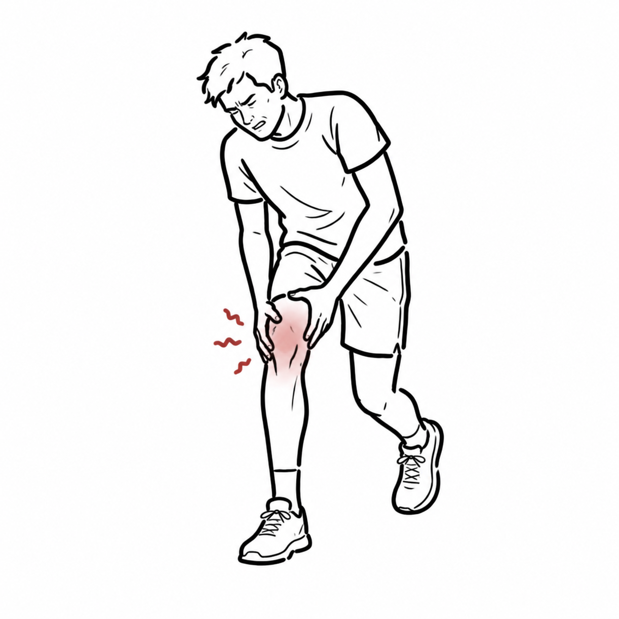
Anterior cruciate ligament (ACL) tear is one of the most common knee injuries, particularly in young athletes involved in pivoting sports (soccer, basketball, skiing). ACL injuries account for ~50% of all knee ligament injuries, with an incidence of ~68 per 100,000 annually, more common in females due to anatomical and biomechanical risk factors. ACL tears cause instability, impaired performance, and predispose to early osteoarthritis if untreated.
Definition
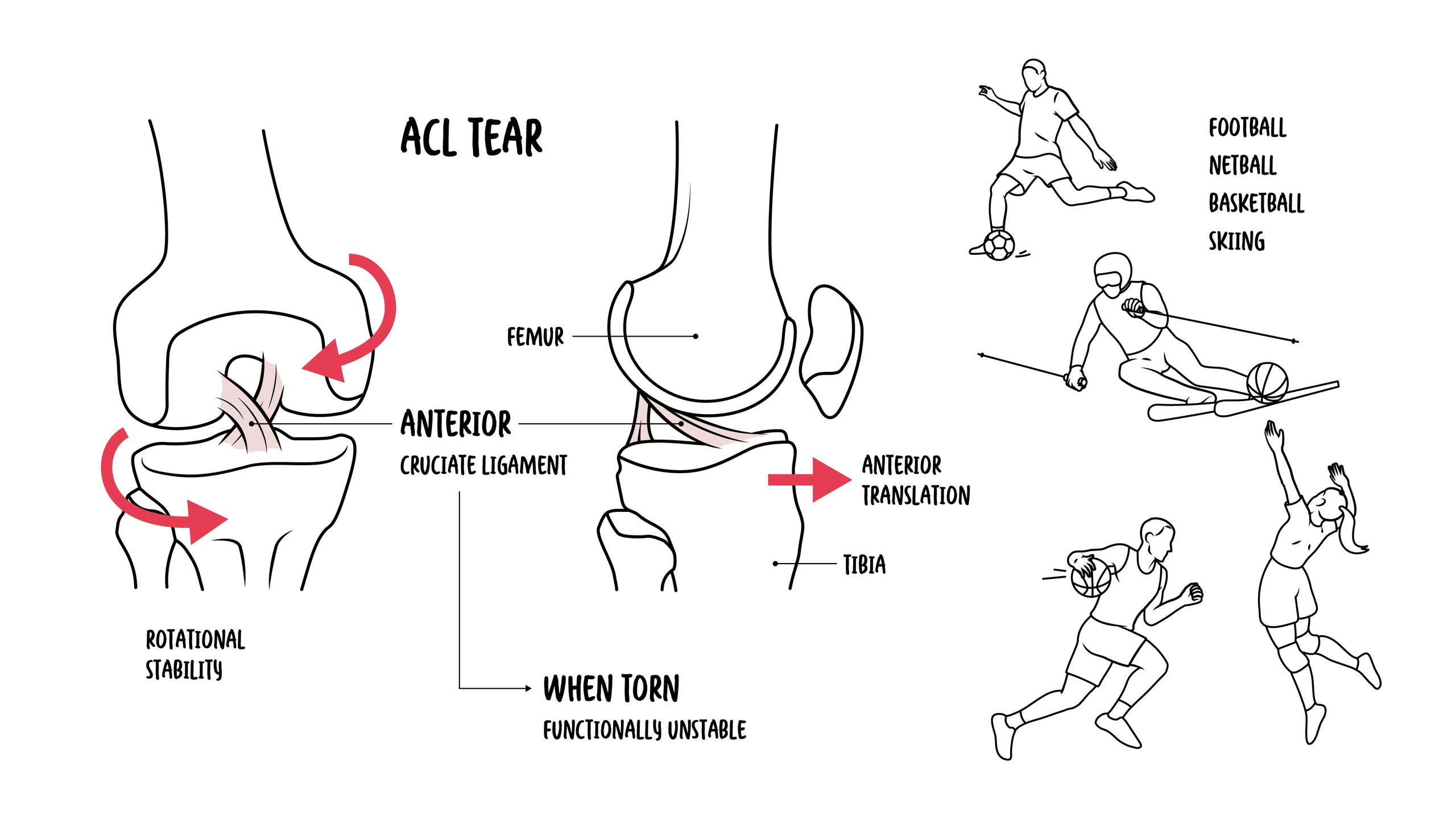
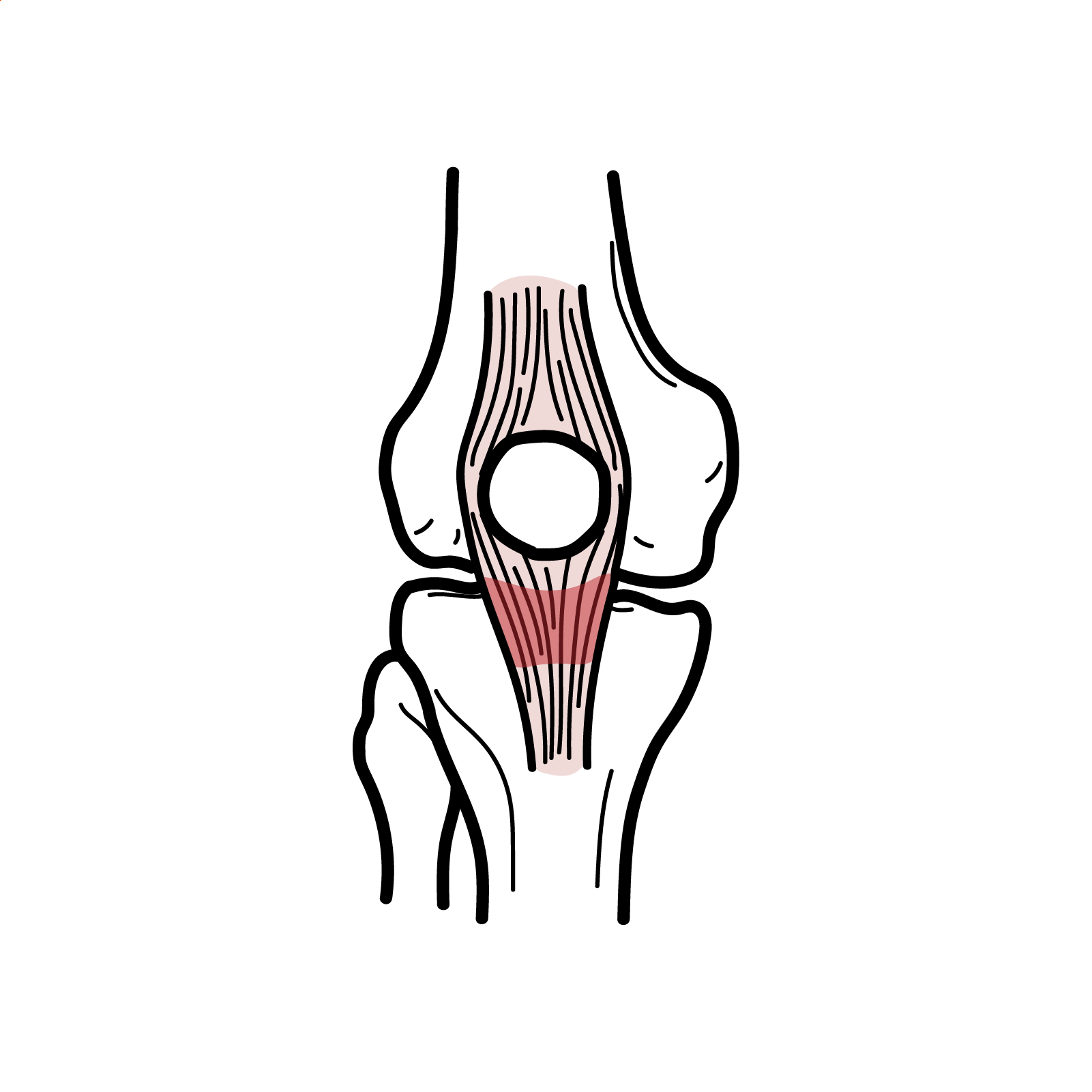
ACL: Intra-articular, extrasynovial ligament preventing anterior tibial translation and providing rotational stability.
Pivot shift: Sudden anterior subluxation of tibia during rotation; clinical sign of ACL deficiency.
Hemarthrosis: Acute intra-articular bleeding causing rapid knee swelling after ligament tear.
Non-contact injury: Injury mechanism where no direct trauma occurs, often from cutting/pivoting movements.
Anatomy & Physiology
- ACL origin: Posterior aspect of lateral femoral condyle.
- Insertion: Anterior intercondylar area of tibial plateau.
- Function: Restricts anterior translation of tibia relative to femur; stabilises knee during rotation and cutting movements.
- Bundles: Anteromedial (tight in flexion) and posterolateral (tight in extension).
ACL is the primary restraint to anterior tibial displacement.
Aetiology & Risk Factors
Aetiology
- Sudden deceleration, pivoting, or change in direction.
- Landing from a jump with knee in valgus/rotation.
- Direct blow to lateral knee causing valgus stress.
Risk Factors
- Female sex (smaller notch width, hormonal effects, neuromuscular control differences).
- High-risk sports: soccer, basketball, netball, skiing.
- Poor conditioning or hamstring–quadriceps imbalance.
- Previous ACL injury.
Pathophysiology
- High-energy force → valgus + external rotation or hyperextension.
- ACL fibres fail → partial or complete tear.
- Immediate hemarthrosis from ligament vascular supply.
- Knee instability → recurrent giving-way episodes.
- Long-term: meniscal tears, chondral damage, osteoarthritis.
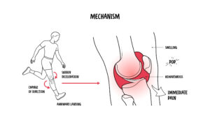
ACL injury is both an acute stabiliser failure and a long-term risk factor for degenerative joint disease.
Clinical Manifestations
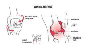
- “Pop” at injury, immediate swelling within 2–3 hours (hemarthrosis), inability to continue playing.
- Instability (“giving way”), difficulty pivoting.
Examination
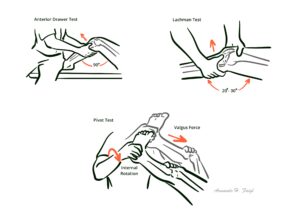
- Positive Lachman test (most sensitive).
- Positive Anterior drawer test.
- Positive Pivot shift test (most specific).
- Reduced ROM due to swelling.
Rapid hemarthrosis + pop + instability strongly suggests ACL tear.
Unhappy Triad of O’Donoghue: ACL tear, MCL tear, Medial meniscus tear.
Diagnosis
- Clinical: History + exam (Lachman = gold standard).
- MRI: Imaging of choice; sensitivity >95%, shows associated meniscal or chondral injuries.
- X-ray: Rule out fractures (Segond fracture = pathognomonic for ACL tear).
| Condition | Differentiating Feature |
| Meniscal tear | Locking/catching, delayed swelling, positive McMurray |
| MCL tear | Medial pain, valgus stress laxity |
| PCL tear | Posterior sag sign, mechanism = dashboard injury |
Treatment
- Acute management: RICE (rest, ice, compression, elevation), analgesia, knee brace, physiotherapy.
- Non-operative: Activity modification + rehab (quads/hamstring strengthening). Suitable for low-demand or older patients.
- Operative: Arthroscopic ACL reconstruction (hamstring/patellar tendon autograft). Indicated in young, active patients, recurrent instability, or associated meniscal/chondral injury.
- Rehabilitation: Gradual return to sport after 9–12 months, with neuromuscular training.
Young + active = surgery; older + low-demand = physiotherapy.
Complications & Prognosis
- Recurrent instability if untreated.
- Secondary meniscal tears.
- Early osteoarthritis.
- Arthrofibrosis (post-surgical).
- Graft failure after reconstruction.
- Good with surgical reconstruction + rehab
- High return to sport but increased long-term OA risk.
Even with reconstruction, ACL injury predisposes to OA.
References
- Griffin LY, Albohm MJ, Arendt EA, et al. Understanding and preventing noncontact ACL injuries. Am J Sports Med. 2006;34(9):1512–32.
- van Eck CF, Schkrohowsky JG, Working ZM, et al. Clinical examination in the diagnosis of ACL tears: a meta-analysis. Knee Surg Sports Traumatol Arthrosc. 2013;21(9):1989–96.
- Frobell RB, Roos HP, Roos EM, et al. Treatment for acute ACL tear: 5-year outcome of the randomized controlled trial. N Engl J Med. 2010;363:331–42.
- Filbay SR, Grindem H. Evidence-based recommendations for rehabilitation after ACL reconstruction. Curr Rev Musculoskelet Med. 2019;12(2):289–96.
- Sanders TL, Maradit Kremers H, Bryan AJ, et al. Incidence of ACL tears and reconstruction: a 20-year population-based study. Am J Sports Med. 2016;44(6):1502–7.






Members only discussions coming soon…