

Clubfoot (Talipes equinovarus)
Overview
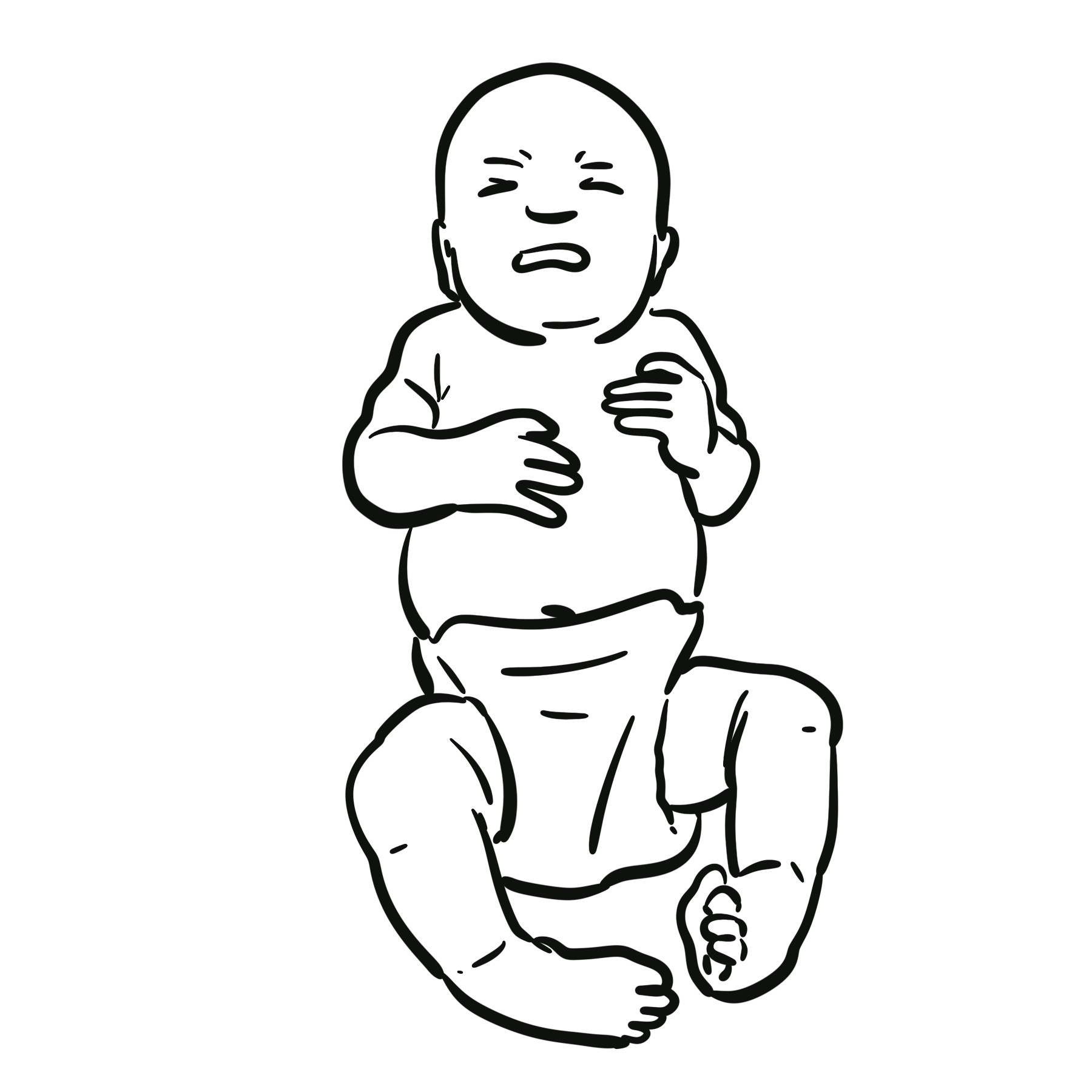
Clubfoot, or congenital talipes equinovarus (CTEV), is a congenital structural deformity in which the foot is fixed in cavus, forefoot adduction, hindfoot varus and ankle equinus (CAVE). It may affect one or both feet and involves abnormal shape and alignment of the bones, joints, muscles, tendons and ligaments below the knee.
Idiopathic clubfoot is the most common form and occurs without another recognised disorder. Non-isolated clubfoot accompanies neuromuscular or syndromic conditions and is often more resistant to correction.
The deformity is painless in infancy but does not resolve spontaneously. Untreated children bear weight on the lateral border or dorsum of the foot, leading to difficulty with footwear, callosities, pain and impaired walking. Early specialist treatment usually produces a plantigrade, flexible and functional foot.
Definition
- Cavus
- High medial arch caused by plantar flexion of the first ray relative to the rest of the forefoot.
- Adductus
- Medial deviation of the forefoot relative to the hindfoot.
- Varus
- Inversion and medial angulation of the hindfoot.
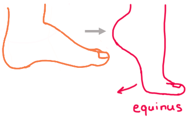
- Equinus
- Fixed plantar flexion at the ankle due to posterior soft-tissue and Achilles tendon tightness.
- Plantigrade foot
- A foot that can rest with the sole flat on the ground during standing and walking.
- Ponseti method
- Sequential manipulation, long-leg casting, usually Achilles tenotomy, and prolonged foot-abduction bracing.
Anatomy & Physiology
Normal foot position depends on coordinated alignment and motion of the talus, calcaneus, navicular, cuboid and forefoot, together with balanced action of the calf, Achilles tendon and intrinsic and extrinsic foot muscles.
- In clubfoot, the talar neck is shortened and the talar head points medially and plantarly.
- The navicular and cuboid are displaced medially; the calcaneus is inverted and adducted beneath the talus.
- Posteromedial soft tissues and the gastrocnemius–soleus–Achilles complex are contracted, maintaining hindfoot varus and ankle equinus.
- The affected foot and calf are commonly smaller, especially in unilateral disease; this difference may persist after successful correction.
Aetiology & Risk Factors
Aetiology
Most cases are idiopathic and probably result from multifactorial genetic and environmental influences. A positive family history increases risk, but no single cause explains most isolated cases.1
Non-isolated clubfoot may occur with neuromuscular or syndromic disorders, including spina bifida and arthrogryposis. These feet are often more rigid and may require longer treatment or additional procedures.1
Risk Factors
- Family history of clubfoot.
- Male sex; clubfoot is more common in boys.
- An associated neuromuscular or congenital syndrome.
- A previous affected child, reflecting familial susceptibility.
Pathophysiology
Abnormal bone morphology and relationships within the tarsus combine with contracted posteromedial soft tissues. The forefoot is pronated relative to the hindfoot, creating cavus, while the whole foot is adducted and the calcaneus remains in varus and equinus beneath the talus.
Successful correction requires the foot to move around the talar head as a coupled unit. The components must therefore be corrected in sequence: cavus first, then adductus and varus, and equinus last.
Trying to force ankle dorsiflexion before the calcaneus has abducted and the hindfoot varus has corrected can create a rocker-bottom deformity. Remember CAVE: Cavus → Adductus → Varus → Equinus.
Clinical Manifestations
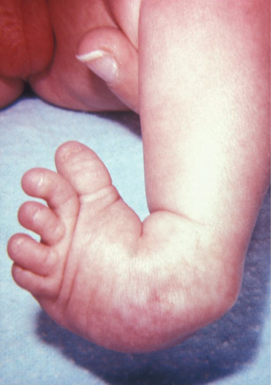
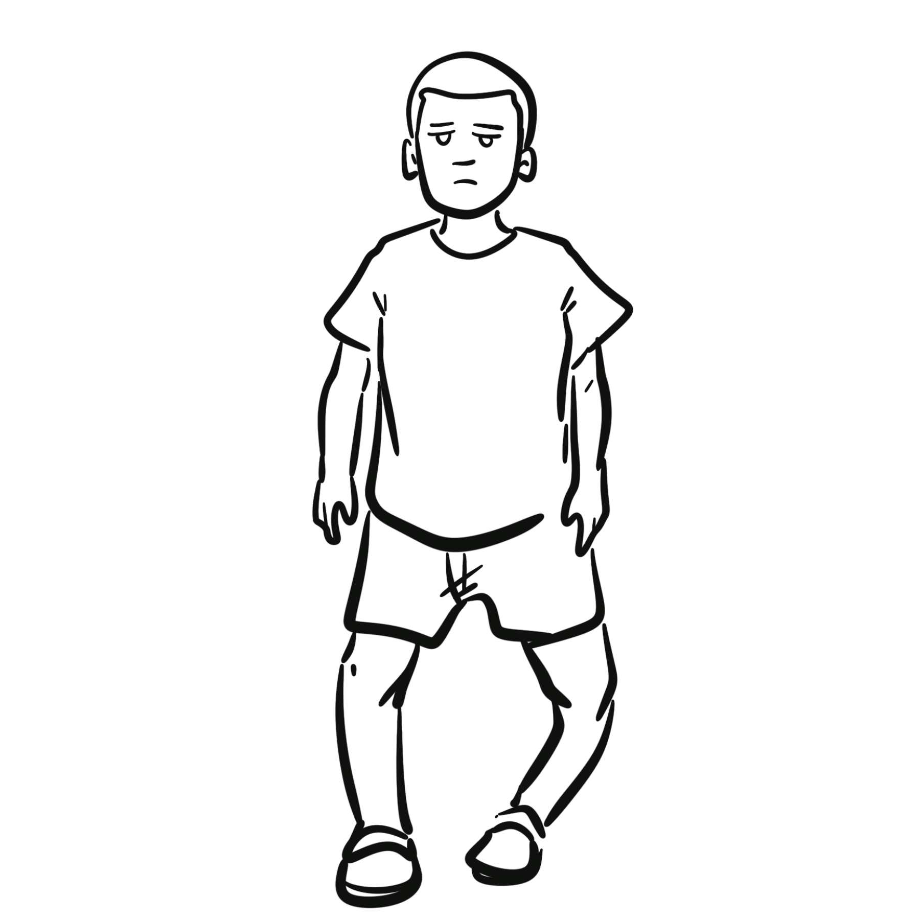
- Foot turned inward and downward at birth, with a high medial arch and deep medial and posterior skin creases.
- Forefoot adduction and supination, hindfoot varus and fixed ankle equinus—the CAVE pattern.
- Rigid or incompletely correctable deformity on gentle manipulation, distinguishing structural clubfoot from positional talipes.
- Shortened Achilles tendon, small heel and underdeveloped calf.
- The affected foot may be shorter and narrower than the unaffected foot.
- Unilateral or bilateral involvement.
- The hips, spine and neurological status should also be examined.
Diagnosis & Investigations
Diagnosis is clinical and is usually evident on newborn examination. The foot is held in equinus and inversion, and the structural deformity cannot be fully corrected with gentle passive movement.2
Investigations
- Prenatal ultrasonography: May detect clubfoot from the second trimester, but false-positive diagnoses occur. Postnatal examination is required for confirmation.
- Plain radiographs: Not routinely required when typical isolated clubfoot is clinically apparent because neonatal tarsal bones are incompletely ossified. Imaging may help with an atypical presentation, uncertain diagnosis or assessment of complex or recurrent deformity.
An antenatal ultrasound diagnosis supports counselling and early referral, but treatment decisions are based on the postnatal clinical examination.
Treatment
The goal is a painless, flexible, plantigrade foot that permits ordinary footwear and normal functional activity. Treatment should begin soon after birth under a clinician trained in clubfoot management, although delayed presentation can still respond to Ponseti treatment.1
Ponseti Method—First-line Treatment
- Manipulation and serial casting: Gentle manipulation and weekly long-leg casts progressively correct the deformity in CAVE order. Initial correction commonly takes approximately 6–8 weeks.
- Percutaneous Achilles tenotomy: Achilles tenotomy is performed when residual equinus persists after cavus, adductus and varus have been corrected. It is required in most infants. A final cast is generally retained for approximately three weeks while the tendon heals.
- Foot-abduction bracing: A foot-abduction brace, commonly called “boots and bar”, maintains the correction. It is worn for approximately 23 hours each day during the first three months and then during sleep—usually 12–14 hours daily—for at least three to four years. Individual protocols may continue until four to five years of age.
- Follow-up: Regular follow-up is required to assess brace fit, skin integrity, foot position and early evidence of recurrence.
The foot-abduction brace does not perform the initial correction. Its essential role is to preserve the correction while the child grows. Poor brace adherence is strongly associated with relapse.1,2
Relapse or Resistant Deformity
- Early recurrence is usually treated with repeat Ponseti manipulation and casting.
- Repeat Achilles tenotomy may be required if equinus persists.
- Dynamic supination in a walking child may be treated with tibialis anterior tendon transfer after passive correction has been restored.
- Limited surgery is reserved for deformity that cannot be corrected or repeatedly relapses despite appropriate non-operative treatment.
- Extensive soft-tissue release is avoided where possible because it can produce a painful, stiff foot later in life.
Complications & Prognosis
Complications
- Relapse, particularly after early discontinuation or inconsistent use of the foot-abduction brace.
- Relapse rates increase with longer follow-up.
- Skin irritation, pressure areas or cast slippage.
- Neurovascular compromise, which is uncommon but requires urgent review.
- Rocker-bottom deformity from incorrect manipulation, especially forced dorsiflexion before correction of hindfoot varus.
- Residual cavus, adductus, varus or equinus.
- Persistent stiffness.
- Dynamic supination during gait.
- Following extensive surgery: scarring, stiffness, pain, overcorrection and later degenerative change.1,2
Prognosis
With timely, correctly performed Ponseti treatment and sustained bracing, most children develop a functional, nearly normal foot and can run, play and wear ordinary shoes.
The treated foot may remain slightly smaller and less mobile, with a smaller calf and occasionally a mildly shorter leg. These differences usually have little functional impact.
Long-term surveillance is important because recurrence can occur throughout growth. Systematic-review data show wide variation in reported relapse, partly because definitions and follow-up durations differ; recurrence becomes more apparent as follow-up lengthens.3
References
- American Academy of Orthopaedic Surgeons. Clubfoot [Internet]. Rosemont (IL): AAOS; [cited 2026 Sep 7]. Available from: https://www.orthoinfo.org/diseases–conditions/clubfoot/
- Hosseinzadeh P. Clubfoot [Internet]. Oakbrook Terrace (IL): Pediatric Orthopaedic Society of North America; [cited 2026 Sep 7]. Available from: https://posna.org/physician-education/study-guide/clubfoot
- Gelfer Y, Wientroub S, Hughes K, Fontalis A, Eastwood DM. Congenital talipes equinovarus: a systematic review of relapse as a primary outcome of the Ponseti method. Bone Joint J. 2019;101-B(6):639–645. doi:10.1302/0301-620X.101B6.BJJ-2018-1421.R1.


Members only discussions coming soon…