

Paediatric Orthopaedics Overview
Biology of the Immature Skeleton
- Physis (Growth Plate): Cartilaginous zone of longitudinal growth. Most susceptible to shear and tension forces (specifically the hypertrophic zone). Damage carries a high risk of growth arrest, limb-length discrepancy, or progressive angular deformity.
- Periosteum: Thick, metabolically active, and mechanically resilient. Limits fracture displacement, provides an intact hinge for closed reduction, and drives rapid periosteal callus formation (non-union is exceptionally rare).
- Remodeling Capacity: Highest in children with substantial remaining skeletal growth (>2 years growth remaining), injuries located near an active physis, and angulation oriented in the plane of joint motion (flexion/extension). Rotational malalignment does not remodel.
- Paediatric-Specific Fracture Patterns:
- Torus / Buckle Fracture: Axial compression failure of metaphyseal cortex without cortical disruption. Stable; managed with simple splinting/removable cast for 3–4 weeks.
- Greenstick Fracture: Incomplete fracture; tensile cortex fractures while the compressive cortex remains intact and plastically deformed. Requires completion of fracture or reduction if significantly angulated.
- Plastic Deformation (Bowing): Microscopic yielding of bone without overt cortical line interruption (often fibula or ulna). Does not recoil spontaneously; may require manual straightening under sedation.
Salter-Harris Classification (Physeal Fractures)
- Type I (Same/Straight across): Fracture plane traverses purely through the hypertrophic zone of the physis. Excellent prognosis; managed with closed reduction and casting.
- Type II (Above): Fracture runs through the physis and exits into the Metaphysis (producing the triangular Thurston-Holland fragment). Most common pattern (~75%); excellent prognosis; closed reduction.
- Type III (Lower): Fracture runs through the physis and exits intra-articularly through the Epiphysis. Carries risk of joint incongruity and physeal bridge; requires anatomic reduction (often ORIF).
- Type IV (Through): Fracture passes vertically through Metaphysis, Physis, and Epiphysis. High risk of permanent growth arrest; requires exact anatomic reduction and internal fixation.
- Type V (Ram / Crush): Compression or crushing injury to the physis. High rate of growth arrest; typically diagnosed retrospectively following premature closure.
Common Hip Disorders by Age
Age Timeline of Paediatric Hip Pathology
- 0 – 6 Months: Developmental Dysplasia of the Hip (DDH)
- 3 – 8 Years: Transient Synovitis, Perthes Disease (Legg-Calvé-Perthes)
- 10 – 16 Years: Slipped Capital Femoral Epiphysis (SCFE), Apophyseal avulsions
Developmental Dysplasia of the Hip (DDH)
- Etiology & Risk Factors: Acetabular dysplasia, subluxation, or frank dislocation. Risk factors: Female sex (female:male 4:1), breech presentation, family history, first-born child, oligohydramnios.
- Physical Examination:
- Neonate to 3 months:
- Barlow Test: Hip flexed and adducted; posterior pressure applied to elicit dislocation/subluxation (checks if hip is dislocatable).
- Ortolani Test: Hip flexed and gently abducted while lifting greater trochanter anteriorly; elicits a “clunk” as head reduces (checks if hip is reducible).
- Infant > 3 months: Soft-tissue contractures develop. Barlow/Ortolani become negative. Key signs are limited hip abduction (<60° when flexed), asymmetric thigh/gluteal skin folds, and Galeazzi sign (unequal knee heights with hips and knees flexed to 90° due to femoral shortening).
- Neonate to 3 months:
- Imaging:
- Age < 4–6 months: Dynamic ultrasound (Graf classification; normal alpha angle > 60°).
- Age > 4–6 months: AP pelvis X-ray (femoral head ossification center appears). Assess Hilgenreiner’s line, Perkin’s line, and Shenton’s line (intact smooth arch between inferior pubic ramus and medial femoral neck).
- Management:
- < 6 months: Pavlik harness (dynamic flexion and abduction; avoid hyperabduction to prevent femoral head osteonecrosis).
- 6–18 months: Closed reduction under GA + hip spica casting for 12 weeks.
- > 18 months or failed closed: Open reduction +/- pelvic (e.g., Salter) or femoral derotation/varus osteotomy.
Legg-Calvé-Perthes Disease (LCPD)
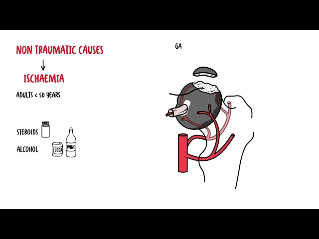
- Pathophysiology: Idiopathic avascular necrosis of the capital femoral epiphysis with subsequent revascularization, resorption, and re-ossification.
- Clinical Presentation: Boys aged 4–8 years presenting with an insidious, mildly painful or painless antalgic limp. Pain frequently referred to the ipsilateral thigh or knee.
- Physical Examination: Reduced internal rotation and abduction of the affected hip. Mild disuse atrophy of thigh musculature.
- Imaging & Stages (Waldenström):
- Initial/Necrotic: Small, sclerotic epiphysis, widening of cartilage space.
- Fragmentation: Resorption of necrotic bone with radiolucent areas and subchondral collapse (crescent sign).
- Re-ossification: New bone formation progressing from periphery inward.
- Healed/Residual: Epiphysis reconstituted; variable residual coxa magna, coxa plana, or coxa breva.
- Management & Prognosis:
- Self-limiting over 2–4 years. Aim is joint preservation and containment of the femoral head within the acetabulum (physiotherapy, abduction bracing, or femoral/pelvic osteotomy if lateral subluxation occurs).
- Strongest prognostic factor is age at presentation (<6 years carries an excellent outcome; >8 years carries higher risk of premature osteoarthritis).
Slipped Capital Femoral Epiphysis (SCFE)
- Pathophysiology: Posterior and inferior displacement of the femoral neck relative to the capital femoral epiphysis through the hypertrophic zone of the physis.
- Demographics: Typically overweight adolescent males (ages 11–14). Consider underlying endocrinopathies (hypothyroidism, growth hormone deficiency, hypopituitarism) if patient is <10 years, >16 years, or underweight.
- Presentation: Dull antalgic limp, groin pain, anterior thigh pain, or isolated referred knee pain. Examination reveals obligate external rotation of the leg during passive hip flexion.
- Classification:
- Stable: Patient can ambulate (with or without crutches). AVN rate < 10%.
- Unstable: Patient unable to bear weight even with crutches. High risk of avascular necrosis (up to 50%).
- Imaging: AP and frog-leg lateral radiographs of both hips.
- Klein’s Line: A line drawn along the superior border of the femoral neck should intersect the lateral capital femoral epiphysis. Failure to intersect = Trethowan’s sign (indicates slip).
- Management:Orthopaedic emergency. Strictly non-weight-bearing immediately upon suspicion. Urgent surgical stabilization via in-situ single percutaneous cannulated screw fixation. Prophylactic pinning of the contralateral normal hip is frequently performed due to 20–40% risk of subsequent bilateral slip.
The Limping Child: Septic Arthritis vs. Transient Synovitis
Differentiating between an urgent septic hip and benign transient synovitis is critical.
Kocher Criteria (Hip Septic Arthritis)
- Non-weight-bearing on affected side
- Oral temperature > 38.5°C
- Erythrocyte sedimentation rate (ESR) > 40 mm/h
- White blood cell count (WBC) > 12.0 x 10^9/L(Fifth adjunct criterion frequently used: C-Reactive Protein [CRP] > 20 mg/L)
- Risk Stratification:
- 1 criterion: ~3% risk of septic arthritis
- 2 criteria: ~40% risk
- 3 criteria: ~93% risk
- 4 criteria: ~99% risk
- Investigation & Management:
- Suspicion of septic arthritis warrants immediate urgent ultrasound to identify joint effusion.
- Effusion with high clinical score mandates urgent needle arthrocentesis (synovial fluid for cell count, Gram stain, culture, and crystals) prior to antibiotic administration where feasible.
- Septic arthritis requires emergent operative drainage and washout (open arthrotomy or arthroscopic decompression) followed by targeted IV antibiotics (most commonly targeting Staphylococcus aureus, Streptococcus pyogenes, and Kingella kingae in children < 4 years).
Lower Limb Alignment & Rotational Variations
Physiological Coronal Alignment Progression
- Birth to 18 months: Physiological Genu Varum (bow legs) up to 15°.
- 18 to 24 months: Neutral axis (straight legs).
- 3 to 4 years: Physiological Genu Valgum (knock knees) reaching peak of ~10–12°.
- 6 to 7 years: Regresses to normal adult physiological valgus (~5–7°).
Pathological Coronal Deformities
- Tibia Vara (Blount Disease): Idiopathic growth arrest of the posteromedial proximal tibial physis, causing progressive, sharp varus angulation. Radiographs show medial metaphyseal beaking, down-sloping, and fragmentation (Langenskiöld stages I–VI). Differentiated from physiologic bowing by asymmetry, progression, and sharp angulation at the proximal tibia rather than a smooth tibial/femoral curve.
- Rickets: Defective mineralization of osteoid (vitamin D deficiency, renal tubular acidosis). Bilateral symmetric bowing, metaphyseal cupping, fraying, and flaring on plain films.
Rotational Profiling (Intoeing)
- Metatarsus Adductus: Medial deviation of the forefoot relative to the hindfoot with a convex lateral foot border. 90% resolve spontaneously by age 1; rigid cases require serial casting.
- Internal Tibial Torsion: Most common cause of intoeing in ages 1–3 years. Foot points medially relative to patella; negative thigh-foot angle (normal is +10° to +15°). Resolves spontaneously by age 4–5.
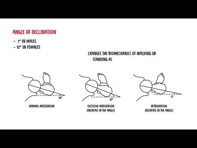
- Increased Femoral Anteversion: Most common cause of intoeing in children > 3 years. Internal rotation of hip is marked (>70°), external rotation is restricted (<20°). Child typically sits in “W” position. Normalizes spontaneously by age 8–10 years; derotation osteotomy reserved only for rare, functionally impaired adolescents.
Congenital Foot & Spine Deformities
Congenital Talipes Equinovarus (CTEV / Clubfoot)
- Anatomical Components (Mnemonic: CAVE):
- C – Cavus: High medial longitudinal arch due to plantarly flexed 1st metatarsal.
- A – Adductus: Medial deviation of forefoot and midfoot at Chopart/Lisfranc joints.
- V – Varus: Inward angulation of hindfoot/calcaneus.
- E – Equinus: Fixed ankle plantarflexion due to tight Achilles tendon.
- Treatment (Ponseti Technique):
- Gold standard. Weekly serial manipulations and long-leg casting.
- Correction order: Correct Cavus first (supinating forefoot to align with hindfoot), then correct Adductus and Varus simultaneously by abducting the foot in supination around a stabilized talar head.
- Percutaneous Achilles tenotomy performed under local anesthesia in ~90% of cases to correct residual Equinus.
- Maintenance: Foot abduction orthosis (boots on a bar) worn 23 hours/day for 3 months, then during nights/naps until age 4–5 years to prevent relapse.
Adolescent Idiopathic Scoliosis (AIS)
- Definition: Structural, lateral curvature of the spine in the coronal plane with Cobb angle >= 10°, accompanied by vertebral axial rotation. Most common in adolescent females during peak growth velocity.
- Assessment:
- Adams Forward Bend Test: Highlights thoracic rib hump or lumbar prominence measured using a scoliometer (angle of trunk rotation >= 7° warrants spinal radiography).
- Skeletal Maturity Grading: Assessed using the Risser sign (ossification of iliac crest apophysis, grades 0 to 5) and status of triradiate cartilage (open vs. closed).
- Treatment Matrix (Based on Cobb Angle & Skeletal Maturity):
- Cobb < 25°: Observation with serial clinical exam and standing long-cassette radiographs every 6 months until skeletal maturity.
- Cobb 25° to 45° in skeletally immature patients (Risser 0–2): Rigid thoracolumbosacral orthosis (TLSO bracing) worn 16–18 hours per day until growth cessation.
- Cobb > 45° to 50°: Surgical intervention (posterior spinal instrumented fusion) to halt progression and correct deformity.



Members only discussions coming soon…