

Septic Arthritis

Overview
Septic arthritis is infection within a joint, usually involving a synovial joint and most often caused by bacteria. Acute bacterial infection of a native joint is a medical and surgical emergency because inflammation can rapidly damage cartilage and may be accompanied by bacteraemia or sepsis. A painful swollen joint warrants urgent evaluation even without fever. Diagnosis centres on prompt joint aspiration for microbiology, while treatment combines source control with appropriately selected antimicrobial therapy.
Definition
Septic arthritis: Infection of a joint space and synovium.
Native joint: A joint without a prosthetic implant; prosthetic joint infection needs a distinct management pathway.
Arthrocentesis: Needle aspiration of synovial fluid for diagnostic testing and sometimes decompression.
Synovial fluid culture: Microbiological test to identify a causative organism and guide antibiotic treatment.
Crystal arthritis: Joint inflammation due to urate or calcium pyrophosphate crystals; crystals do not exclude concurrent infection.

Anatomy & Physiology
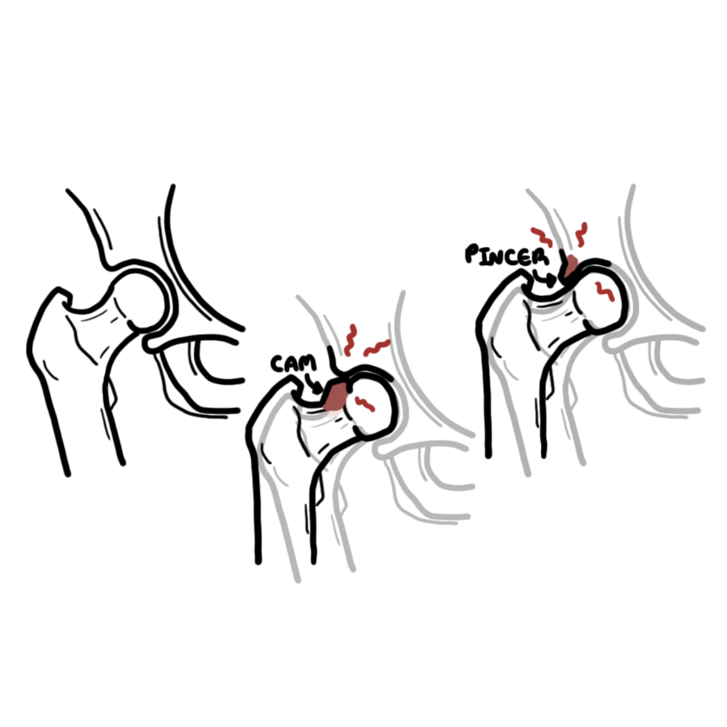
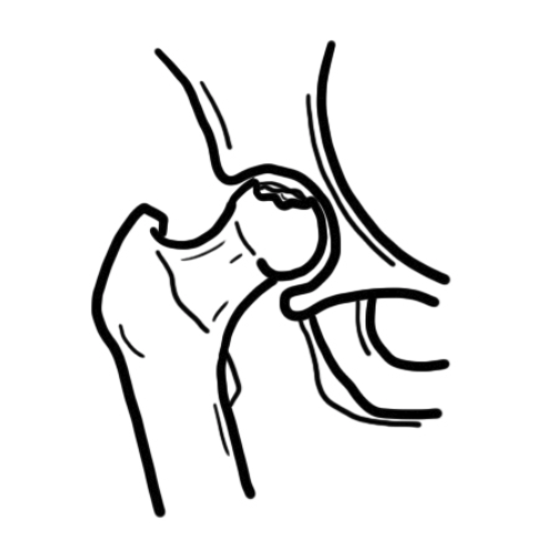
Synovial joints have cartilage-covered bone surfaces enclosed by a capsule lined with vascular synovium. Synovial fluid lubricates the joint and nourishes cartilage. Organisms can enter from the bloodstream, through an injury or procedure, or from an adjacent focus of infection. The hip is a deep joint, so an effusion or infection may be less obvious on examination; ultrasound can help identify fluid and guide aspiration.
Aetiology & Risk Factors
| Organism or setting | Clinical relevance |
|---|---|
| Staphylococcus aureus | A leading cause of acute bacterial native-joint infection across age groups; consider local methicillin resistance patterns. |
| Streptococci | Another common cause; skin, soft tissue or systemic infection may coexist. |
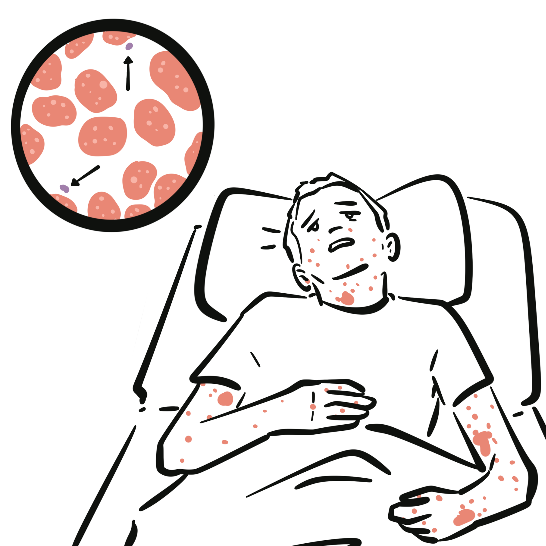
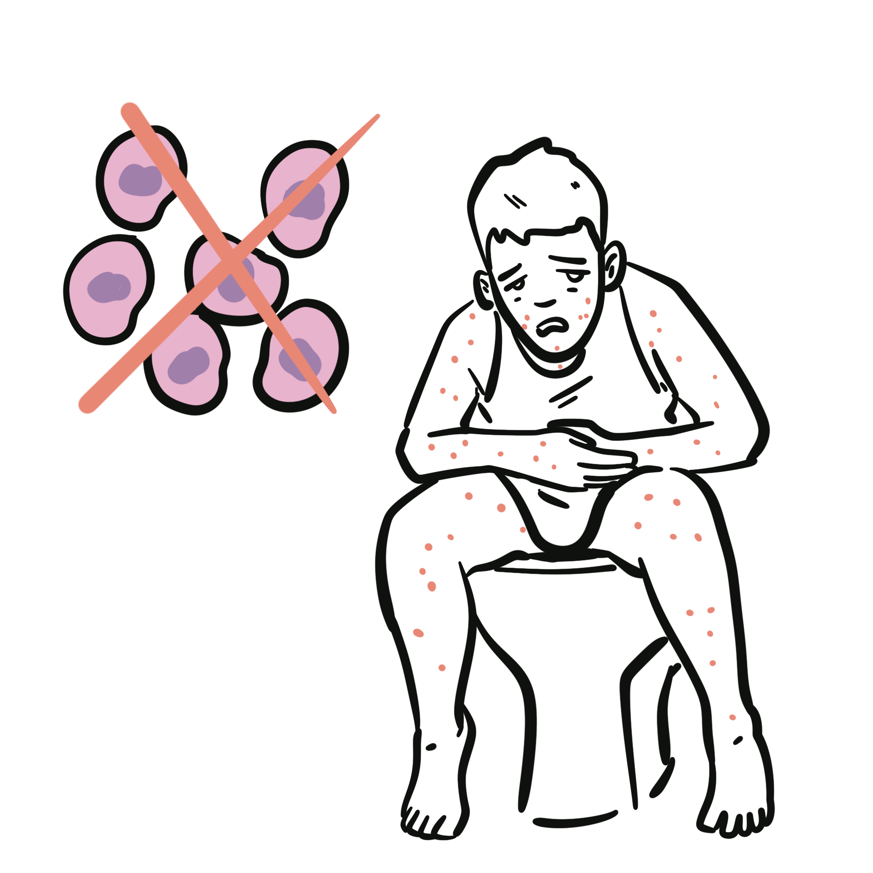
| Neisseria gonorrhoeae | Consider in sexually active people with compatible exposure; disseminated infection may include tenosynovitis and rash. |
| Children | S. aureus remains important; Kingella kingae is relevant in young children, and vaccination status affects other organisms. |
| Other pathogens | Gram-negative organisms, mycobacteria and fungi are considered according to host factors, exposures and a more indolent course. |
Predisposing factors include older age, diabetes, rheumatoid arthritis or other damaged joints, immunosuppression, skin infection, bacteraemia, injection drug use, penetrating trauma and recent joint injection or surgery. A prosthetic joint raises concern for implant-associated infection and requires a separate specialist pathway.
Pathophysiology
Microorganisms reach the synovium most often via haematogenous spread. Rapid bacterial proliferation and the host inflammatory response increase joint fluid and pressure. Neutrophils and inflammatory mediators release enzymes that damage cartilage; delayed drainage or antimicrobial treatment increases the risk of permanent loss of joint function. Infection may extend into adjacent bone or soft tissue, and bacteria in blood can cause systemic sepsis.
Gross and Microscopic Pathology
In acute bacterial infection the synovium is inflamed and fluid may appear turbid or purulent. Synovial fluid is commonly neutrophil-rich, but appearance and cell count cannot independently confirm or exclude infection. With progression, cartilage and subchondral bone can be damaged. Specific microbiological identification relies on Gram stain, culture or selected molecular tests; a negative initial Gram stain does not rule out septic arthritis.
Clinical Manifestation
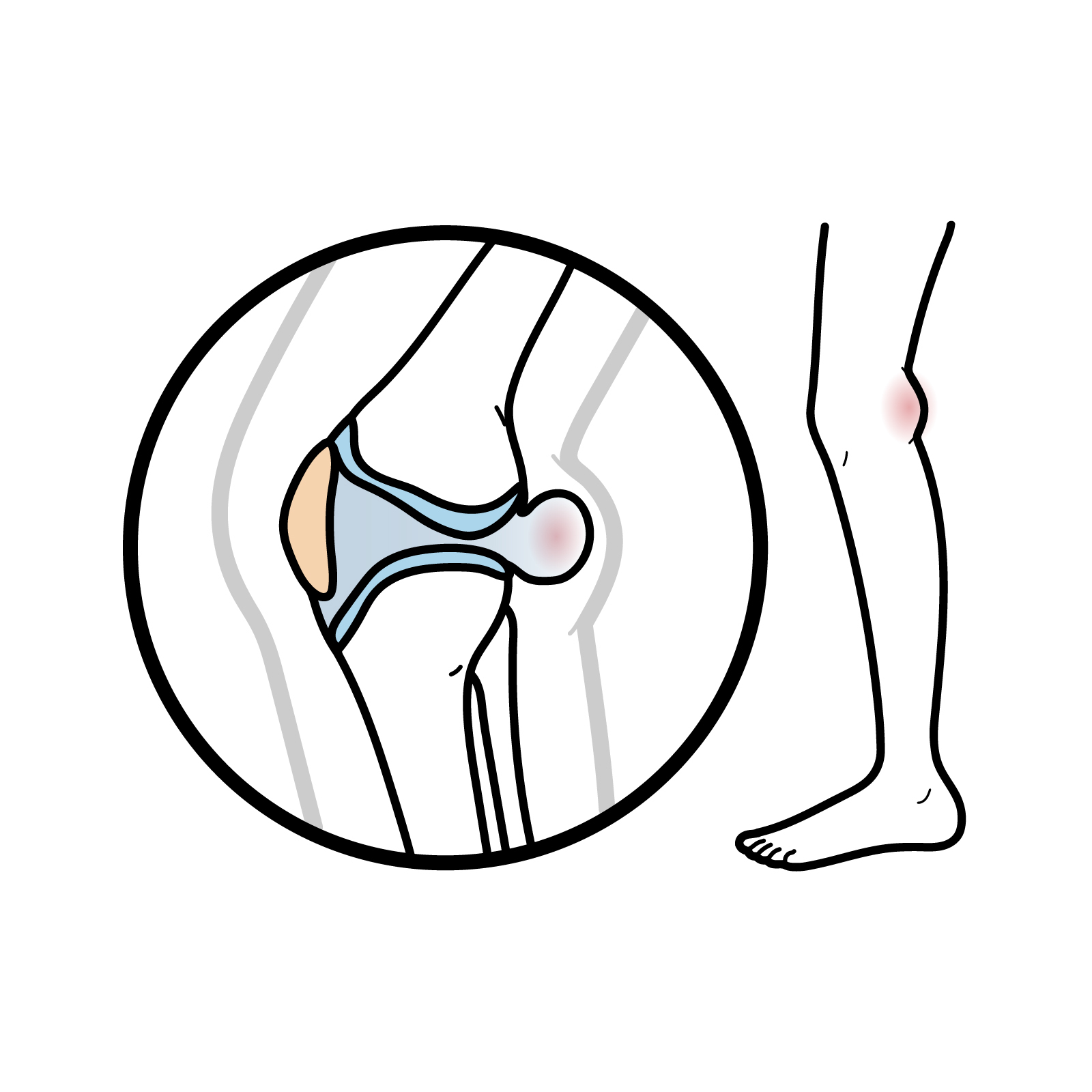
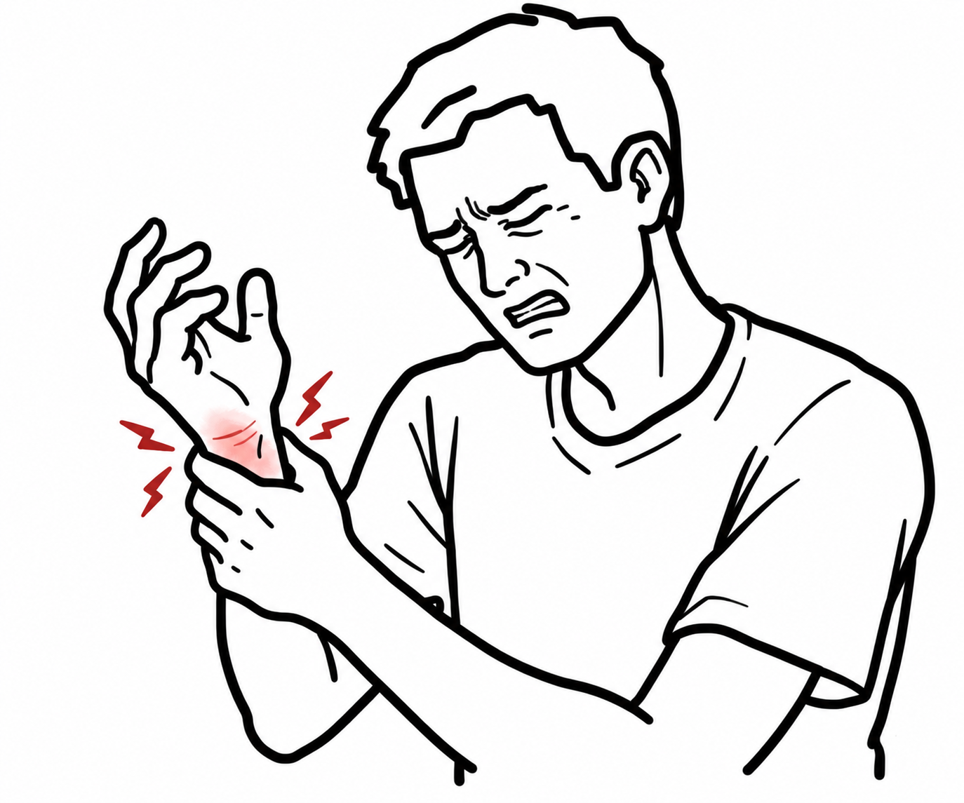
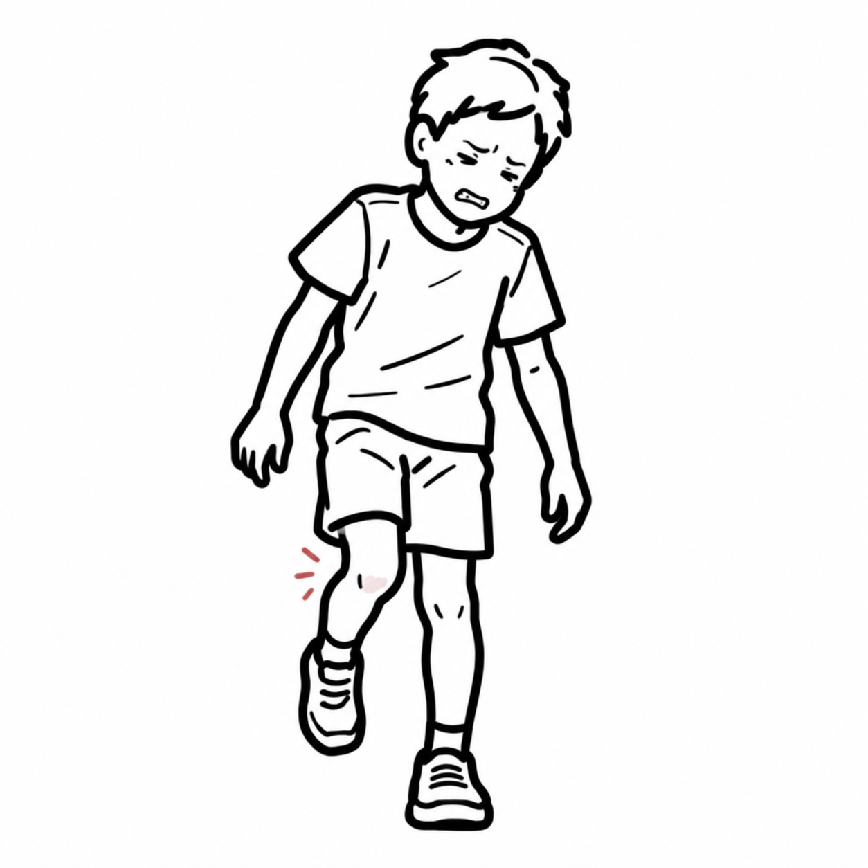
- Acute severe joint pain, swelling, warmth, effusion and marked pain on movement; the knee is commonly affected, but any joint can be involved.
- Reduced range of motion and inability to bear weight, particularly with lower-limb or hip involvement.
- Fever, rigors or malaise may occur, but absence of fever does not exclude infection.
- Most cases involve one joint; multiple joints can be affected, especially in systemic infection or vulnerable patients.
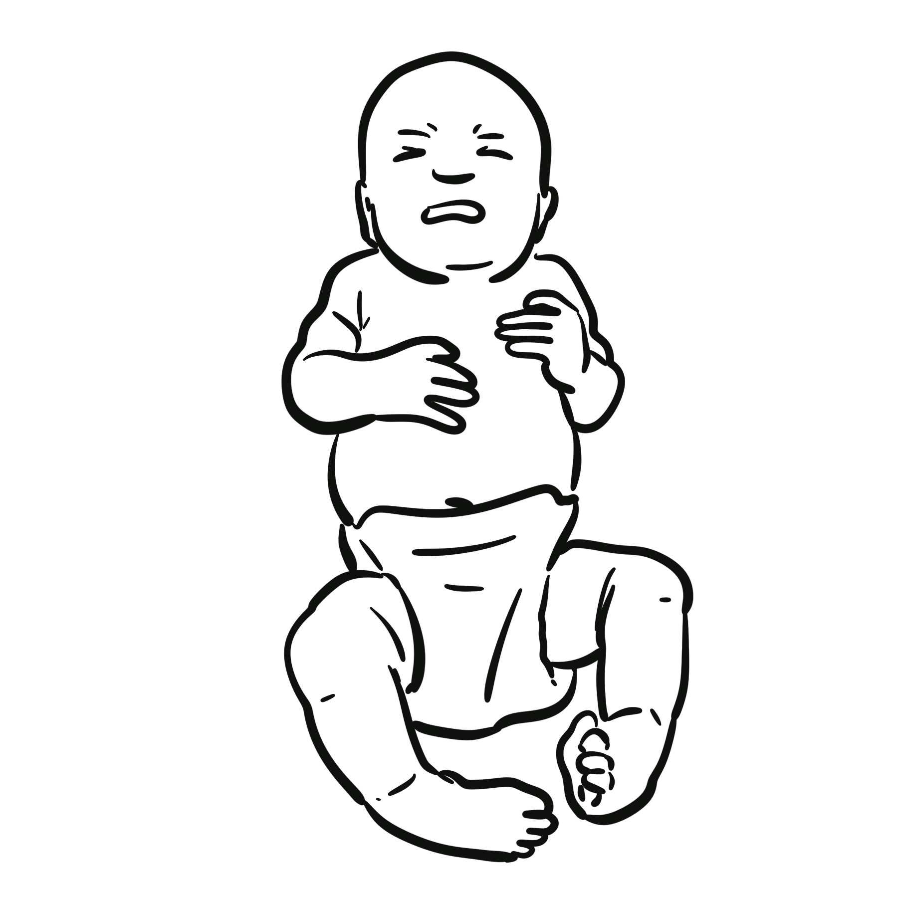
- The hip or shoulder may be painful without striking superficial redness or swelling. In children, limp, refusal to bear weight or irritability may predominate.
A hot swollen joint is septic arthritis until urgent assessment has addressed the possibility. Gout or calcium pyrophosphate crystals can coexist with infection.

Diagnosis
Arrange urgent hospital assessment and aspiration of an accessible affected joint. If the patient is stable, obtain synovial and indicated blood cultures before empirical antibiotics when this will not cause a harmful delay. In sepsis or septic shock, give prompt antibiotics and resuscitation under the sepsis pathway; do not wait for aspiration.
| Test | Interpretation and limitation |
|---|---|
| Joint aspiration | Send fluid for Gram stain and culture, cell count with differential, and crystal analysis. Culture is central to organism identification; a negative Gram stain or culture does not necessarily exclude infection. |
| Synovial white cell count | A high count, especially above 50,000/µL, supports infection but is not diagnostic. Lower counts, including below 25,000/µL, reduce probability but cannot rule it out, particularly with immunosuppression or early infection. |
| Crystal analysis | Urate or calcium pyrophosphate crystals support crystal arthritis, but infection can coexist. |
| Blood cultures | Obtain when febrile, septic or bacteraemia is suspected, ideally before antibiotics when feasible. |
| Full blood count, CRP, ESR and renal function | Assess inflammation, organ function and a baseline for monitoring; normal or modest inflammatory markers do not safely exclude infection. |
| Ultrasound | Detects effusion and guides aspiration of a difficult joint, especially the hip. |
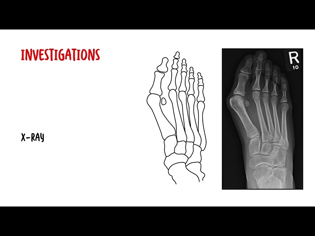
| Plain radiographs; MRI when indicated | Radiographs assess other pathology and provide a baseline. MRI can assess adjacent osteomyelitis, abscess or difficult deep-joint infection. |
Differential Diagnosis
- Gout or calcium pyrophosphate deposition disease.
- Inflammatory arthritis flare, including rheumatoid or psoriatic arthritis.
- Traumatic haemarthrosis or fracture.
- Reactive or viral arthritis.
- Cellulitis, bursitis or adjacent osteomyelitis.
- Transient synovitis in a child with hip pain; infection must first be considered when the child is unwell or cannot bear weight.
Classification
| Clinical category | Key distinction |
|---|---|
| Acute bacterial native-joint septic arthritis | Rapid presentation; bacterial identification and urgent drainage plus antibiotics are priorities. |
| Gonococcal arthritis | Associated with disseminated N. gonorrhoeae infection; consider sexual history and testing at relevant mucosal sites. |
| Indolent infection | Mycobacterial or fungal infection may present more gradually and needs targeted investigation and treatment. |
| Prosthetic joint infection | Biofilm and implanted material change diagnostic criteria and management; follow a separate specialist protocol. |
Treatment
- Initial care: urgent orthopaedic and infectious diseases or rheumatology input, analgesia, assessment for sepsis and supportive care.
- Source control: aspirate and drain the infected joint. Arthroscopic lavage is commonly used for large joints; open surgery or repeat drainage may be needed depending on joint, stage and clinical response.
- Antibiotics: after appropriate samples in a stable patient, start empirical therapy covering likely organisms according to local resistance, age, exposure and Gram stain. Start immediately under a sepsis pathway if unstable. Narrow therapy when cultures and susceptibilities return.
- Route and duration: individualise intravenous-to-oral switch and total course according to organism, source control, response and whether osteomyelitis is present; monitor symptoms and CRP rather than using a fixed regimen for every patient.
- Recovery: begin mobilisation and rehabilitation once infection is controlled and drains are removed, to limit stiffness and contracture.
NSAIDs may help pain if appropriate, but they do not treat the infection. Antimicrobial treatment and adequate joint drainage are the core interventions.
Complications & Prognosis
Complications
- Permanent cartilage destruction, reduced range of motion and post-infectious osteoarthritis.
- Bacteraemia, sepsis or septic shock; spread to adjacent bone or soft tissue.
- Recurrent or persistent infection requiring further drainage or revised antimicrobial treatment.
- In children, hip or growth plate involvement can result in growth disturbance or deformity.
- Treatment harms include antibiotic adverse effects, line complications and operative risks.
Prognosis
Early recognition, microbiological sampling, effective antibiotics and drainage improve the chance of preserving joint function. Outcomes vary with age, comorbidity, causative organism, joint involved, associated sepsis and treatment delay. Some patients have persistent pain or disability despite eradication of infection; severe systemic illness can be fatal. Follow-up should assess clinical response, inflammatory markers and recovery of movement.
References
- Ravn C, Neyt J, Benito N, Abreu MA, Achermann Y, Bozhkova S, et al. Guideline for management of septic arthritis in native joints (SANJO). J Bone Jt Infect. 2023;8(1):29-37. doi:10.5194/jbji-8-29-2023.
- Woods CR, Bradley JS, Chatterjee A, Kronman MP, Arnold SR, Robinson J, et al. Clinical practice guideline by the Pediatric Infectious Diseases Society (PIDS) and the Infectious Diseases Society of America (IDSA): 2023 guideline on diagnosis and management of acute bacterial arthritis in pediatrics. J Pediatric Infect Dis Soc. 2024;13(1):1-59. doi:10.1093/jpids/piad089.
- Mathews CJ, Weston VC, Jones A, Field M, Coakley G. Bacterial septic arthritis in adults. Lancet. 2010;375(9717):846-855. doi:10.1016/S0140-6736(09)61595-6.
- Carpenter CR, Schuur JD, Everett WW, Pines JM. Evidence-based diagnostics: adult septic arthritis. Acad Emerg Med. 2011;18(8):781-796. doi:10.1111/j.1553-2712.2011.01121.x.



Members only discussions coming soon…