

Otitis Media
Overview
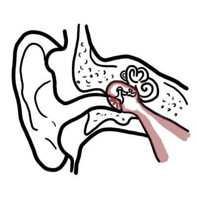
Otitis media is an inflammatory disease of the middle ear cleft, which encompasses the Eustachian tube, tympanic cavity, mastoid antrum, and mastoid air cells. It represents a disease continuum ranging from acute suppurative bacterial infection to sterile fluid effusion and chronic destructive mucosal changes. The underlying pathophysiology is driven by upper respiratory tract infections and Eustachian tube dysfunction, which generate negative middle ear pressure, leading to fluid accumulation and subsequent microbial superinfection.
Epidemiologically, otitis media is one of the most common childhood conditions worldwide and the leading cause for pediatric outpatient visits and oral antibiotic prescriptions. Approximately 80% of children experience at least one episode of acute otitis media by 3 years of age, with a peak incidence occurring between 6 and 24 months of age. Incidence declines significantly after age 7 due to maturation of Eustachian tube anatomy and immune development. Unmanaged or recurrent disease remains a major cause of acquired conductive hearing loss, speech delay, and potential intracranial complications.
Definition
Acute Otitis Media (AOM): Sudden onset of acute signs and symptoms of middle ear inflammation (e.g., otalgia, fever, irritability) accompanied by the presence of a middle ear effusion and a distinctly bulging tympanic membrane.
Otitis Media with Effusion (OME / “Glue Ear”): Presence of non-purulent fluid in the middle ear space without active signs or symptoms of acute inflammation; key cause of fluctuating conductive hearing loss in young children.
Chronic Suppurative Otitis Media (CSOM): Persistent purulent discharge (otorrhea) through a chronic, non-healing tympanic membrane perforation lasting longer than 2 to 6 weeks.
Myringotomy & Grommet Insertion: Surgical creation of a small incision in the tympanic membrane to drain middle ear fluid, often accompanied by insertion of a tympanostomy tube (grommet) to equalize middle ear pressure.
Pneumatic Otoscopy: The gold standard bedside diagnostic technique using a rubber bulb attachment on an otoscope to assess tympanic membrane mobility in response to positive and negative pressure changes.
What to notice
Detailed image description
Classification
- Acute Otitis Media (AOM)
- Acute bacterial or viral infection of middle ear fluid with severe local inflammation.
- Characteristics: Rapid onset, severe otalgia, fever, and an opaque, hyperemic, strongly bulging tympanic membrane.
- Otitis Media with Effusion (OME)
- Fluid accumulation in the middle ear without acute infection.
- Characteristics: Absence of acute systemic or local symptoms; presenting features include conductive hearing loss, speech delay, and an amber or dull retracted tympanic membrane with air-fluid levels or bubbles.
- Chronic Suppurative Otitis Media (CSOM)
- Persistent middle ear and mastoid inflammation with chronic otorrhea and perforated tympanic membrane.
- Characteristics: Painless purulent discharge, persistent conductive hearing loss, and underlying structural mucosal disease.
- Bullous Myringitis
- A painful subtype of acute otitis media characterized by fluid-filled blisters (bullae) forming on the outer surface of the tympanic membrane.
- Characteristics: Severe, sudden-onset ear pain; historically linked to Mycoplasma pneumoniae, though most commonly caused by standard AOM bacterial pathogens (Streptococcus pneumoniae).
The crucial distinction between Acute Otitis Media (AOM) and Otitis Media with Effusion (OME) is that OME lacks signs of acute infection (no severe pain, fever, or marked bulging of the tympanic membrane). Antibiotics are NOT indicated for OME.
Aetiology & Risk Factors
Aetiology
- Bacterial Pathogens
- Streptococcus pneumoniae: Most common bacterial cause
- Non-Typeable Haemophilus influenzae: Second most common bacterial cause
- Moraxella catarrhalis: high rate of spontaneous resolution (> 75%).
- Streptococcus pyogenes (Group A Strep): Less common (~3–5%), but carries a high rate of spontaneous tympanic membrane perforation and mastoiditis in older children.
- Viral Pathogens
- Primary Respiratory Viruses: Respiratory Syncytial Virus (RSV), Rhinovirus, Influenza Virus, Adenovirus, and Enterovirus.
- Viruses act as primary triggers by causing nasal/Eustachian tube mucosal swelling or co-infecting middle ear fluid alongside bacteria.
- Anatomical Factors
- Young age (Eustachian tubes in infants are shorter, wider, and oriented more horizontally (~10 degrees) compared to adults (~45 degrees)), cleft palate, and Down Syndrome.
- Environmental & Behavioral Factors
- Day-care attendance
- Exposure to secondhand tobacco smoke
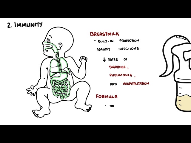
- Absence of exclusive breastfeeding (< 6 months)
- Pacifier use beyond 6 months of age
- Supine bottle feeding.
- Host Factors
- Male sex
- Family history of recurrent AOM
- Uderlying immunodeficiency
- Atopic rhinitis.
Infant Eustachian tubes are shorter, wider, and more horizontal, allowing nasopharyngeal secretions and pathogens to easily reflux into the middle ear space during swallowing or crying.
Pathophysiology
- Eustachian Tube Obstruction: Viral upper respiratory infection causes mucosal inflammation and swelling of the Eustachian tube lining.
- Negative Middle Ear Pressure: Functional obstruction prevents normal ventilation, leading to absorption of trapped nitrogen and oxygen by middle ear mucosa, generating negative pressure.
- Fluid Accumulation & Inoculation: Negative pressure draws transudative fluid into the tympanic cavity and aspirates nasopharyngeal secretions containing respiratory viruses and bacteria into the middle ear.
- Suppuration & Tissue Pressure: Microbial multiplication triggers an acute neutrophilic inflammatory response. Purulent exudate fills the unyielding bony cavity, placing high mechanical tension on the tympanic membrane, causing severe pain and reduced ossicular mobility.
The primary event initiating acute otitis media is Eustachian tube dysfunction following a viral upper respiratory tract infection.
Clinical Manifestations
- Otalgia
- Otorrhea:
- Conductive Hearing Loss
- Fever
- Poor feeding, anorexia, vomiting, and diarrhea (especially in infants due to shared autonomic responses).
Classic Otoscopic Findings
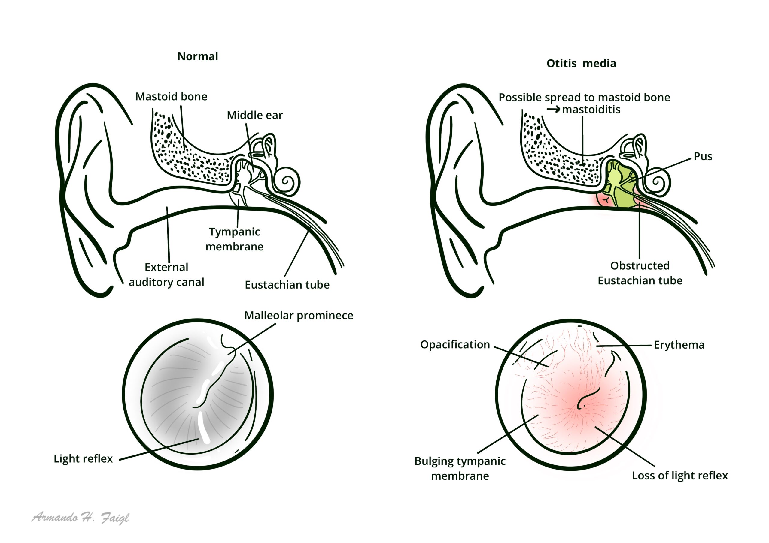
- Bulging Tympanic Membrane: Most specific otoscopic sign; loss of normal concave contour with outward displacement of the pars flaccida/pars tensa.
- Erythema & Opacification: Intense redness and loss of translucency; tympanic membrane appears dull, cloudy, or yellowish-white.
- Loss of Anatomical Landmarks: Obscuration of the light reflex (cone of light) and the handle/short process of the malleus.
- Impaired Mobility: Absence of tympanic membrane movement during pneumatic otoscopy.
A distinctly bulging tympanic membrane is the single most reliable otoscopic indicator for differentiating Acute Otitis Media from simple viral congestion or Otitis Media with Effusion.
Diagnosis
Diagnostic Criteria for AOM
Diagnosis requires meeting all three of the following clinical elements:
- Acute Onset: Rapid onset of signs and symptoms of middle ear inflammation within 48 hours.
- Middle Ear Effusion (MEE): Confirmed by bulging tympanic membrane, limited/absent mobility on pneumatic otoscopy, air-fluid level, or otorrhea.
- Middle Ear Inflammation: Marked tympanic membrane erythema OR acute otalgia interfering with normal sleep/activity.
Bedside & Diagnostic Modalities
- Pneumatic Otoscopy: Essential clinical tool; demonstrates reduced or absent tympanic membrane compliance when positive or negative pressure is applied.
- Tympanometry: Objective measure of middle ear compliance. Demonstrates a Type B (flat) tympanogram in the presence of middle ear fluid.
- Acoustic Reflectometry: Non-invasive screening tool measuring reflected sound waves to detect middle ear fluid.
- Microbiological Culture: Not routinely performed. Reserved for cases with spontaneous otorrhea, recurrent treatment failure, or immunocompromised hosts requiring tympanocentesis.
Treatment
Analgesia (First-Line for All Patients)
- Paracetamol (Acetaminophen)
- Ibuprofen
Watchful Waiting / Delayed Prescribing Strategy
- Indicated for low-risk children to minimize unnecessary antibiotic exposure
Immediate Antibiotic Indications
- Empiric immediate oral antibiotic therapy is mandatory for:
- All infants aged < 6 months.
- Children aged 6 to 23 months with bilateral AOM or severe symptoms.
- Any patient presenting with severe illness (severe otalgia, fever >= 39.0°C, or duration > 48 hours).
- Patients with otorrhea, craniofacial malformations, or immunocompromise.
Pharmacological Regimens
- First-Line Antibiotic: High-dose Amoxicillin oral (80–90 mg/kg/day divided into 2 doses for 5 to 10 days). High dosing overcomes intermediate penicillin resistance in S. pneumoniae.
- Second-Line / Treatment Failure (at 48–72 hours): Amoxicillin-clavulanate (Augmentin)
Surgical Management
- Tympanostomy Tube Insertion (Grommets)
- Recurrent AOM (defined as >= 3 distinct episodes in 6 months OR
- >= 4 episodes in 12 months with at least 1 in the preceding 6 months) OR
- persistent OME lasting > 3 months with documented hearing loss
High-dose Amoxicillin (80–90 mg/kg/day) is the first-line antibiotic choice for Acute Otitis Media. If symptoms fail to improve after 48–72 hours, switch to Amoxicillin-clavulanate to cover beta-lactamase producing organisms.
Complications & Prognosis
Intratemporal Complications
- Acute Mastoiditis: Most common suppurative complication
- Tympanic Membrane Perforation: Spontaneous rupture due to ischemic pressure necrosis; usually heals spontaneously within 2 to 6 weeks.
- Facial Nerve Palsy (CN VII)
- Labyrinthitis: Spread of infection to the inner ear causing acute sensorineural hearing loss, vertigo, nystagmus, and nausea.
- Petrositis (Gradenigo Syndrome): Extension of infection to the petrous apex; characterized by the classic triad of deep retro-orbital pain (CN V involvement), abducens nerve palsy (CN VI involvement), and persistent otorrhea.
Intracranial Complications
- Meningitis
- Epidural / Subdural Abscess & Brain Abscess
- Lateral / Sigmoid Sinus Thrombosis: Septic thrombophlebitis presenting with “picket-fence” spiking fevers, papilledema, and neck tenderness along the internal jugular vein.
Post-auricular redness, tenderness, and forward displacement of the pinna in a child with otitis media indicates Acute Mastoiditis and requires emergency contrast CT scan and ENT admission.
- Most uncomplicated AOM cases resolve spontaneously or with targeted antibiotics within 7 to 10 days.
- Fluid persistence (OME) after successful treatment of AOM is common and expected: ~70% at 2 weeks, ~40% at 1 month, and ~10% at 3 months.
References
- Lieberthal AS, Carroll AE, Chonmaitree T, Ganiats TG, Hoberman A, Jackson MA, et al. The diagnosis and management of acute otitis media. Pediatrics. 2013;131(3):e964-e999. doi:10.1542/peds.2012-3488
- Rosenfeld RM, Shin JJ, Schwartz SR, Coggins R, Gagnon L, Hackell JM, et al. Clinical practice guideline: Otitis media with effusion (update). Otolaryngol Head Neck Surg. 2016;154(1 Suppl):S1-S41. doi:10.1177/0194599815623467
- Schilder AG, Chonmaitree T, Cripps AW, Rosenfeld RM, Casselbrant ML, Haggard MP, Venekamp RP. Otitis media. Nat Rev Dis Primers. 2016;2:16063. doi:10.1038/nrdp.2016.63



Members only discussions coming soon…