

Leukocyte Adhesion Deficiency
Overview
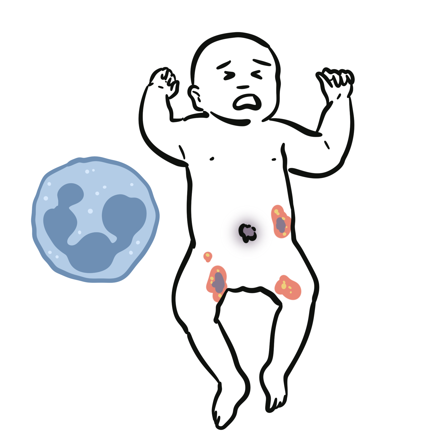
Leukocyte adhesion deficiency (LAD) is a rare primary immunodeficiency characterised by recurrent bacterial and fungal infections, impaired wound healing, and absence of pus formation due to defective leukocyte adhesion and migration. It results from genetic defects in leukocyte integrins or signalling pathways, preventing neutrophil extravasation from blood vessels into tissues. LAD has an estimated incidence of 1 per 100,000–200,000 live births. The most common and severe form is LAD type 1 (CD18/β2 integrin deficiency).
Definition
Integrin (CD18/β2): Cell surface protein mediating firm adhesion of leukocytes to endothelium.
Chemotaxis: Directed movement of immune cells toward sites of infection/inflammation.
Pus: Accumulation of neutrophils at infection site; absent in LAD due to failure of migration.
Omphalitis: Infection of the umbilical stump; common early presentation in LAD.
What to notice
Detailed image description
Anatomy & Physiology
- Normal neutrophil migration:
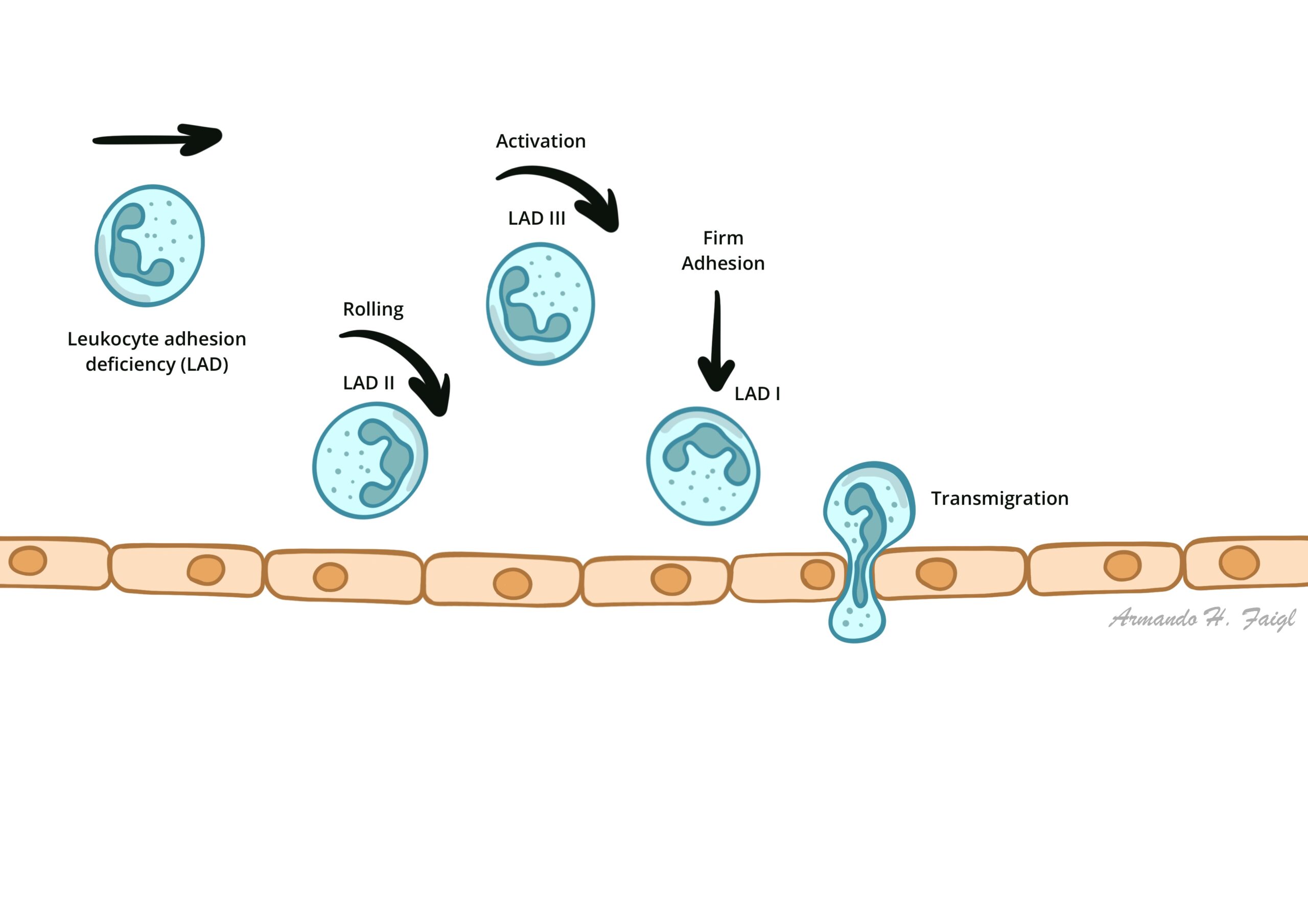
- Rolling (selectins) → adhesion (integrins: LFA-1/CD18) → diapedesis → chemotaxis.
- In LAD: defective adhesion (type 1), defective rolling (type 2), or defective signalling (type 3) → neutrophils cannot exit bloodstream.
- Neutrophil count normal/high in blood, but no neutrophils at infection site.
Neutrophilia + recurrent infections without pus strongly suggests LAD.
Aetiology
- LAD-1: AR mutation in ITGB2 gene (β2 integrin/CD18 deficiency).
- LAD-2: AR defect in GDP-fucose transporter → impaired selectin ligand synthesis → defective rolling.
- LAD-3: AR defect in KINDLIN-3 → impaired integrin activation affecting leukocytes and platelets.
Pathophysiology
- Genetic mutation (ITGB2, GDP-fucose transporter, KINDLIN-3).
- Defective adhesion/rolling/signalling.
- Neutrophils cannot migrate to infected tissues.
- Result: recurrent bacterial infections, impaired wound healing, no pus despite high neutrophil counts.
- In LAD-3, defective platelet integrins → bleeding tendency.
LAD = “neutrophils stuck in the blood.”
Clinical Manifestations
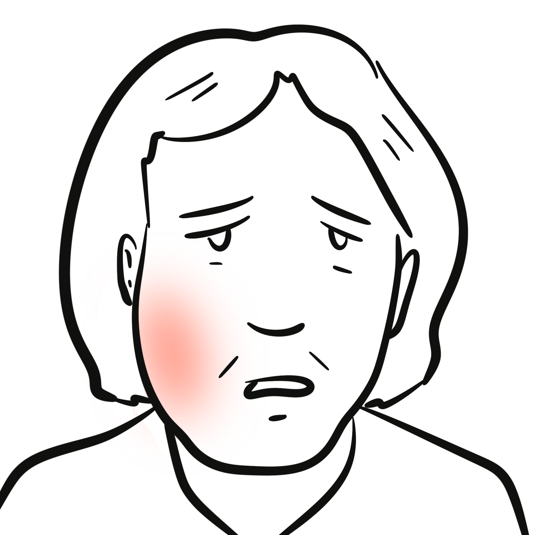
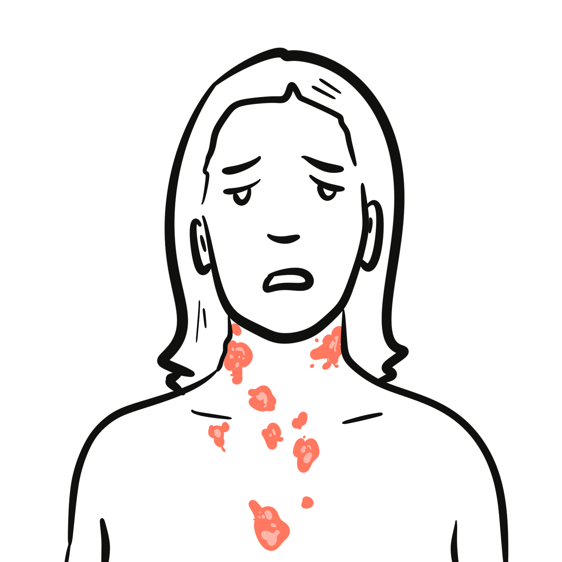
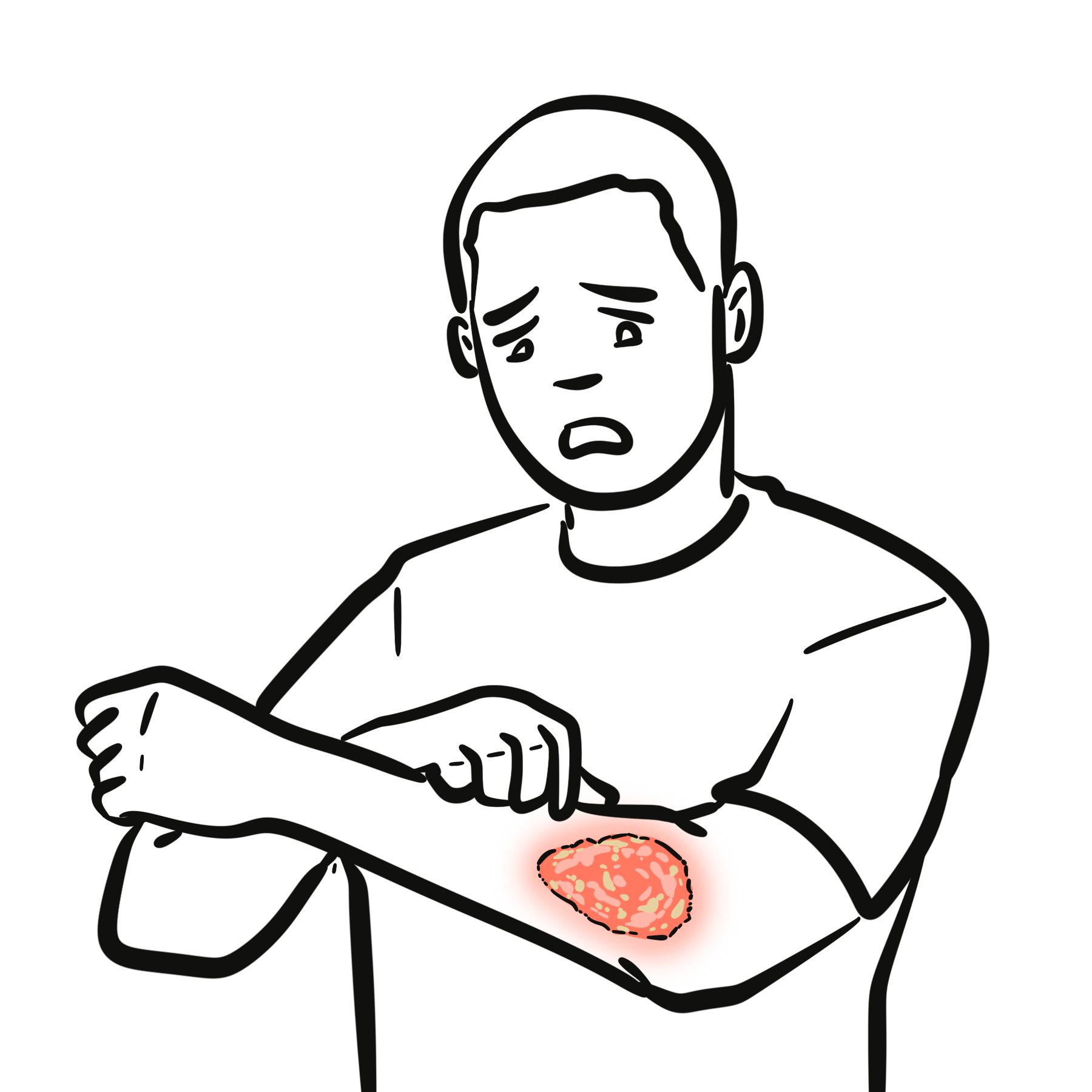
- Recurrent bacterial and fungal infections (skin, mucosa, respiratory tract).
- Omphalitis (umbilical stump infection) in neonates.
- Delayed separation of umbilical cord (>3 weeks).
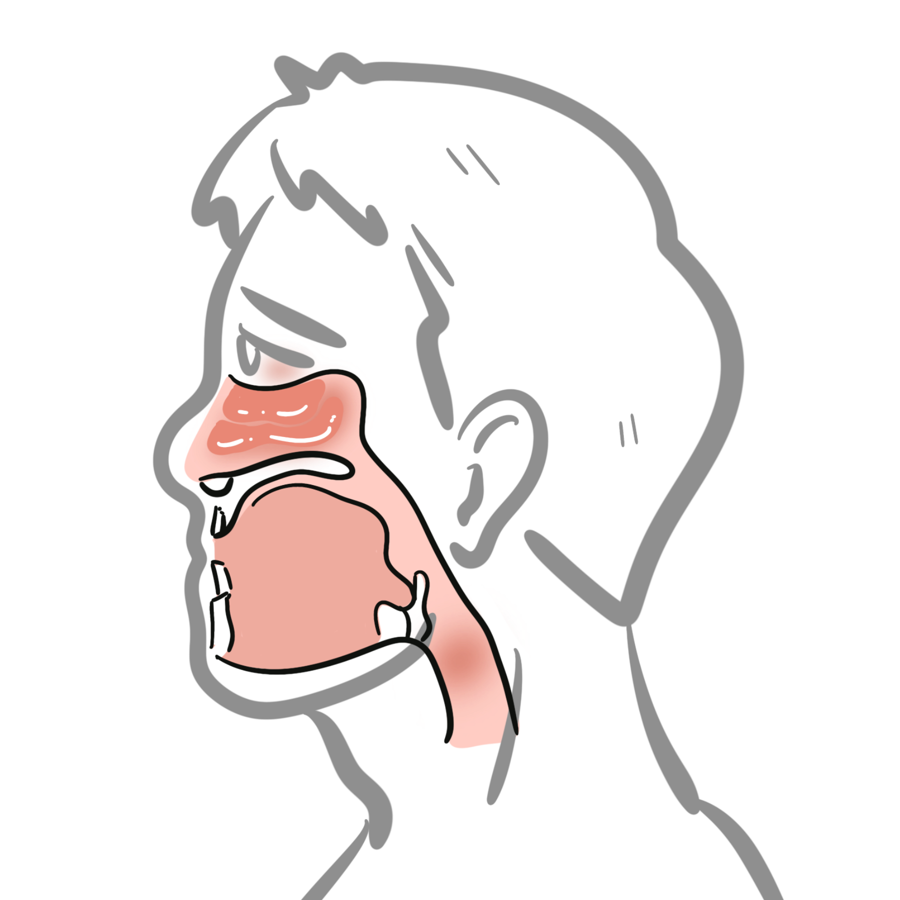
- Poor wound healing, severe gingivitis, periodontitis.
- Absence of pus formation at infection sites.
- Persistent neutrophilia on blood counts.
- LAD-3: bleeding/bruising due to platelet dysfunction.
Exam buzzword = “Recurrent infections, delayed cord separation, no pus, high neutrophils.”
Diagnosis
- CBC: Persistent neutrophilia.
- Flow cytometry: Absence/reduction of CD18/CD11 expression (LAD-1).
- Functional assays: Rolling/adhesion assays for LAD-2/LAD-3.
- Genetic testing: Confirms subtype.
Differential Diagnosis
| Condition | Differentiating Feature |
| Chronic granulomatous disease | Neutrophils reach tissues but fail to kill → pus present |
| Severe congenital neutropenia | Very low neutrophil count, not migration defect |
| Chediak–Higashi | Giant granules in neutrophils, partial albinism |
Classification
| Type | Molecular Defect | Key Features |
| LAD-1 | CD18 (β2 integrin) deficiency | Severe bacterial infections, no pus, neutrophilia |
| LAD-2 | GDP-fucose transporter defect | Infections, growth retardation, mental delay |
| LAD-3 | KINDLIN-3 defect | Infections + bleeding tendency (platelet defect) |
Treatment
- Prompt and aggressive antibiotics/antifungals.
- Supportive wound care and oral hygiene.
- HSCT = curative for LAD-1 and LAD-3.
- Fucose supplementation may help in LAD-2.
- Avoid live vaccines in severely immunocompromised cases.
HSCT is the only curative option for most LAD forms.
Complications & Prognosis
- Recurrent severe bacterial sepsis.
- Chronic oral disease → periodontitis, tooth loss.
- Failure to thrive in severe cases.
- Prognosis: poor without HSCT in severe LAD-1 and LAD-3; supportive care may allow survival in milder cases.
Most severe cases die in early childhood without transplant.
References
- Etzioni A, Harlan JM. Cell adhesion molecules in leukocyte adhesion deficiency. Semin Hematol. 2000;37(4):373–81.
- Almarza Novoa E, Kasbekar S, Thrasher AJ, et al. Leukocyte adhesion deficiency: Molecular basis, clinical presentation, and therapeutic approaches. Front Immunol. 2018;9:1031.
- Anderson DC, Springer TA. Leukocyte adhesion deficiency: An inherited defect in the Mac-1, LFA-1, and p150,95 glycoproteins. Annu Rev Med. 1987;38:175–94.
- Fischer A, Notarangelo LD, Neven B, et al. Primary immunodeficiency diseases: An update on the classification from the International Union of Immunological Societies Expert Committee. J Clin Immunol. 2020;40:24–64.
- Kinashi T. Intracellular signalling controlling integrin activation in lymphocytes. Nat Rev Immunol. 2005;5(7):546–59.





Members only discussions coming soon…