

Limping Child
OVERVIEW
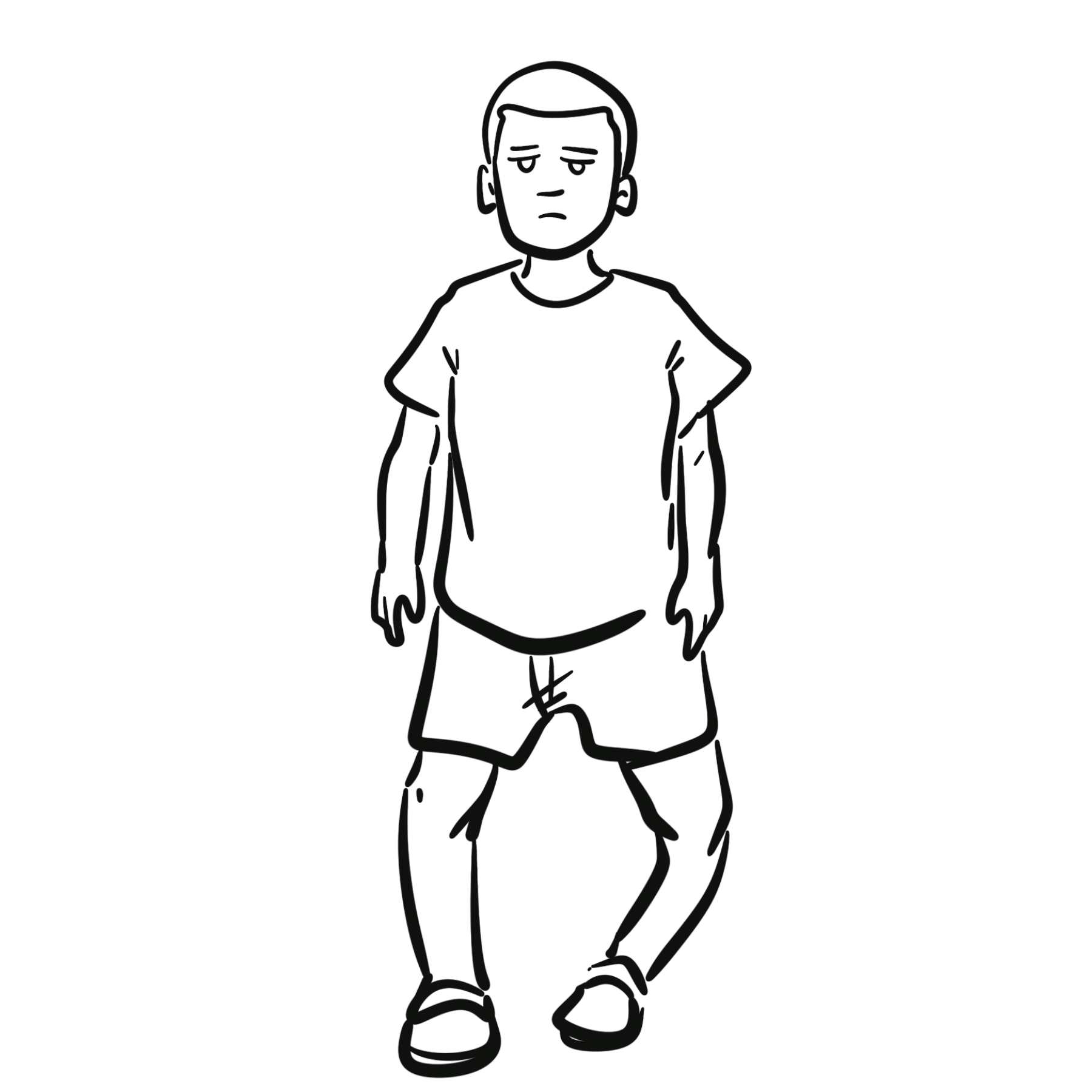
Definition: A limp is an abnormal gait pattern characterized by asymmetry, reduced stance phase, or deviation from normal biomechanics due to pain, weakness, structural deformity, or neuromuscular dysfunction.
Gait Types:
- Antalgic Gait (Most Common): Shortened stance phase on the affected lower extremity to minimize weight-bearing pain.
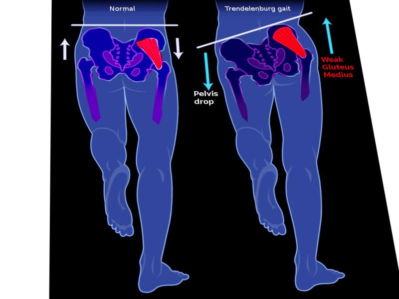
- Trendelenburg Gait: Pelvic drop toward the unaffected side during the stance phase of the affected limb, caused by abductor muscle weakness or hip instability.
- Circumduction Gait: Swinging the leg outward in a semi-circle to clear the floor, indicating limb length discrepancy or joint stiffness.
Clinical Significance: The priority in pediatric evaluation is rapid stratification by age and the urgent exclusion of joint infection, bone infection, and adolescent femoral head displacement before considering self-limiting viral conditions.
Knee and thigh pain in a child or adolescent is hip pathology until proven otherwise. Irritation of the obturator and femoral nerves frequently refers pain strictly to the distal thigh or knee joint, leading to dangerous diagnostic delays in slipped capital femoral epiphysis and septic arthritis.
TRIAGE & ALGORITHM
Step 1: Emergency Red Flag Screening
- Septic Arthritis / Acute Osteomyelitis:
- Inability or refusal to bear any weight, high fever (> 38.5°C), systemic toxicity, severe pain with gentle passive range of motion -> Immediate orthopedic consultation, ultrasound, and emergency joint aspiration.
- Slipped Capital Femoral Epiphysis:
- Overweight or rapidly growing adolescent (age 10–16 years) with an antalgic limp and knee, thigh, or groin pain -> Immediate non-weight-bearing protocol to prevent complete avascular necrosis of the femoral head.
- Non-Accidental Injury:
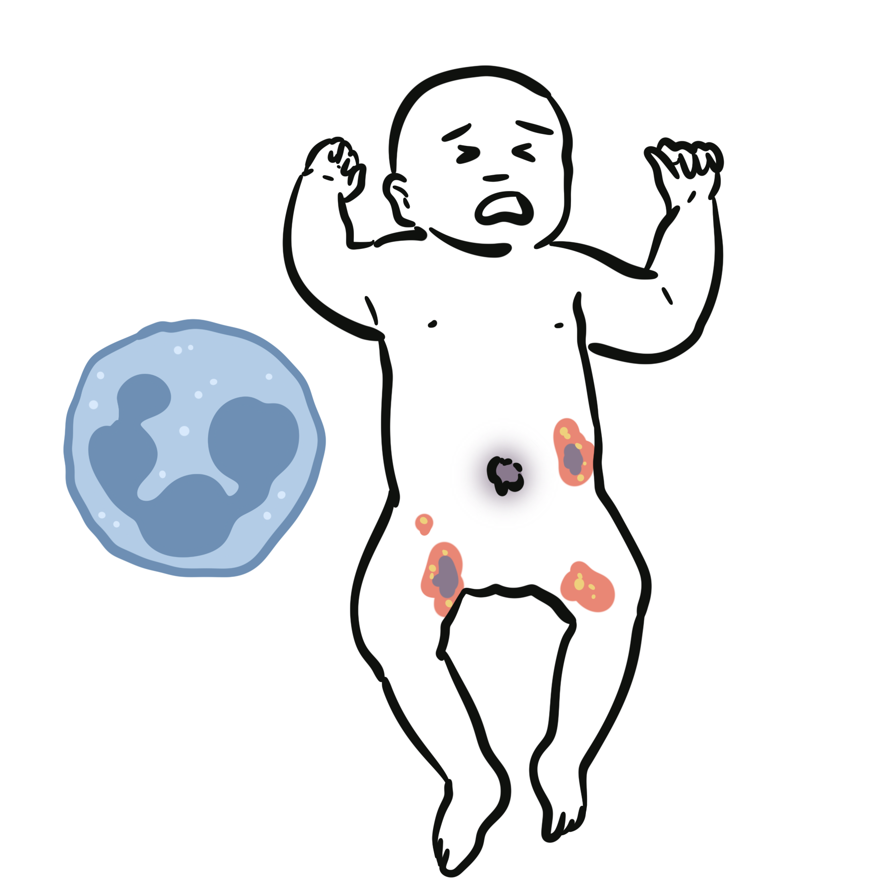
- Limp or refusal to walk in an infant or non-ambulatory child, unexplained spiral fractures, inconsistent history, bruising on soft tissue areas (ears, neck, torso).
- Malignancy (Leukemia, Bone Sarcomas):
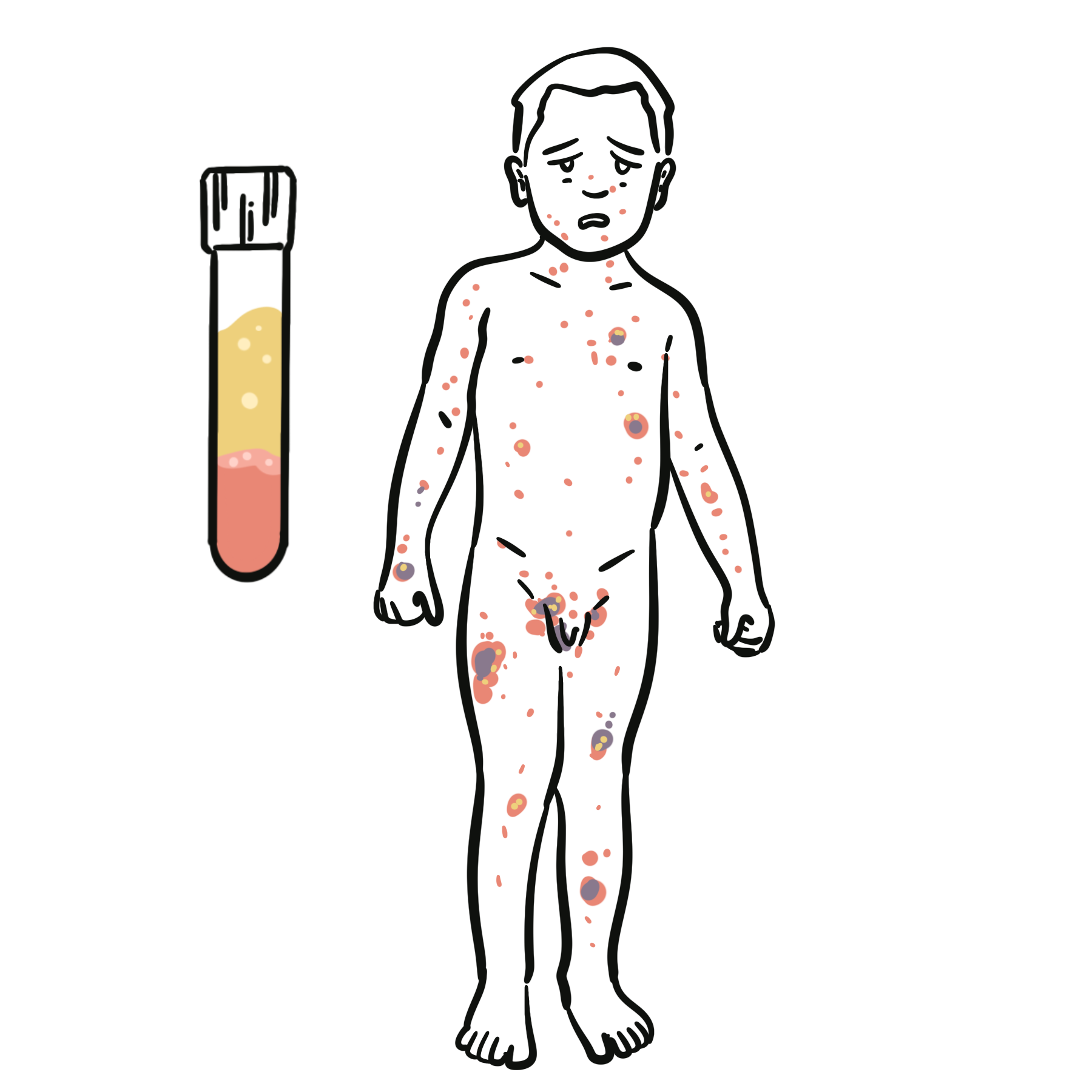
- Constant pain waking the child from sleep at night, unexplained fevers, weight loss, pallor, petechiae, hepatosplenomegaly.
Step 2: Age-Stratified Clinical Approach
- Toddler (Aged 1 to 3 Years):
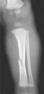
- Toddler fracture (undisplaced spiral fracture of the distal tibia), transient synovitis, septic arthritis, osteomyelitis.
- Young Child (Aged 4 to 8 Years):
- Transient synovitis (most common), Legg-Calve-Perthes disease, osteomyelitis, juvenile idiopathic arthritis.
- Adolescent (Aged > 9 to 10 Years):
- Slipped capital femoral epiphysis, apophysitis (Osgood-Schlatter, Sinding-Larsen-Johansson), osteochondritis dissecans, primary bone tumors (osteosarcoma, Ewing sarcoma).
Step 3: Diagnostic Decision Branching (Kocher Criteria for Hip Pain)
- Apply Kocher criteria:
- Inability to bear weight.
- Temperature > 38.5°C.
- Erythrocyte sedimentation rate > 40 mm/h.
- White blood cell count > 12.0 x 10^9/L.
- 4 of 4 criteria met: -> > 90% probability of Septic Arthritis -> Urgent ultrasound-guided arthrocentesis and operative washout.
- 0 to 1 criterion met: -> High probability of Transient Synovitis -> Outpatient rest, non-steroidal anti-inflammatory drugs, and clinical review in 48 hours.
A Toddler Fracture is a non-displaced spiral or oblique fracture of the distal tibial shaft caused by a minor twisting fall. Standard early radiographs can appear entirely normal. If a toddler presents with an acute refusal to bear weight and localized distal tibial tenderness, place the limb in an immobilization cast and repeat radiography in 10 to 14 days to identify periosteal callus formation.
DIFFERENTIAL DIAGNOSIS
Infectious & Emergent Entities:
- Septic Arthritis:
- Distinguishing features: Rapid onset of severe localized joint pain; child holds hip in flexion, abduction, and external rotation; refusal to tolerate even gentle passive movement; high inflammatory markers; synovial white blood cell count > 50,000 x 10^6/L with > 90% polymorphs.
- Acute Hematogenous Osteomyelitis:
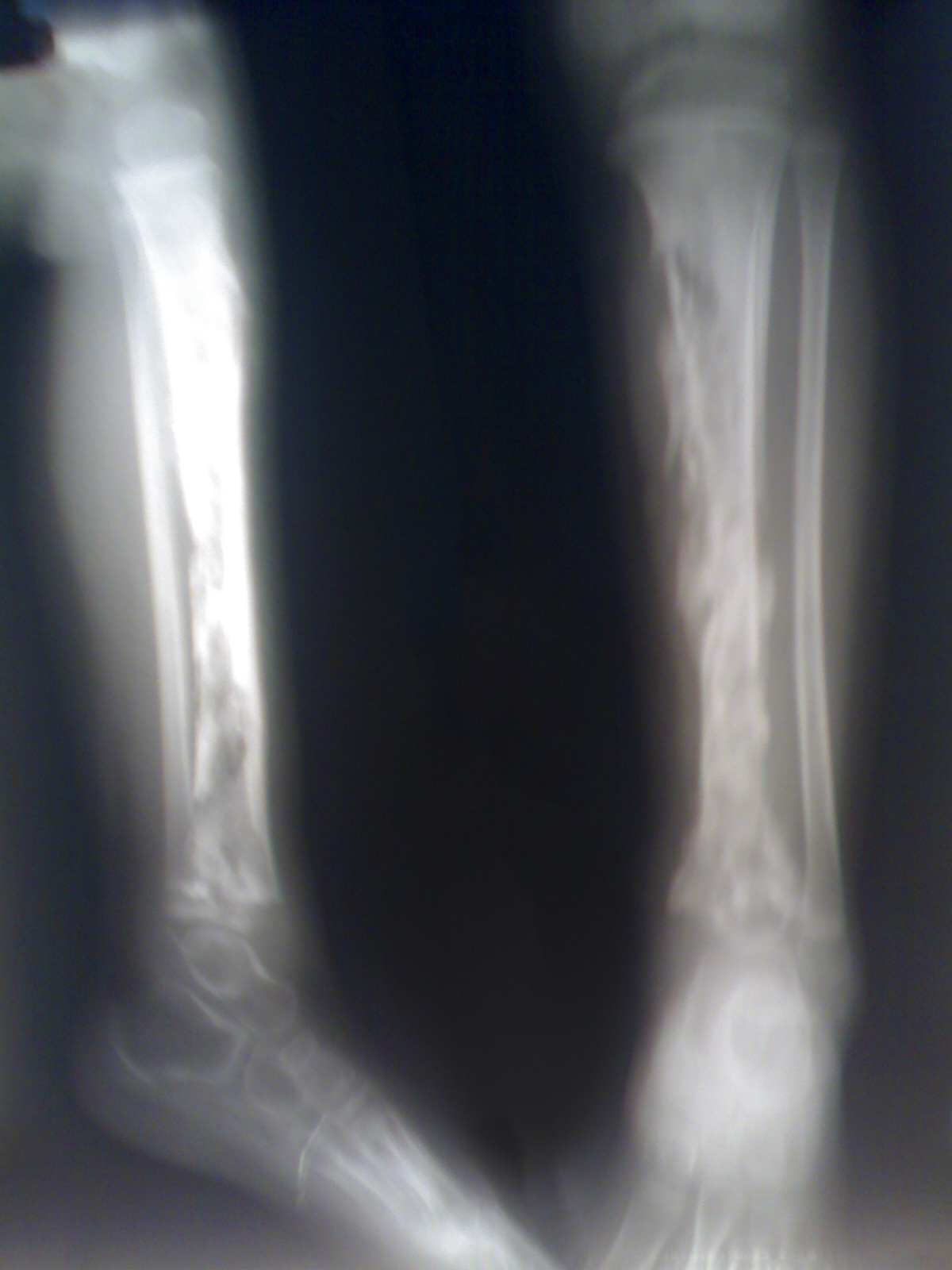
- Distinguishing features: Focal bone tenderness over the metaphysis (distal femur, proximal tibia); localized swelling and warmth; refusal to bear weight; high fever; subperiosteal abscess on advanced imaging.
- Discitis / Vertebral Osteomyelitis:
- Distinguishing features: Young child refusing to walk or sit; lumbar spine tenderness, loss of lumbar lordosis; child bends with knees rather than spine when picking up objects from the floor.
Non-Infectious Hip Pathologies:
- Transient Synovitis (“Irritable Hip” – Most Common Cause Overall):
- Distinguishing features: Age 3 to 8 years; sudden onset antalgic limp following a viral upper respiratory infection 1 to 2 weeks prior; child is non-toxic, afebrile or low-grade; weight-bearing is preserved (reluctant but possible); self-limiting within 7 to 10 days.
- Slipped Capital Femoral Epiphysis:
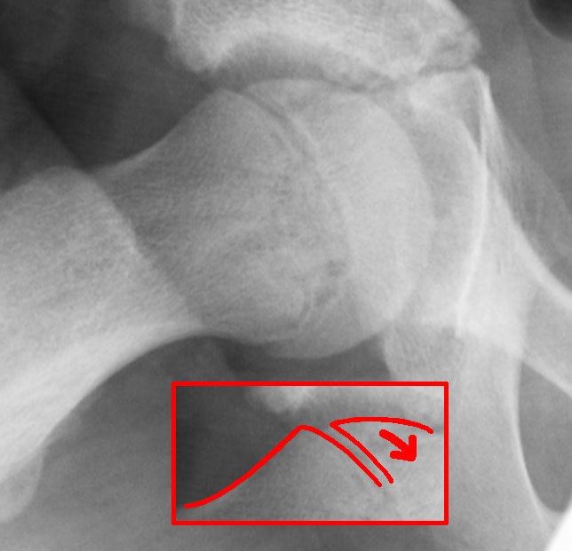
- Distinguishing features: Overweight boy aged 11 to 14 years; antalgic limp with pain referred to medial thigh or knee; obligate external rotation of the hip during passive flexion; radiography shows displacement of the femoral epiphysis relative to the metaphysis.
- Legg-Calve-Perthes Disease (Idiopathic Avascular Necrosis of Femoral Epiphysis):
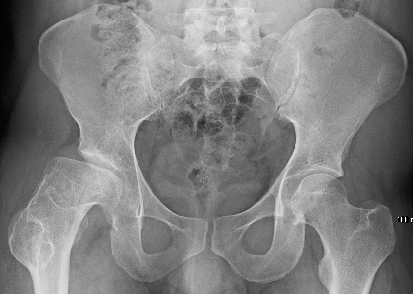
- Distinguishing features: Boys aged 4 to 8 years; insidious onset of a painless or mildly aching limp; decreased hip abduction and internal rotation; radiographs demonstrate femoral epiphysis fragmentation, flattening, and sclerosis.
Traumatic & Overuse Disorders:
- Toddler Fracture:
- Distinguishing features: Child aged 9 months to 3 years learning to walk; refusal to bear weight after a minor fall; pain elicited by gentle axial torsion of the tibia; normal or subtle faint hairline fracture on plain film.
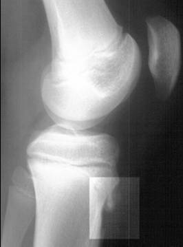
- Traction Apophysitis (Osgood-Schlatter / Sever Disease):
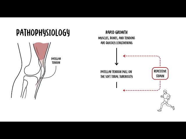
- Distinguishing features: Active adolescents; localized point tenderness over the tibial tuberosity (Osgood-Schlatter) or posterior calcaneus (Sever disease), aggravated by jumping and running.
Inflammatory & Hematological Conditions:
- Juvenile Idiopathic Arthritis:
- Distinguishing features: Chronic limp, joint swelling persisting for > 6 weeks; morning stiffness lasting > 30 minutes that improves with movement throughout the day.
- Acute Lymphoblastic Leukemia:
- Distinguishing features: Deep, unremitting bone pain waking child from sleep, non-articular bony tenderness, cytopenias on blood count, hepatosplenomegaly, and lymphadenopathy.
In slipped capital femoral epiphysis, passive flexion of the affected hip causes obligatory external rotation. This physical examination finding is practically pathognomonic and should prompt immediate non-weight-bearing immobilization and urgent bilateral hip radiography.
INVESTIGATIONS
First-Line / Bedside & Emergency Diagnostics:
- Plain Radiography (Anatomical Region-Specific):
- Pelvis / Hip: Anteroposterior pelvis and bilateral frog-leg lateral views (essential to detect posterior slippage in slipped capital femoral epiphysis and femoral head changes in Perthes disease).
- Lower Leg / Foot: Complete orthogonal views of the tibia, fibula, and foot if a toddler fracture or localized bony tenderness is suspected.
- Laboratory Panel:
- Full blood count: Evaluates leukocytosis (infection) and cytopenias (malignancy).
- Inflammatory markers: Erythrocyte sedimentation rate and C-reactive protein (differentiates transient synovitis from septic arthritis/osteomyelitis).
- Blood cultures: Mandatory prior to antibiotic administration whenever fever and joint pain co-exist.
- Targeted Joint Ultrasound:
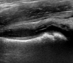
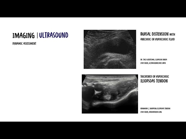
- Modality of choice for detecting joint effusion in the hip (capsular distension > 2 mm compared to the contralateral normal hip confirms an effusion); facilitates ultrasound-guided diagnostic arthrocentesis.
Targeted / Specialized Advanced Imaging:
- Magnetic Resonance Imaging:
- Modality of choice for detecting early acute osteomyelitis (demonstrates bone marrow edema within 24 hours when plain radiographs remain normal), soft tissue abscesses, early Perthes disease, and discitis.
- Diagnostic Arthrocentesis (Synovial Fluid Analysis):
- Synovial fluid cell count, gram stain, and culture are required whenever the Kocher criteria suggest a high probability of septic arthritis.
Plain radiographs can remain completely normal for the first 10 to 14 days of acute hematogenous osteomyelitis. Do not rule out bone infection based on a normal initial plain film; if localized metaphyseal tenderness and elevated inflammatory markers persist, obtain an urgent magnetic resonance imaging scan.
CRITICAL MANAGEMENT
Septic Arthritis Emergency Decompression Protocol:
- Emergency Operative Washout: Urgent arthroscopic or open arthrotomy washout by pediatric orthopedics within hours to prevent irreversible chondral degradation and avascular necrosis.
- Parenteral Antibiotic Therapy:
- Administer intravenous Flucloxacillin 50 mg/kg (up to 2 g) every 6 hours (or Cefazolin; add Vancomycin if methicillin-resistant Staphylococcus aureus is suspected).
- In children aged < 2 to 3 years: Include coverage for Kingella kingae with Cefotaxime 50 mg/kg or Ceftriaxone.
- Antibiotics must be administered only after synovial fluid and blood cultures are obtained.
Slipped Capital Femoral Epiphysis Protocol:
- Strict Non-Weight-Bearing: Patient must be placed in a wheelchair or bed immediately—no walking allowed.
- Urgent Surgical Stabilization:In situ single central percutaneous cannulated screw fixation across the physis to prevent displacement progression and osteonecrosis.
Transient Synovitis Management:
- Reassurance, strict bed rest, and mobilization as tolerated.
- Non-steroidal anti-inflammatory drugs: Oral Ibuprofen 10 mg/kg every 8 hours for 5 to 7 days.
- Mandatory follow-up: Clinical re-evaluation within 48 to 72 hours; if fever develops, pain worsens, or symptoms persist > 7 to 10 days, repeat inflammatory markers and radiography.
Toddler Fracture Protocol:
Undisplaced tibial spiral fractures: Immobilization in an above-knee or below-knee fiberglass cast for 3 to 4 weeks, with complete resolution expected and no long-term deformity.
In children under 3 to 4 years of age, Kingella kingae is the leading bacterial cause of septic arthritis and osteomyelitis. It is a fastidious organism that rarely grows on standard agar plates; always inoculate synovial fluid directly into blood culture bottles and request dedicated polymerase chain reaction testing.


Members only discussions coming soon…