

Biliary Atresia
Overview
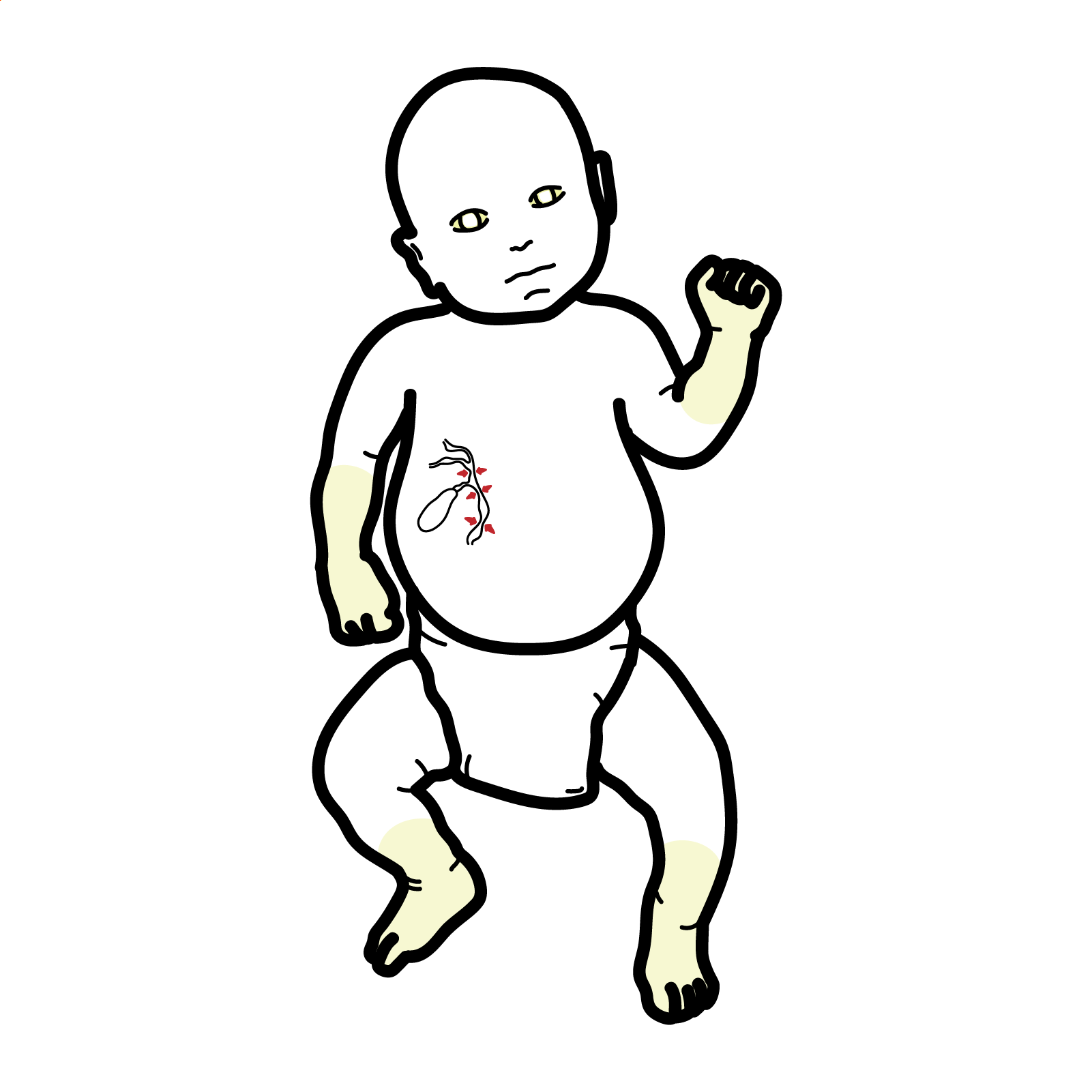
Biliary atresia is a progressive fibro-inflammatory disorder of infancy in which the extrahepatic bile ducts—and variably the intrahepatic ducts—become obliterated. This prevents bile from draining from the liver, producing conjugated hyperbilirubinaemia, progressive hepatic fibrosis and, without treatment, cirrhosis and liver failure.
Affected infants often appear well during the first weeks of life. Persistent jaundice may therefore be mislabelled as physiological jaundice or breast-milk jaundice. The most important early clues are pale or acholic stools, dark urine and persistent jaundice beyond 2 weeks of age.
Biliary atresia is a time-critical diagnosis. Early referral and Kasai hepatoportoenterostomy offer the best chance of restoring bile drainage and preserving the native liver.
Definition
- Biliary atresia
- Progressive fibro-inflammatory obliteration of the extrahepatic biliary tree presenting during early infancy with obstructive cholestasis.
- Kasai hepatoportoenterostomy
- Surgery in which the obliterated extrahepatic ducts are excised and a Roux-en-Y intestinal limb is connected to the porta hepatis to permit bile drainage from microscopic residual ductules.
- Acholic stool
- Pale, white, grey or clay-coloured stool caused by absent or markedly reduced bile pigment entering the intestine.
Physiological and breast-milk jaundice produce unconjugated hyperbilirubinaemia. Conjugated jaundice is always pathological and requires prompt investigation.
Anatomy & Physiology
Bile is produced by hepatocytes and transported through:
- Intrahepatic canaliculi and small bile ducts
- Right and left hepatic ducts
- Common hepatic duct
- Common bile duct
- Duodenum
The gallbladder stores and concentrates bile between feeds. Bile entering the intestine:
- assists fat digestion and absorption
- enables absorption of vitamins A, D, E and K
- carries bilirubin, cholesterol and other substances for excretion
- gives stool its characteristic brown colour
In biliary atresia, obstruction prevents bile from entering the intestine. Conjugated bilirubin refluxes into the bloodstream and is excreted in urine, producing dark urine. Lack of intestinal bile pigment produces pale stools. Retained bile acids injure hepatocytes and stimulate inflammation, fibrosis and progressive portal hypertension.
Aetiology & Risk Factors
Aetiology
Biliary atresia is not usually caused by failure of the ducts to form. Most evidence supports an acquired or evolving perinatal fibro-inflammatory injury in a genetically susceptible infant.
Proposed contributors include:
- dysregulated immune-mediated bile-duct injury
- abnormal ductal development or morphogenesis
- perinatal viral triggers
- genetic susceptibility
- environmental or toxic exposures
No single infectious, genetic or environmental cause explains most cases. Biliary atresia is not caused by parental behaviour, feeding practices or routine medications.
Clinical phenotypes
Isolated biliary atresia is the most common phenotype and occurs without major congenital abnormalities.
Biliary atresia splenic malformation syndrome may be associated with polysplenia or asplenia, situs abnormalities, interrupted inferior vena cava, preduodenal portal vein, intestinal malrotation and congenital heart disease.
Cystic biliary atresia may produce a cystic structure near the porta hepatis and mimic a choledochal cyst, but the intrahepatic and extrahepatic ducts do not communicate normally.
Risk factors
There are no reliable maternal or neonatal risk factors that can be used to exclude or predict biliary atresia. It usually occurs sporadically.
Biliary atresia is generally not considered a simple inherited disorder. Recurrence in siblings is uncommon, and identical twins may be discordant.
Pathophysiology
- Inflammation targets the biliary epithelium.
- Extrahepatic bile ducts become progressively damaged and replaced by fibrous tissue.
- Bile flow from the liver to the intestine becomes obstructed.
- Conjugated bilirubin and bile acids accumulate in the liver and bloodstream.
- Retained bile acids cause hepatocellular injury and ductular proliferation.
- Progressive fibrosis leads to biliary cirrhosis and portal hypertension.
- Reduced bile delivery to the intestine causes fat malabsorption and deficiency of vitamins A, D, E and K.
- Ongoing cholestasis, malnutrition and recurrent cholangitis accelerate liver injury.
The process may continue after technically successful Kasai surgery because the underlying intrahepatic cholangiopathy is not completely corrected.
The Kasai procedure creates a route for bile drainage but does not replace a normal biliary tree or cure the underlying liver disease. A child can clear jaundice after surgery and still develop portal hypertension or require transplantation later.
Clinical Manifestations
Early presentation
Most infants are born at term and initially appear healthy. Typical features develop during the first weeks of life:
- jaundice persisting beyond 2 weeks
- yellow discolouration of the skin and sclera
- pale, white, grey or clay-coloured stools
- dark yellow or brown urine
- hepatomegaly
- firm liver edge
- initially preserved feeding and general appearance
Stool colour may fluctuate early in the disease. A history of occasional yellow or pigmented stools does not reliably exclude biliary atresia.
Progressive disease
- poor weight gain or growth faltering
- irritability
- abdominal distension
- splenomegaly
- ascites
- easy bruising or bleeding from vitamin K deficiency
- pruritus
- muscle and subcutaneous fat wasting
- prominent abdominal veins
- gastrointestinal bleeding from portal hypertension
- recurrent bacterial cholangitis following Kasai surgery
Features of associated congenital abnormalities
- cardiac murmur or cyanosis
- abnormal cardiac position
- splenic abnormalities
- intestinal malrotation
- abnormal abdominal organ arrangement
The combination of persistent jaundice, dark urine and pale stools should be treated as neonatal cholestasis until proven otherwise.
Stool-colour cards and parent photographs can improve recognition of acholic stool. Stool colour should be directly inspected whenever possible because descriptions such as “cream,” “mustard” or “light yellow” may be interpreted differently.
Diagnosis
Biliary atresia should be considered in every infant with conjugated hyperbilirubinaemia. The investigation must proceed rapidly because no single non-invasive test reliably excludes the disease and diagnostic delay may reduce the benefit of Kasai surgery.
1. Confirm cholestasis immediately
Measure total bilirubin and direct or conjugated bilirubin. A direct bilirubin concentration greater than 1 mg/dL, approximately 17 μmol/L, is abnormal and warrants evaluation, regardless of the total bilirubin concentration.
Any infant who remains jaundiced at 2 weeks should have fractionated bilirubin measured. Earlier testing is required when there are pale stools, dark urine, hepatomegaly, poor feeding, illness or another clinical concern.
Do not wait for jaundice to become severe. Infants with biliary atresia may have only modest visible jaundice while significant fibrosis is already developing.
2. Assess severity and identify immediately treatable causes
An elevated gamma-glutamyl transferase supports an obstructive or high-GGT cholestatic process but is not diagnostic. A prolonged INR may reflect vitamin K deficiency or impaired hepatic synthetic function. Administer parenteral vitamin K and reassess; failure to correct increases concern for significant liver dysfunction.
Undertake targeted testing for alternative treatable causes according to the presentation, particularly blood and urine cultures when infection is possible, newborn-screen results, alpha-1 antitrypsin phenotype or genotype, congenital-infection testing when indicated, and metabolic or genetic testing when the phenotype suggests another disorder. Testing for uncommon disorders should not delay biliary imaging or referral.
3. Abdominal ultrasonography
An absent, small or irregular gallbladder and a triangular-cord sign increase suspicion. However, a normal-appearing gallbladder or otherwise nondiagnostic ultrasound does not exclude biliary atresia.
4. Liver biopsy
Percutaneous liver biopsy is highly useful when the diagnosis remains uncertain and can distinguish an obstructive biliary pattern from many hepatocellular causes of neonatal cholestasis.
Features supporting biliary atresia include:
- bile-duct proliferation
- bile plugs
- portal oedema
- portal inflammation
- progressive portal fibrosis
Histology can be less typical very early in the disease and must be interpreted alongside age, stool colour, biochemistry and imaging.
5. Cholangiography
Definitive assessment of extrahepatic bile-duct patency requires cholangiography. When suspicion remains high, the infant should proceed promptly to an experienced paediatric hepatobiliary centre for operative cholangiography and Kasai hepatoportoenterostomy during the same procedure if biliary atresia is confirmed.
Failure of contrast to enter a patent extrahepatic biliary system confirms obstruction compatible with biliary atresia.
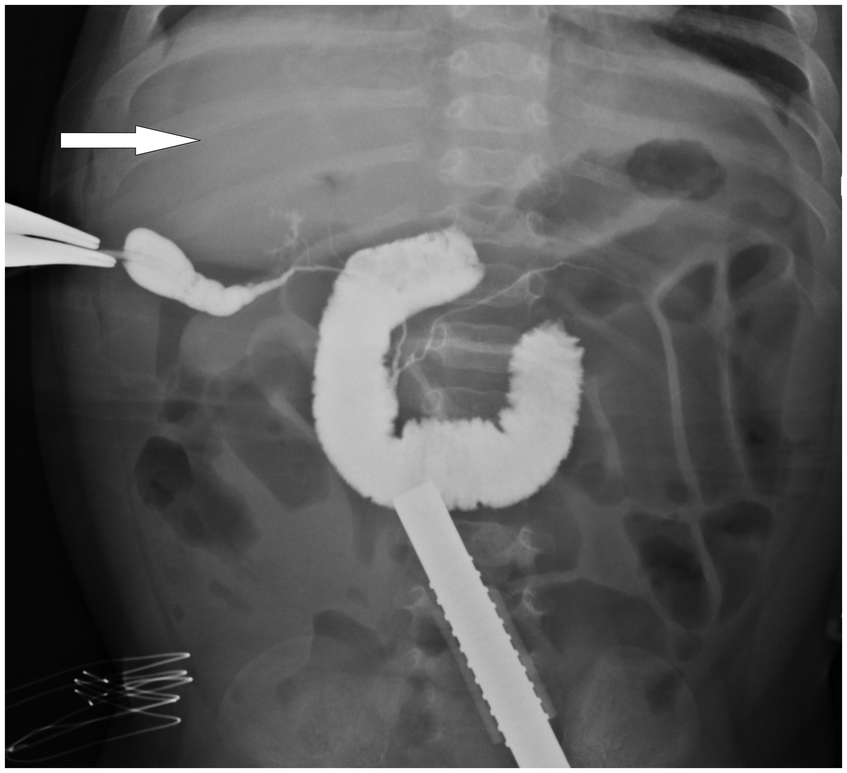
Intraoperative cholangiography in biliary atresia. Source: Gu C, Sun J, Ding L, Li B, Zhang Y, Jiang G. Frontiers in Pediatrics (2025). CC BY 4.0.
What to notice
Detailed image description
Approved diagnostic figure for Biliary Atresia. Original source: https://www.frontiersin.org/journals/pediatrics/articles/10.3389/fped.2025.1541455/full Direct source asset: https://www.frontiersin.org/files/Articles/1541455/xml-images/fped-13-1541455-g001.webp Licence: Creative Commons Attribution 4.0 International (CC BY 4.0). No modifications. Retrieved 20 August 2026.
The investigation is not finished when ultrasound or scintigraphy is “inconclusive.” In a cholestatic infant with pale stools, uncertainty should accelerate specialist assessment rather than provide reassurance.
Key differential diagnoses
| Differential | Distinguishing considerations |
|---|---|
| Neonatal hepatitis | Hepatocellular inflammation without extrahepatic duct obliteration; numerous infectious, metabolic and genetic causes |
| Alpha-1 antitrypsin deficiency | Abnormal phenotype or genotype; may closely resemble biliary atresia clinically and histologically |
| Choledochal cyst | Cystic biliary dilatation with demonstrable communication with the biliary tree; cystic biliary atresia may mimic it |
| Alagille syndrome | Cholestasis, characteristic facies, cardiac disease, butterfly vertebrae, posterior embryotoxon and paucity of intrahepatic ducts |
| Progressive familial intrahepatic cholestasis | Genetic cholestasis; some forms have low or normal GGT despite marked cholestasis |
| Congenital infection | Growth restriction, rash, thrombocytopenia, microcephaly, hearing or ocular abnormalities |
| Galactosaemia or tyrosinaemia | Systemic illness, hypoglycaemia, coagulopathy or metabolic abnormalities; newborn screening is important |
| Sepsis or urinary tract infection | Acutely unwell infant, inflammatory response and positive cultures |
| Cystic fibrosis–associated cholestasis | Abnormal newborn screening or sweat testing and associated pancreatic or respiratory features |
| Inspissated bile syndrome | Often associated with haemolysis, prematurity, parenteral nutrition or severe illness; ducts remain anatomically patent |
Classification
Anatomical classification
- Type I: obstruction of the common bile duct
- Type II: obstruction at the common hepatic duct
- Type III: obstruction at the porta hepatis
Type III is the most common form.
Clinical classification
- Isolated or perinatal biliary atresia
- Biliary atresia splenic malformation syndrome
- Biliary atresia associated with other congenital abnormalities
- Cystic biliary atresia
The anatomical type is less important clinically than early diagnosis, surgical expertise, restoration of bile drainage and the severity of intrahepatic disease.
Treatment
Immediate referral
Suspected biliary atresia requires urgent referral to a centre with paediatric hepatology, paediatric hepatobiliary surgery, interventional radiology and pathology expertise, a liver-transplant pathway and specialist multidisciplinary support. Serial outpatient observation of conjugated jaundice is inappropriate.
Kasai hepatoportoenterostomy
The Kasai procedure is the initial operation for most infants without advanced irreversible liver disease. The surgeon excises the fibrotic extrahepatic biliary remnant, exposes the porta hepatis and connects a Roux-en-Y jejunal limb directly to the liver hilum, allowing microscopic residual ductules to drain bile.
The procedure should be performed as early as possible. Outcomes are generally better when surgery occurs within the first 30–60 days of life, although age alone should not automatically exclude an infant from specialist surgical assessment.
Successful bile drainage is suggested by falling serum bilirubin, return of pigmented stool, reduced jaundice and improved growth and nutritional status.
The Kasai procedure is not curative. Even after jaundice clears, lifelong monitoring is required for cholangitis, fibrosis, portal hypertension and nutritional complications.
Nutritional management
- energy-dense breast milk or formula
- additional calories
- medium-chain triglycerides
- water-miscible preparations of fat-soluble vitamins
- monitoring of vitamin levels, INR and bone health
- nasogastric feeding when oral intake is inadequate
- specialist dietetic review
Weight alone may overestimate nutritional status when hepatomegaly, splenomegaly or ascites is present. Length, mid-upper-arm circumference and skinfold measurements may provide additional information.
Cholangitis
Ascending bacterial cholangitis is common after Kasai surgery. Suspect it with fever, irritability, poor feeding, recurrent or worsening jaundice, pale stools, abdominal discomfort or increasing bilirubin and liver enzymes. Obtain cultures where feasible and commence prompt intravenous antibiotics according to local microbiological guidance.
Liver transplantation
Liver transplantation is indicated when there is:
- failure to establish bile drainage after Kasai surgery
- progressive liver failure
- refractory ascites
- recurrent or severe cholangitis
- recurrent variceal bleeding
- severe portal hypertension
- hepatopulmonary syndrome
- refractory pruritus
- severe growth failure or malnutrition
- deteriorating quality of life
- hepatic malignancy
Primary liver transplantation may be considered when an infant presents late with advanced decompensated cirrhosis or when Kasai surgery is unlikely to provide meaningful benefit.
A “successful” Kasai usually means effective bile drainage and clearance of jaundice. It does not mean that the liver has returned to normal or that transplantation will never be required.
Complications & Prognosis
Complications
- Progressive biliary fibrosis and cirrhosis
- Portal hypertension
- Splenomegaly and hypersplenism
- Oesophageal or gastric varices
- Gastrointestinal bleeding
- Ascites
- Recurrent bacterial cholangitis
- Fat and fat-soluble vitamin malabsorption
- Vitamin K–deficiency bleeding
- Metabolic bone disease and fractures
- Growth failure and sarcopenia
- Pruritus
- Hepatopulmonary syndrome
- Portopulmonary hypertension
- Hepatic decompensation
- Hepatocellular carcinoma, rarely
- Need for liver transplantation
Prognosis
Outcome depends on age and liver condition at surgery, surgical expertise, restoration of bile drainage, bilirubin response after Kasai surgery, frequency of cholangitis, severity of fibrosis and portal hypertension, nutritional status and access to liver transplantation.
A substantial proportion of infants achieve useful bile drainage following Kasai surgery, allowing survival with the native liver for years or decades. However, most patients eventually develop chronic liver complications, and many require transplantation during childhood or later life.
Survival into adulthood is now expected for most children when Kasai surgery, specialist follow-up and liver transplantation are available.
Falling bilirubin after Kasai is encouraging, but bilirubin alone does not measure the full burden of disease. A child with normal bilirubin may still develop clinically significant portal hypertension.
References
- Fawaz R, Baumann U, Ekong U, et al. Guideline for the evaluation of cholestatic jaundice in infants: joint recommendations of NASPGHAN and ESPGHAN. J Pediatr Gastroenterol Nutr. 2017;64(1):154–168. doi:10.1097/MPG.0000000000001334.
- Bezerra JA, Wells RG, Mack CL, et al. Biliary atresia: clinical and research challenges for the twenty-first century. Hepatology. 2018;68(3):1163–1173. doi:10.1002/hep.29905.
- National Institute of Diabetes and Digestive and Kidney Diseases. Biliary atresia. Accessed August 20, 2026. https://www.niddk.nih.gov/health-information/liver-disease/biliary-atresia
- National Institute of Diabetes and Digestive and Kidney Diseases. Treatment for biliary atresia. Accessed August 20, 2026. https://www.niddk.nih.gov/health-information/liver-disease/biliary-atresia/treatment
- Bezerra JA, Spino C, Magee JC, et al. Use of corticosteroids after hepatoportoenterostomy for bile drainage in infants with biliary atresia: the START randomized clinical trial. JAMA. 2014;311(17):1750–1759. doi:10.1001/jama.2014.2623.
- Gu C, Sun J, Ding L, Li B, Zhang Y, Jiang G. Partial quadrate lobectomy improves early outcomes of laparoscopic Kasai surgery in type III biliary atresia. Front Pediatr. 2025;13:1541455. doi:10.3389/fped.2025.1541455.
Members only discussions coming soon…