

Allergic Conjunctivitis

Overview
Allergic conjunctivitis is inflammation of the conjunctiva caused by hypersensitivity to an allergen. It usually presents with bilateral ocular itching, redness, watering and conjunctival swelling, often alongside allergic rhinitis, asthma or atopic dermatitis.1,2
Seasonal and perennial allergic conjunctivitis are common and generally do not threaten vision. Vernal keratoconjunctivitis and atopic keratoconjunctivitis are more severe forms that may involve the cornea, produce scarring and cause permanent visual impairment.1,3
Marked ocular pain, significant photophobia, reduced visual acuity, corneal opacity or staining, an abnormal pupil, or a unilateral red eye in a contact-lens wearer should not be attributed to uncomplicated allergy. These findings require urgent assessment for keratitis, uveitis, acute angle closure or another sight-threatening disorder.1,4
Definition
- Allergic conjunctivitis
- Conjunctival inflammation caused by an immune-mediated response to an environmental allergen.
- Chemosis
- Oedematous swelling of the conjunctiva.
- Papilla
- A raised conjunctival lesion with a central vascular core, commonly seen on the tarsal conjunctiva in allergic disease.
- Keratoconjunctivitis
- Inflammation involving both the conjunctiva and cornea, with a greater risk of impaired vision.
- Limbal Horner–Trantas dots
- Small collections of eosinophils and epithelial debris around the corneal limbus, classically associated with vernal keratoconjunctivitis.
Anatomy & Physiology
The conjunctiva is a thin, vascular mucous membrane covering the anterior sclera and lining the inner eyelids. It forms part of the ocular surface together with the corneal epithelium, tear film and eyelid margins.
The tear film lubricates the ocular surface, removes particulate material and contains antimicrobial and immunological proteins. Blinking distributes the tear film and helps clear allergens through the lacrimal drainage system.
The conjunctiva contains mast cells, lymphocytes and other immune cells. Its direct exposure to airborne particles makes it particularly susceptible to allergic inflammation. The normally avascular cornea is less commonly involved; corneal signs therefore suggest more severe disease rather than uncomplicated seasonal or perennial conjunctivitis.
Classification
| Form | Typical pattern | Distinguishing features | Risk to vision |
|---|---|---|---|
| Acute allergic conjunctivitis | Sudden reaction after substantial allergen exposure | Rapid itching, chemosis and eyelid swelling; often resolves within hours | Low |
| Seasonal allergic conjunctivitis | Recurs during pollen seasons | Bilateral itching and watering with allergic rhinitis | Low |
| Perennial allergic conjunctivitis | Persistent or recurrent throughout the year | Usually milder symptoms associated with dust mites, mould or animal dander | Low |
| Vernal keratoconjunctivitis | Severe, often seasonal disease in children or young people | Giant upper-tarsal papillae, ropy mucus, limbal disease and possible shield ulcer | Significant |
| Atopic keratoconjunctivitis | Chronic disease, usually with atopic dermatitis | Eyelid eczema, conjunctival scarring and corneal involvement | Significant |
| Contact lens-associated papillary conjunctivitis | Associated with contact lenses, prostheses or exposed sutures | Lens intolerance, mucus and upper-tarsal papillae | Usually low, but infection must be excluded |
Aetiology & Risk Factors
Aetiology
Most allergic conjunctivitis results from a type I, immunoglobulin E-mediated hypersensitivity reaction to an environmental allergen. Common triggers include:
- grass, tree and weed pollens
- house-dust mites
- animal dander
- mould spores
- occupational allergens
- cosmetics or topical preparations.
Seasonal allergic conjunctivitis follows exposure to allergens that vary throughout the year, particularly pollens.
Perennial allergic conjunctivitis is associated with allergens present year-round, such as house-dust mites, mould and animal dander.2
Vernal and atopic keratoconjunctivitis have more complex inflammatory mechanisms involving mast cells, eosinophils, T lymphocytes and chronic ocular-surface inflammation.
Contact lens-associated papillary conjunctivitis is driven by mechanical irritation and an inflammatory response to deposits or material on a contact lens, ocular prosthesis or exposed suture.3,5
Risk Factors
- allergic rhinitis
- asthma
- atopic dermatitis
- personal or family history of atopy
- predictable seasonal allergen exposure
- warm, dry or windy conditions with high pollen levels
- occupational exposure to airborne allergens
- contact-lens wear
- poor contact-lens hygiene or infrequent lens replacement
- ocular prostheses or exposed ocular sutures
- frequent eye rubbing
- prolonged exposure to preserved topical eye preparations.
Vernal keratoconjunctivitis occurs predominantly in children and young people, particularly boys living in warm climates.
Atopic keratoconjunctivitis more commonly begins in late adolescence or adulthood and is strongly associated with atopic dermatitis.2,3
Pathophysiology
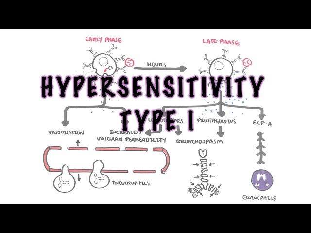
Initial exposure to an allergen promotes sensitisation and production of allergen-specific immunoglobulin E. This immunoglobulin binds to mast cells in the conjunctiva.
On re-exposure, allergen cross-linking of mast-cell–bound immunoglobulin E causes rapid mast-cell degranulation. Histamine and other mediators produce vasodilatation, increased vascular permeability, sensory-nerve stimulation and lacrimation. This explains the characteristic itching, conjunctival injection, chemosis and watering.
A later inflammatory phase recruits eosinophils, basophils and T lymphocytes. Recurrent or persistent inflammation can produce papillary hypertrophy, epithelial injury and ocular-surface dysfunction.
In vernal and atopic keratoconjunctivitis, chronic eosinophilic inflammation may damage the corneal epithelium. This can cause punctate keratitis, epithelial defects, shield ulcers, scarring and secondary infection.
Itching strongly supports an allergic mechanism, but severe pain, pronounced photophobia or reduced vision suggests that inflammation has extended beyond the conjunctiva—or that an alternative sight-threatening diagnosis is present.
Clinical Manifestations
Typical symptoms include:
- intense ocular itching
- bilateral redness
- watering or clear discharge
- burning or mild grittiness
- eyelid swelling
- transient blurred vision caused by tearing or discharge
- associated sneezing, nasal itching or rhinorrhoea.
Symptoms are commonly bilateral, although one eye may initially be more affected. Thick purulent discharge, severe pain and persistent reduction in vision are atypical for uncomplicated allergic conjunctivitis.
Clinical Examination
- bilateral conjunctival injection
- chemosis
- watery or stringy mucoid discharge
- eyelid oedema
- papillae on the tarsal conjunctiva
- allergic eyelid skin changes
- signs of associated rhinitis or eczema.
Visual acuity, pupils and corneal appearance should be normal in uncomplicated seasonal or perennial disease.
A normal visual acuity and clear, non-staining cornea support uncomplicated disease. Preauricular lymphadenopathy, follicles and a recent respiratory illness favour viral conjunctivitis.
Features of Severe Allergic Eye Disease
Vernal keratoconjunctivitis
- giant cobblestone papillae beneath the upper eyelid
- thick, ropy mucus
- limbal inflammation
- Horner–Trantas dots
- marked photophobia
- punctate epithelial keratitis
- corneal shield ulcer.
Atopic keratoconjunctivitis
- chronic conjunctival inflammation
- eyelid eczema, thickening or fissuring
- blepharitis
- conjunctival scarring
- corneal epithelial disease
- keratoconus
- cataract or secondary infection.
Contact lens-associated papillary conjunctivitis typically causes itching, mucus, reduced lens tolerance, excessive lens movement and papillary hypertrophy beneath the upper eyelid.5
Simple seasonal or perennial allergic conjunctivitis is itchy and uncomfortable but should not cause persistent visual loss or marked corneal pain. Corneal change, reduced vision or significant photophobia requires urgent ophthalmic assessment.
Diagnosis & Investigations
Allergic conjunctivitis is usually a clinical diagnosis based on characteristic itching, bilateral redness and watering, an exposure pattern, and associated atopic disease. There is no single diagnostic test required for routine seasonal or perennial disease.1,4
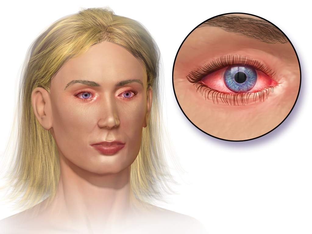
Medical illustration of allergic conjunctivitis. Credit: BruceBlaus / Blausen Medical, via Wikimedia Commons, licensed under CC BY 3.0.
What to notice
Detailed image description
Source: https://commons.wikimedia.org/wiki/File:Blausen_0013_AllergicConjunctivitis.png Creator: BruceBlaus / Blausen Medical Licence: Creative Commons Attribution 3.0 (CC BY 3.0): https://creativecommons.org/licenses/by/3.0/ Modifications: None.
Targeted Investigations
Routine laboratory testing is unnecessary in typical disease. Consider targeted investigation when the diagnosis is uncertain, disease is severe or recurrent, or identification of a trigger would alter management:
- skin-prick testing or serum allergen-specific immunoglobulin E for suspected immediate hypersensitivity
- patch testing for suspected contact allergy
- conjunctival scraping or cytology in selected specialist cases
- microbiological sampling when infection is suspected
- corneal imaging or topography when keratoconus is suspected.
A positive allergy test demonstrates sensitisation but does not prove that the allergen is responsible for the ocular symptoms; results must be interpreted alongside the clinical history.
Important Differential Diagnoses
| Differential diagnosis | Distinguishing features |
|---|---|
| Viral conjunctivitis | Follicles, watery discharge, preauricular lymphadenopathy, infectious contact or respiratory prodrome |
| Bacterial conjunctivitis | Purulent or mucopurulent discharge with eyelids stuck together; itching is less prominent |
| Dry-eye disease | Burning, fluctuating vision and foreign-body sensation rather than dominant itching |
| Blepharitis | Eyelid-margin inflammation, crusting and meibomian-gland dysfunction |
| Contact or toxic conjunctivitis | Temporal relationship to cosmetics, topical medicines, preservatives or lens solutions |
| Microbial keratitis | Contact-lens use, pain, photophobia, reduced vision, corneal opacity or epithelial defect |
| Anterior uveitis | Photophobia, pain, ciliary injection, small or irregular pupil and anterior-chamber inflammation |
| Acute angle-closure glaucoma | Severe pain, headache, nausea, reduced vision, corneal haze and a fixed mid-dilated pupil |
| Episcleritis or scleritis | Sectoral deep redness; scleritis causes severe boring pain and globe tenderness |
| Ocular foreign body | Usually unilateral, with exposure history, focal discomfort or corneal staining |
A unilateral painful red eye in a contact-lens wearer is microbial keratitis until adequately assessed. Remove the lens, do not restart lens wear and arrange urgent ophthalmic evaluation.
Treatment
Treatment depends on disease severity, subtype and the presence of corneal involvement.
General Measures
Initial measures include:
- minimise exposure to confirmed allergens when practical
- apply a cold compress to closed eyelids
- use preservative-free lubricating drops to dilute allergens and support the ocular surface
- avoid rubbing the eyes
- wash hands and face after allergen exposure
- remove contact lenses during active symptoms
- manage associated allergic rhinitis, asthma and atopic dermatitis.
Complete elimination of environmental allergens is often unrealistic. Broad, burdensome avoidance measures should be reserved for clinically relevant, confirmed triggers.
Pharmacological Treatment
- Topical dual-action antihistamine and mast-cell–stabilising preparations are generally preferred for seasonal and perennial allergic conjunctivitis because they provide rapid symptom relief and reduce subsequent mast-cell activation.
- Mast-cell stabilisers work best when commenced before predictable allergen exposure and used regularly, as their preventive effect is not immediate.
- Oral second-generation antihistamines may help when allergic rhinitis or widespread allergic symptoms coexist. However, systemic antihistamines can worsen ocular dryness in some patients.6
- Topical vasoconstrictors provide short-term reduction in redness but do not treat the underlying inflammation. Prolonged use may cause rebound hyperaemia and should be avoided.
- Topical antibiotics have no role in uncomplicated allergic conjunctivitis.
Severe or Refractory Disease
Refer for ophthalmic assessment when there is:
- corneal involvement
- reduced visual acuity
- marked photophobia or pain
- suspected vernal or atopic keratoconjunctivitis
- significant papillary disease
- failure to respond to appropriate first-line treatment
- uncertainty about the diagnosis.
Topical corticosteroids may rapidly suppress severe inflammation, but should be prescribed and monitored by an appropriately trained clinician. Potential complications include raised intraocular pressure, glaucoma, cataract, delayed epithelial healing and worsening of herpetic or other ocular infection.2,3
Topical ciclosporin or tacrolimus may be used as steroid-sparing treatment for chronic vernal or atopic keratoconjunctivitis under specialist supervision.3,6
Allergen immunotherapy may reduce ocular symptoms when clinically important immunoglobulin E-mediated sensitisation is confirmed and symptoms remain substantial despite standard treatment, particularly when allergic rhinitis also requires treatment.6
A steroid eye drop may make several inflammatory red-eye disorders feel better temporarily while worsening undiagnosed herpetic or microbial keratitis. Exclude infection and corneal disease before using topical ocular corticosteroids.
Complications & Prognosis
Complications
Uncomplicated seasonal and perennial allergic conjunctivitis rarely produce permanent ocular injury. Potential complications of severe, chronic or inadequately treated disease include:
- punctate epithelial keratitis
- corneal epithelial defects
- shield ulcer
- corneal scarring or neovascularisation
- secondary microbial keratitis
- conjunctival fibrosis
- keratoconus associated with chronic eye rubbing
- reduced visual acuity
- sleep, school or work impairment
- contact-lens intolerance.
Treatment-related complications include preservative-induced ocular-surface toxicity, rebound hyperaemia from vasoconstrictors, and corticosteroid-associated glaucoma, cataract or infection.
Prognosis
Seasonal and perennial allergic conjunctivitis usually respond well to allergen reduction, cold compresses, lubricants and topical anti-allergy treatment. Symptoms may recur while exposure continues.
Vernal keratoconjunctivitis often improves after puberty or early adulthood, but active corneal disease can impair vision. Atopic keratoconjunctivitis may persist for many years and requires long-term ophthalmic follow-up to prevent corneal scarring and other sight-threatening complications.2,3
Prognosis is less favourable when there is persistent corneal involvement, delayed specialist assessment, chronic eye rubbing, poor treatment adherence or unsupervised use of topical corticosteroids.
References
1. American Academy of Ophthalmology. Conjunctivitis Preferred Practice Pattern® [Internet]. San Francisco: American Academy of Ophthalmology; 2023 [cited 2026 Aug 11]. Available from: https://www.aao.org/education/preferred-practice-pattern/conjunctivitis-ppp-2023
2. Australasian Society of Clinical Immunology and Allergy. Allergic conjunctivitis [Internet]. Sydney: ASCIA; [cited 2026 Aug 11]. Available from: https://www.allergy.org.au/patients/allergic-rhinitis-hay-fever-and-sinusitis/allergic-conjunctivitis
3. College of Optometrists. Atopic keratoconjunctivitis (AKC) [Internet]. Version 17. London: College of Optometrists; 2025 [updated 2025 Feb 18; cited 2026 Aug 11]. Available from: https://www.college-optometrists.org/clinical-guidance/clinical-management-guidelines/atopickeratoconjunctivitis_akc
4. Azari AA, Arabi A. Conjunctivitis: a systematic review. J Ophthalmic Vis Res. 2020;15(3):372–395. doi:10.18502/jovr.v15i3.7456
5. College of Optometrists. Contact lens-associated papillary conjunctivitis, giant papillary conjunctivitis [Internet]. Version 11. London: College of Optometrists; 2025 [updated 2025 Oct 14; cited 2026 Aug 11]. Available from: https://www.college-optometrists.org/clinical-guidance/clinical-management-guidelines/cl-associatedpapillaryconjunctivitis_clapc_giantpa
6. Leonardi A, Silva D, Formigo DP, Bozkurt B, Sharma V, Allegri P, et al. Allergic conjunctivitis management: update on ophthalmic solutions. Curr Allergy Asthma Rep. 2024;24:289–308. doi:10.1007/s11882-024-01150-0




Members only discussions coming soon…