

Peripheral Arterial Disease (Chronic Lower Limb Ischaemia)
Overview
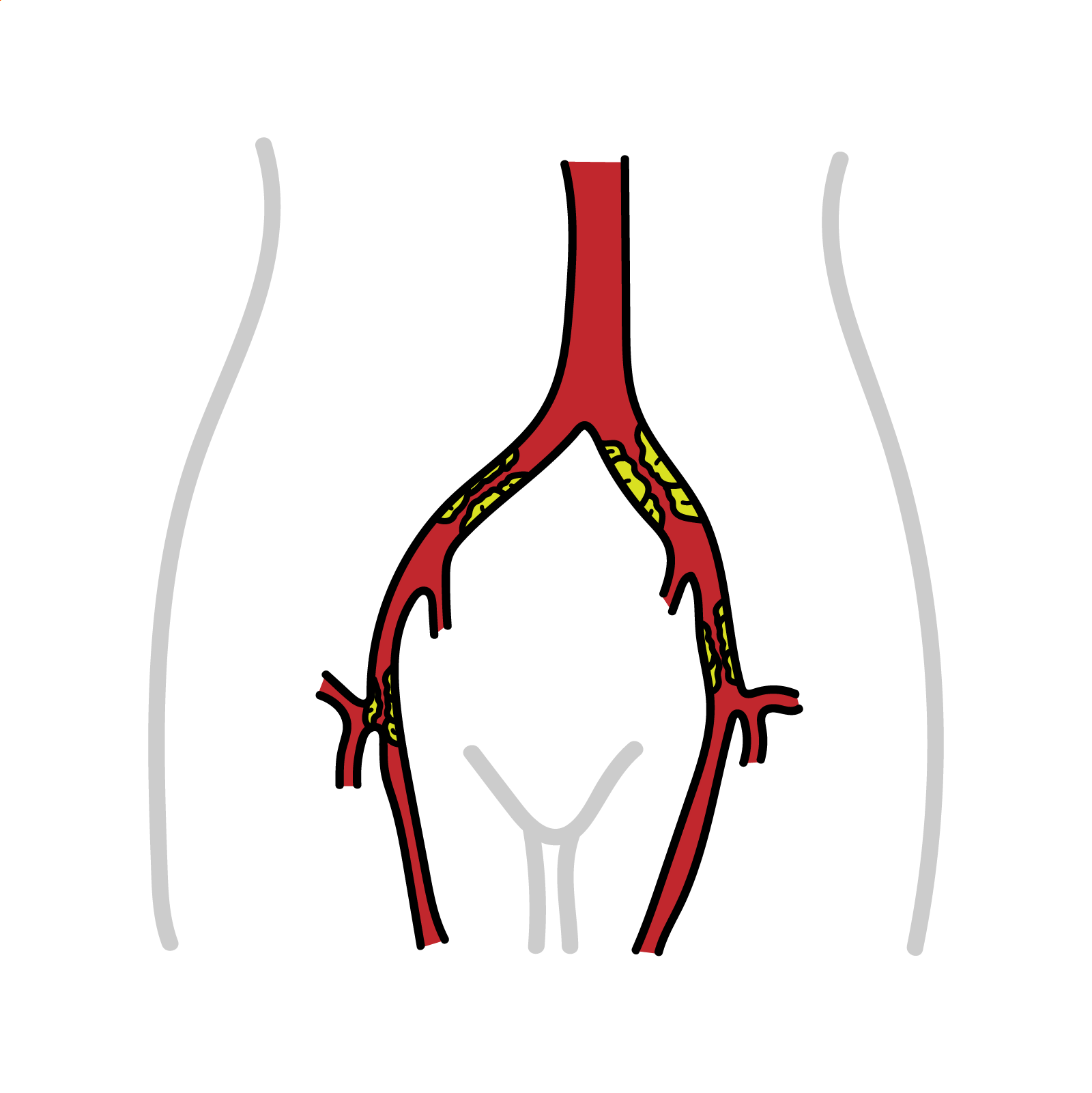
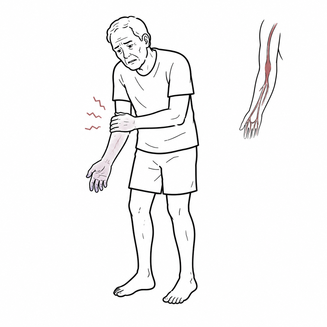
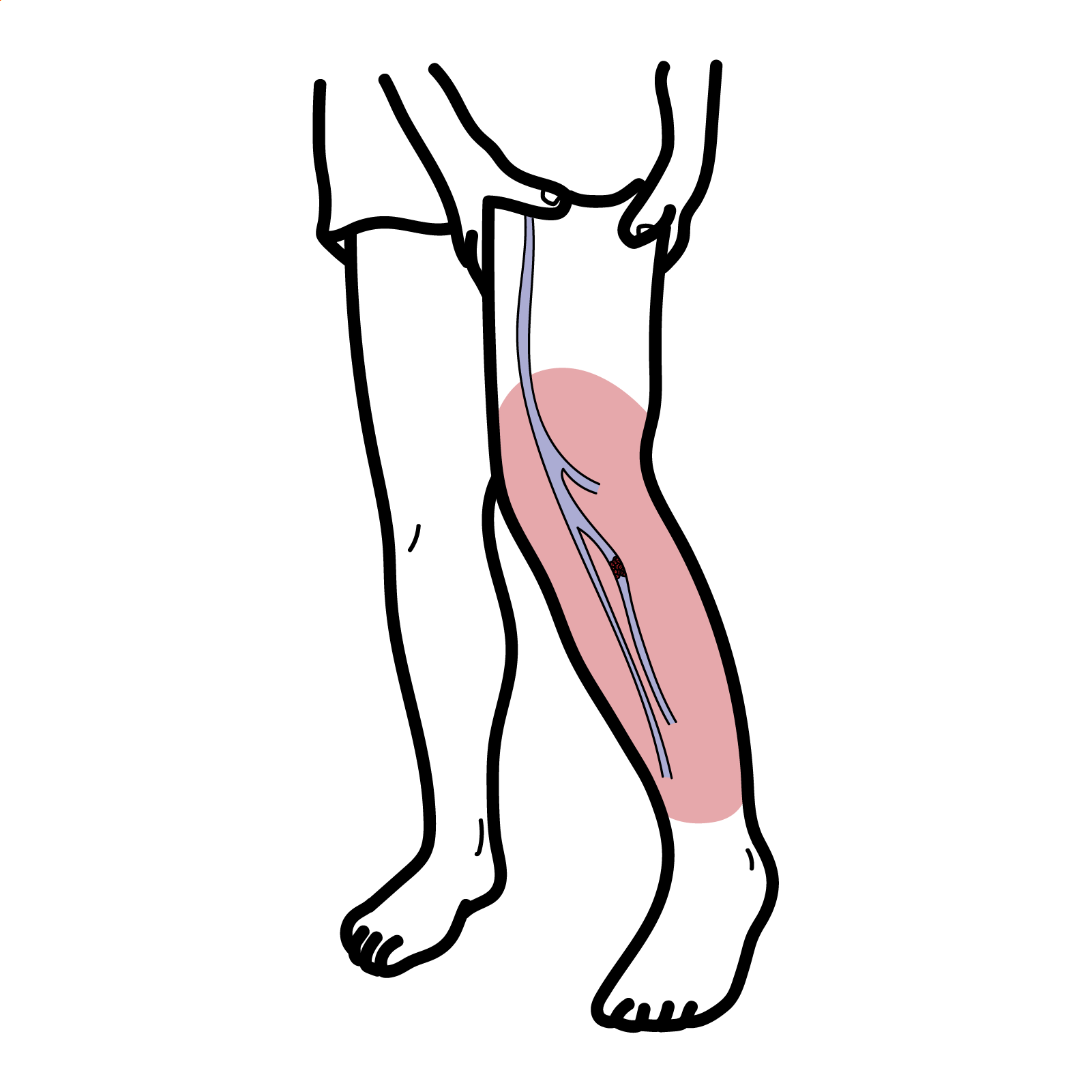
Peripheral arterial disease (PAD) is most commonly caused by atherosclerotic narrowing or occlusion of the arteries supplying the lower limbs. It may be asymptomatic, produce exertional intermittent claudication, or progress to chronic limb-threatening ischaemia (CLTI) with rest pain, ulceration or gangrene. Acute limb ischaemia (ALI) results from a sudden reduction in limb perfusion and is a vascular emergency.
PAD affects more than 200 million adults worldwide, with prevalence increasing markedly with age. It is an important marker of systemic atherosclerosis and is associated with increased risks of myocardial infarction, stroke, cardiovascular death, limb revascularisation and amputation.[1–4]
Definition
Ankle-Brachial Index (ABI): Ratio of ankle to brachial systolic blood pressure, determined using Doppler ultrasound flow.
Claudication: Pain, ache, or cramp in muscles that increases with walking or leg exertion in a predictable manner and resolves with rest.
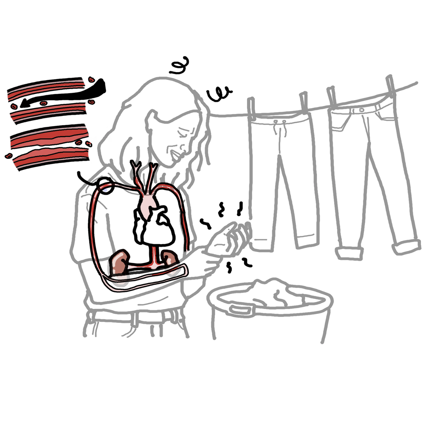
Peripheral arterial disease: Atherosclerosis leading to narrowing of the major arteries distal to the aortic arch. It can involve both the upper and lower extremities
Acute Limb ischemia: Sudden decrease in the blood flow to a limb, resulting in a potential threat to the viability of the extremity
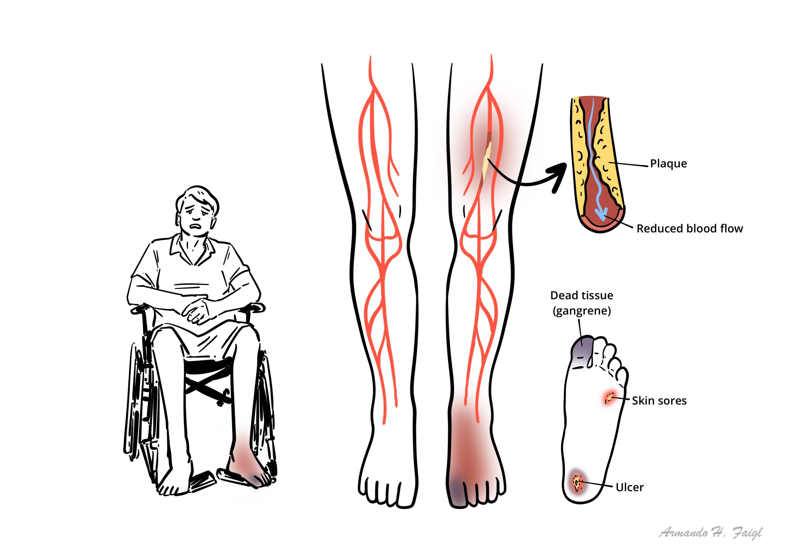
Critical Limb ischaemia: severe obstruction of the arteries which markedly reduces blood flow to the extremities (hands, feet and legs) and has progressed to the point of severe pain and even skin ulcers, sores, or gangrene.
What to notice
Detailed image description
PAD is both a limb disease and a marker of systemic cardiovascular disease. Many patients with objectively confirmed PAD do not report classical intermittent claudication.
Classification
Clinical presentation classification
- Asymptomatic PAD: Objective evidence of PAD without recognised limb symptoms.
- Chronic symptomatic PAD: Exertional limb symptoms, including intermittent claudication.
- Chronic limb-threatening ischaemia: Chronic rest pain, non-healing ulceration or gangrene due to arterial disease.
- Acute limb ischaemia: Sudden reduction in limb perfusion threatening limb viability.
| FONTAINE STAGE CLASSIFICATION OF PVD | |
| Stage I | Asymptomatic, decreased pulses, ABI <0.9 |
| Stage II | Intermittend claudication |
| Stage III | Daily rest pain |
| Stage IV | Focal tissue necrosis |
Anatomy & Physiology
Lower-limb arterial anatomy
- The abdominal aorta divides into the right and left common iliac arteries.
- Each common iliac artery divides into:
- Internal iliac artery, which supplies the pelvis and gluteal region.
- External iliac artery, which continues as the common femoral artery below the inguinal ligament.
- The common femoral artery divides into:
- Profunda femoris artery, supplying the deep thigh muscles.
- Superficial femoral artery, continuing through the adductor canal.
- The superficial femoral artery becomes the popliteal artery after passing through the adductor hiatus.
- The popliteal artery divides into:
- Anterior tibial artery, continuing as the dorsalis pedis artery.
- Tibioperoneal trunk.
- Posterior tibial artery.
- Peroneal artery.
- The posterior tibial artery divides into the medial and lateral plantar arteries within the foot.
Major arterial pulse points
- Femoral pulse: Immediately inferior to the inguinal ligament, approximately midway between the anterior superior iliac spine and pubic symphysis.
- Popliteal pulse: Deep within the popliteal fossa, best palpated with the knee slightly flexed.
- Posterior tibial pulse: Posterior and inferior to the medial malleolus.
- Dorsalis pedis pulse: On the dorsum of the foot, lateral to the extensor hallucis longus tendon.
Ankle–brachial index calculation
- ABI is calculated for each leg using:
Higher ankle systolic pressure from the dorsalis pedis or posterior tibial artery ÷ higher brachial systolic pressure
A patient may have adequate blood flow at rest but develop symptoms during exercise because the arterial circulation cannot increase flow sufficiently to meet increased oxygen demand.
Aetiology & Risk Factors
Aetiology
Atherosclerotic PAD
- Atherosclerosis is the most common cause of chronic lower-limb PAD.
- Common sites include:
- Distal abdominal aorta and iliac arteries.
- Superficial femoral and popliteal arteries.
- Tibial and pedal arteries, particularly in diabetes and chronic kidney disease.
Non-atherosclerotic causes
- Thromboangiitis obliterans.
- Large-, medium- or small-vessel vasculitis.
- Popliteal artery entrapment syndrome.
- Cystic adventitial disease.
- Fibromuscular dysplasia.
- Radiation-induced arterial disease.
- External compression by a tumour or musculoskeletal structure.
- Congenital arterial abnormalities.
Major modifiable risk factors
- Cigarette smoking.
- Diabetes mellitus.
- Hypertension.
- Dyslipidaemia.
- Physical inactivity.
- Obesity and adverse dietary patterns.
Non-modifiable and associated risk factors
- Increasing age, particularly ≥65 years.
- Family history of premature atherosclerotic disease.
- Chronic kidney disease, particularly end-stage kidney disease.
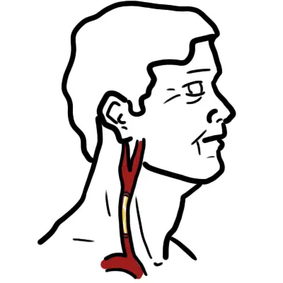
- Established coronary, carotid or cerebrovascular disease.
- Polyvascular atherosclerotic disease.
- Previous lower-limb revascularisation.
- Previous acute limb ischaemia.
- Frailty and malnutrition.
The most important risk factors for PAD are cigarette smoking and diabetes mellitus. Hypertension, dyslipidemia, and elevated homocysteine levels also play significant roles.
Claudication in a young athletic patient without conventional cardiovascular risk factors should prompt consideration of popliteal artery entrapment or cystic adventitial disease rather than assuming atherosclerosis.
Pathophysiology
- Endothelial injury develops
- Smoking, hypertension, hyperglycaemia and abnormal lipids impair normal endothelial function.
- Nitric oxide availability decreases and vascular inflammation increases.
- Lipoproteins accumulate within the arterial wall
- Low-density lipoprotein particles enter and are retained within the intima.
- Oxidised lipids stimulate recruitment of monocytes and inflammatory cells.
- Atherosclerotic plaques form
- Monocytes differentiate into macrophages and ingest lipids, forming foam cells.
- Smooth-muscle cells migrate and produce a fibrous cap.
- Plaques may become calcified and progressively narrow the arterial lumen.
- Arterial resistance increases
- Blood flow may remain adequate at rest.
- Vasodilatory reserve is reduced and exercise-related increases in blood flow become limited.
- Exercise-induced ischaemia produces claudication
- Muscle oxygen demand exceeds supply during walking.
- Anaerobic metabolism and metabolite accumulation produce pain, cramping and fatigue.
- Symptoms settle when exercise stops and metabolic demand falls.
- Chronic ischaemia affects skeletal muscle
- Mitochondrial dysfunction, muscle-fibre changes, denervation and reduced muscle strength may develop.
- Functional impairment may therefore persist even after arterial blood flow is improved.
- Severe perfusion failure causes CLTI
- Resting blood flow becomes inadequate.
- Ischaemic rest pain, poor wound healing, ulceration and tissue necrosis develop.
- Plaque rupture or thrombosis may cause acute occlusion
- Sudden cessation of blood flow produces ALI.
- Nerve and muscle tissue rapidly become ischaemic.
- Irreversible tissue injury develops
- Prolonged severe ischaemia causes muscle necrosis, nerve injury, gangrene, infection and limb loss.[1,4]
Claudication occurs when exercise demand exceeds available arterial blood flow.Rest pain develops when perfusion is inadequate even without exercise
Clinical Manifestation
Asymptomatic and atypical PAD
- PAD may be detected through:
- An abnormal ABI.
- Reduced peripheral pulses.
- Arterial calcification on imaging.
- Investigation of another vascular condition.
- Many patients describe atypical symptoms rather than classical claudication.
- Reduced activity, frailty, arthritis or neuropathy may prevent patients from exercising sufficiently to develop claudication.
Intermittent claudication
- Reproducible muscular discomfort triggered by walking or exertion.
- Common descriptions include:
- Cramping.
- Aching.
- Tightness.
- Heaviness.
- Weakness or fatigue.
- Symptoms generally resolve within approximately 10 minutes of rest.
- Symptoms recur at a relatively predictable walking distance.
- Pain does not usually begin while sitting or lying down.
| Localisation of symptoms | ||
| Disease level | Typical symptoms | Important examination findings |
|---|---|---|
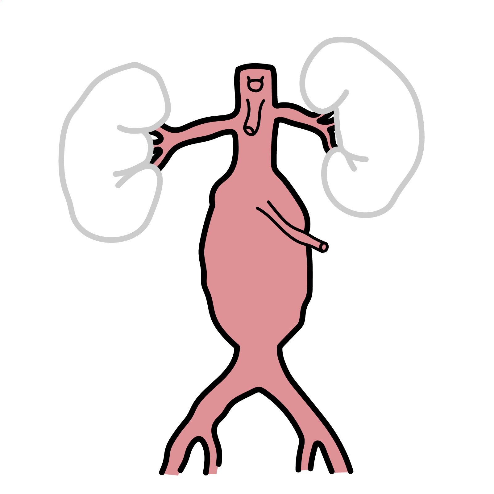
| Aortoiliac | Buttock, hip or thigh claudication; erectile dysfunction may occur | Reduced or absent femoral pulses; femoral or abdominal bruit |
| Femoropopliteal | Calf claudication | Femoral pulse may be preserved; popliteal and pedal pulses reduced |
| Infrapopliteal | Foot discomfort, rest pain or distal tissue loss | Absent pedal pulses; toe or forefoot ulceration |
| Multilevel disease | Symptoms involving several muscle groups | Multiple reduced pulses and extensive trophic changes |
Leriche syndrome triad: Buttock or thigh claudication, Erectile dysfunction and Reduced or absent femoral pulses.
Chronic limb-threatening ischaemia
- Ischaemic rest pain:
- Commonly affects the forefoot or toes.
- Often worse at night.
- Worsened by leg elevation.
- Temporarily relieved by hanging the leg over the bed or standing.
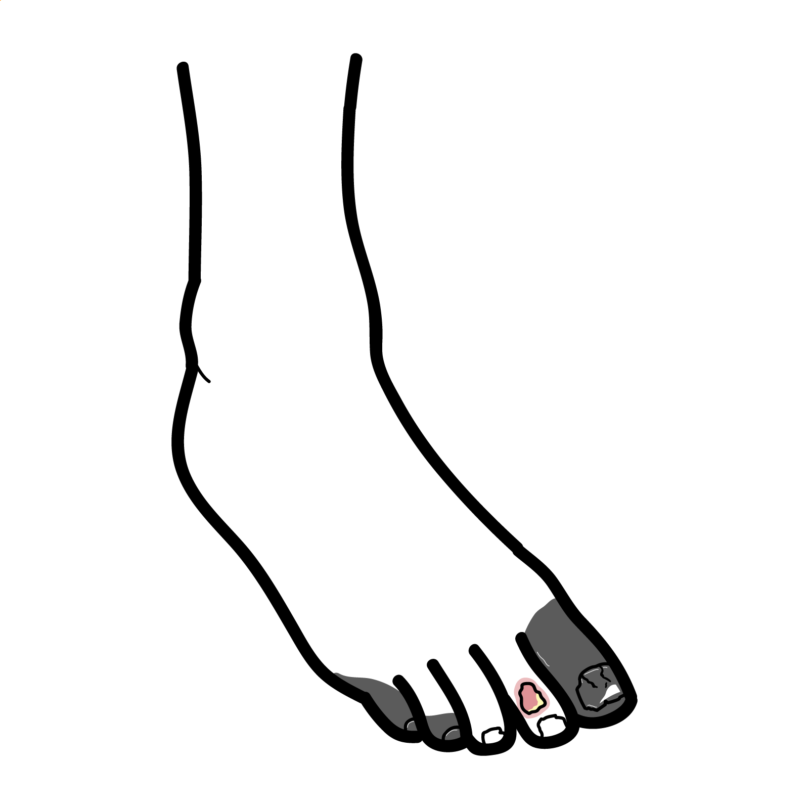
- Non-healing arterial ulcer.
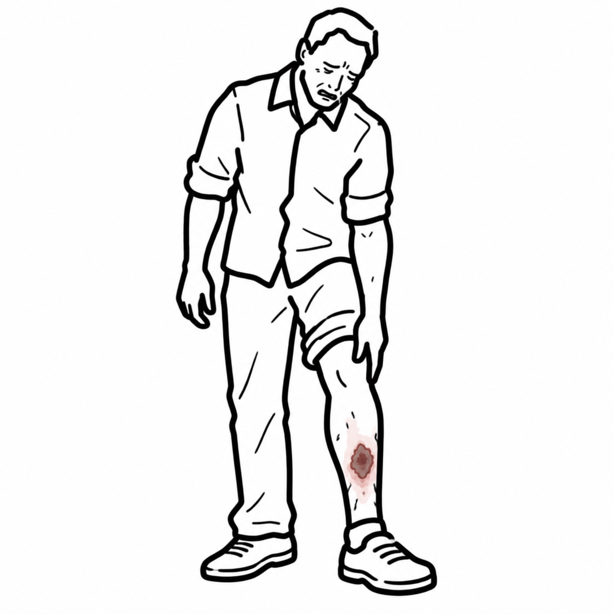
- Digital or forefoot gangrene.
- Recurrent tissue infection.
- Reduced walking ability or inability to bear weight because of ischaemic pain.
Clinical Examination in PAD
- Reduced or absent femoral, popliteal, posterior tibial or dorsalis pedis pulses.
- Asymmetry between the limbs.
- Femoral or abdominal arterial bruits.
- Cool skin.
- Prolonged capillary refill.
- Pallor on elevation.
- Shiny, thin or atrophic skin.
- Reduced distal hair growth.
- Thickened or brittle toenails.
- Calf muscle wasting.
- Non-healing distal ulceration.
- Reduced sensation from associated diabetic neuropathy.
Buerger test
- Elevate the patient’s legs to approximately 45 degrees.
- Observe for pallor developing in the affected foot.
- Lower the legs and sit the patient upright.
- Observe for delayed return of colour followed by dependent rubor.
- A markedly reduced Buerger angle indicates severe arterial insufficiency.
- The test is supportive but is less reliable than objective physiological testing
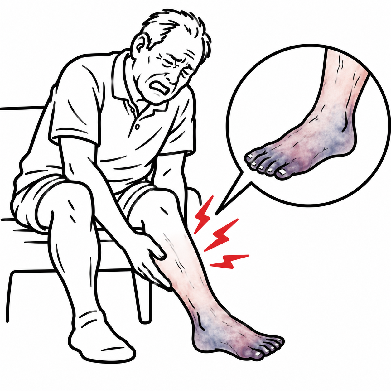
ACUTE LIMB ISCHAEMIA – The 6 P’s
- Pain (absent in 20% of cases)
- Pulse
- Pallor (within a few hours becomes mottled cyanosis)
- Pressure (pain on stretch, firm, tense)
- Paresthesia
- Paralysis
Diagnosis
Ankle–brachial index
- When peripheral artery disease is suspected, the test most commonly used to evaluate for arterial insufficiency is the ankle-brachial index (ABI). Normally, blood pressures in the large arteries of the legs and arms are similar. In PVD this is different.
- ABI should be measured in both legs.
- Exercise treadmill ABI is recommended when exertional symptoms are suggestive but resting ABI is >0.90 and ≤1.40.
| Resting ABI | Interpretation |
| ≤0.90 | Abnormal and diagnostic of PAD in the appropriate clinical setting |
| 0.91–0.99 | Borderline |
| 1.00–1.40 | Normal |
| >1.40 | Non-compressible arteries |
Duplex ultrasonography
- Combines anatomical imaging with assessment of blood-flow velocity.
- Can identify:
- Arterial stenosis.
- Occlusion.
- Aneurysm.
- Graft or stent failure.
CT angiography
- Provides detailed anatomical assessment of stenoses and occlusions.
- Useful when planning endovascular or surgical revascularisation.
Catheter angiography
- Invasive reference anatomical investigation.
- Usually performed when:
| DIFFERENCES BETWEEN TYPES OF CLAUDICATION | |||
| Arterial – Intermittent (atherosclerosis) | Neurogenic (Lumbar spinal stenosis) | Venous (Deep vein thrombosis) | |
| Pain | Pain is in the muscle of the calf, thigh or buttock | Pain is in whole leg ca be associated with tingling and numbness | Involvement of whole leg. Patients may describe feeling their leg is going burst |
| Unilateral or Bilateral | Unilateral in femoropoliteal disease. Bilateral in aortic-iliac disease | Bilateral | Most commonly unilateral |
| Provoking facotrs | Gradual onset after walking “claudication distance” | Comes on suddenly on standing or walking | Gradual onset after beginning to walk |
| Relieving factors | Pain is relieved by rest | Relieved by sitting down, bending over and stopping walking | Relief on elevating the leg |
| Unable to straighten legs | |||
| Pulses/ABIs | Absent/reduced pulses | Absent/reduced pulses | Normal |
| Skin and tissue changes | Mottled skin, Gangere | Absent | Cyanosed, Oedematous |
Classic exertional symptoms with a normal resting ABI should be investigated using an exercise ABI. ABI >1.40 suggests non-compressible arteries rather than excellent circulation.
Use toe pressures or toe–brachial index when diabetes or chronic kidney disease produces a falsely elevated ABI.
In diabetic vessels there are calcification of arteries. This means it can give false ABI readings. Thus different sites of compression (toes) are used.
Ankle-brachial index is the ratio of the ankle blood pressure to the highest brachial systolic pressure. Ankle blood pressure is obtained by inflating a blood pressure cuff above the ankle and detecting the return of the dorsalis pedis or posterior tibial artery pulse by Doppler ultrasonography as the cuff is slowly deflated.
Treatment
Management of Chronic Limb Ischaemia
- Smoking cessation
- Lipid lowering therapy – Statin, etc
- Control Blood Pressure
- Antiplatelet therapy (to reduce risk of cardiovascular events)
- Symptomatic PAD should generally receive single antiplatelet therapy.
- Foot care
- Structured exercise therapy
- Cilostazol
- May improve walking distance and claudication symptoms.
- Acts through phosphodiesterase-3 inhibition.
- Common adverse effects include:
- Headache.
- Palpitations.
- Diarrhoea.
- Contraindicated in heart failure of any severity.
- Pentoxifylline is not recommended because meaningful benefit has not been demonstrated
Revascularisation may restore arterial flow, but exercise therapy remains important because skeletal muscle dysfunction and deconditioning also contribute to walking limitation.
Revascularisation
- Surgical embolectomy or thrombectomy.
- Catheter-directed thrombolysis.
- Mechanical or aspiration thrombectomy.
- Endovascular angioplasty or stenting.
- Surgical bypass.
- Hybrid surgical and endovascular procedures.
Complications & Prognosis
- Increased cardiovascular risk: MI, Ischaemic stroke, Heart failure.
- Chronic limb-threatening ischaemia
- Acute limb ischaemia
- Non-healing arterial ulcer
- Osteomyelitis.
- Minor or major amputation
- Acute reperfusion complications
- Compartment syndrome.
- Hyperkalaemia
- Acute kidney injury
- Cardiac arrhythmia
- Treatment complications
- Arterial dissection or perforation.
- Distal embolisation.
- Graft or stent thrombosis.
- Restenosis.
An ischaemic ulcer or gangrene should be considered limb-threatening until proven otherwise.
- PAD is associated with increased long-term risks of myocardial infarction, stroke and cardiovascular death.
- Cardiovascular events frequently determine long-term prognosis, even in patients whose limb symptoms are mild.
- Claudication often remains stable but can cause substantial functional limitation and reduced quality of life.
- Only a minority of patients with uncomplicated claudication progress directly to major amputation.
- Prognosis is substantially worse once CLTI develops.
- Historical studies report approximately 25–35% mortality and major amputation rates approaching 30% within one year of CLTI presentation, although outcomes vary according to comorbidity, treatment access and revascularisation options.[1,5]
References
- Gornik HL, Aronow HD, Goodney PP, et al. 2024 ACC/AHA/AACVPR/APMA/ABC/SCAI/SVM/SVN/SVS/SIR/VESS guideline for the management of lower extremity peripheral artery disease. Circulation. 2024;149(24).
- Mazzolai L, Teixido-Tura G, Lanzi S, et al. 2024 ESC Guidelines for the management of peripheral arterial and aortic diseases. Eur Heart J. 2024;45(36):3538-3700.
- Criqui MH, Matsushita K, Aboyans V, et al. Lower extremity peripheral artery disease: contemporary epidemiology, management gaps, and future directions: a scientific statement from the American Heart Association. Circulation. 2021;144(9).
- Golledge J. Update on the pathophysiology and medical treatment of peripheral artery disease. Nat Rev Cardiol. 2022;19(7):456-474.
- Conte MS, Bradbury AW, Kolh P, et al. Global vascular guidelines on the management of chronic limb-threatening ischemia. Eur J Vasc Endovasc Surg. 2019;58(1 Suppl).e33.
- Nordanstig J, Behrendt CA, Baumgartner I, et al. European Society for Vascular Surgery 2024 clinical practice guidelines on the management of asymptomatic lower limb peripheral arterial disease and intermittent claudication. Eur J Vasc Endovasc Surg. 2024;67(1):9-96.
- Twine CP, Kakkos SK, Aboyans V, et al. European Society for Vascular Surgery 2023 clinical practice guidelines on antithrombotic therapy for vascular diseases. Eur J Vasc Endovasc Surg. 2023;65(5):627-689.
- Anand SS, Bosch J, Eikelboom JW, et al. Rivaroxaban with or without aspirin in patients with stable peripheral or carotid artery disease. Lancet. 2018;391(10117):219-229.
- Bonaca MP, Bauersachs RM, Anand SS, et al. Rivaroxaban in peripheral artery disease after revascularization. N Engl J Med. 2020;382(21):1994-2004.
- Song P, Rudan D, Zhu Y, et al. Global, regional, and national prevalence and risk factors for peripheral artery disease in 2015: an updated systematic review and analysis. Lancet Glob Health. 2019;7(8).

Members only discussions coming soon…