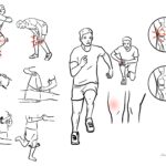
Meniscal Tear


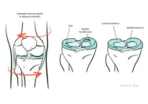
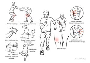
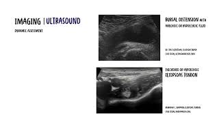
Medial and lateral knee joint menisci serve to transfer load and absorb shock, aid joint stability and provide lubrication. The meniscus is the most commonly injured structure in the knee joint. Acute tears occurs after axial loading combined with rotation. People may complain of a sensation of clicking or catching of the knee with motion. Clinical findigns include positive joint line tenderness, effusion, and positive Mc’Murrary test are important physical exam findings. MRI can help classify location and morphology of meniscal tears.
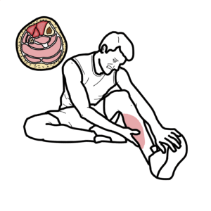
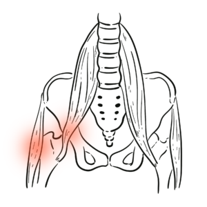
Meniscus: The menisci are C-shaped fibrocartilaginous disks in the knee that serve to transfer load and absorb shock, aid joint stability and provide lubrication.
Arthroscopy: is the examination of the inside of a joint, using a special illuminating instrument inserted through a small incision or ‘portal’.
Meniscectomy: is the surgical removal of all or part of a torn meniscus
The knee joint is a synovial joint, in that it is encapsulated by the synovial membrane which produced synovial fluid. As the knee is a modified hinge joint it is structured to perform two principal actions, flexion (bending) and extension (straightening). The muscles which act at the knee are predominantly the quadriceps (extension) and the hamstrings (flexion). The knee can also do a bit of internal and external rotation.
The knee joint is made up of three main bones:
Cartilage The ends of the bones are covered by articular cartilage. Between these bone articulation there is also the meniscus, another type of cartilage. The menisci are C-shaped fibrocartilaginous disks in the knee (lateral more O-shaped)
The meniscus
Function:
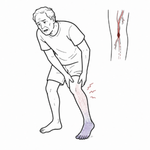
Blood supply The menisci receive blood from branches of the geniculate arteries. The peripheral 1/3 of the meniscus is vascular
The medial meniscus is firmly attached to the joint capsule along its entire peripheral edge. The lateral meniscus there is a region posterolaterally where it is not firmly attached.
The medial meniscus has less mobility than the lateral meniscus and is more susceptible to tearing when trapped between the femoral condyle and tibial plateau.
The lateral meniscus is larger – greater share of the lateral compartment pressure than the medial meniscus carries for the medial compartment.
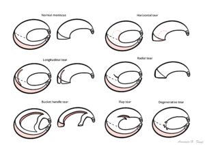
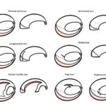
Meniscal tears can be classified either by etiology or by their arthroscopic and MRI appearance.
Etiologic classification divides tears into either acute tears or degenerative tears.
Arthroscopic and MRI appearance:
Acute tears For younger patients vertical longitudinal (bucket-handle) are more common. Acute tears occurs after axial loading combined with rotation.
Degenerative tears In older patients, complex and degenerative tears are more common.
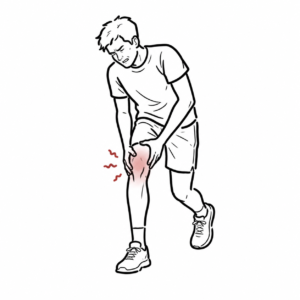
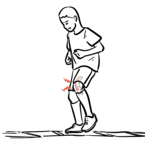
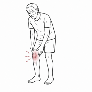
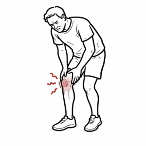
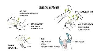
Clinical Presentation
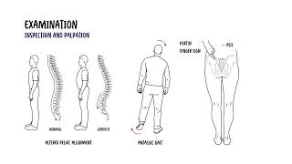
Examination
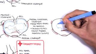
The medial meniscus is more frequently torn, partly because of the different shape (less mobility) but also because of its attachment to the medial collateral ligament.
O’Donohgue Triad (unhappy triad) Injury to the ACL, MCL and Medial meniscus.
Immediate conservative measures include the RICE regimen:
Small stable meniscus tears often become asymptomatic and do not need to be treated surgically. Those causing persistent symptoms should be assessed with arthroscopy.
Longer term measures include
NSAIDs inhibit COX1/COX2, enzymes responsible for converting arachadonic acid to Prostagladins, ThromoxaneA2. Prostaglandins promote inflammation, fever and pain. Thromboxane A2 causes platelet aggregation (blood clotting). Side effects: Increased BP, Dyspepsia, Peptic ulcer, duodenal ulcer, liver damage (rare and long-term), acute kidney injury, tinnitus (high-dose) and allergy. Contraindications: Pre-existing renal dysfunction, cardiac failure, severe hepatic dysfunction, history of GI bleeding, allergy, elderly >65.
Surgical Treatment
Post surgical rehabilitation is an important part of treatment.
Complications of surgery including septic arthritis.
UpToDate
Oxford Handbook of Clinical Medicine
Best Practice
RACGP Meniscal tear – Presentation, diagnosis and management, 2012
Discussion