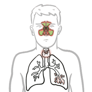
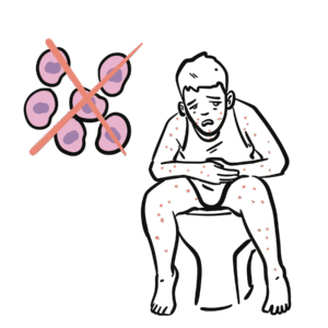
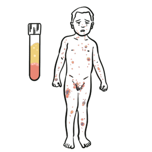
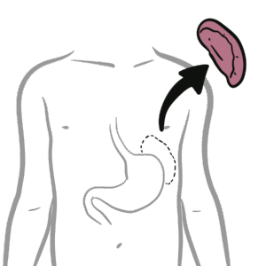
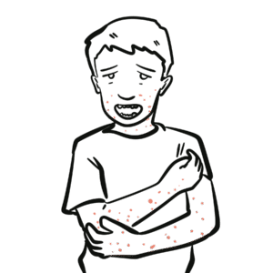
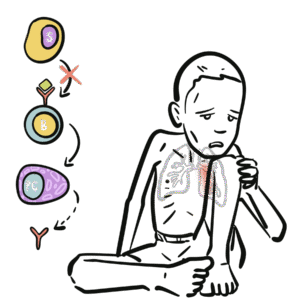
0:00 Hypersensitivity reactions are exaggerated or inappropriate immune response to 0:20 benign 0:21 antigen. 0:22 It is the immune response and not the antigen that is actually harmful. 0:29 An external antigen such as a drug, pollen, or food can elicit an inappropriate 0:35 immune 0:35 response. 0:36 However, an inappropriate immune response to an internal antigen, such as your 0:42 own skin 0:43 or neuron, would be called an autoimmune reaction. 0:48 Hypersensitivity reactions can be the mechanism of diseases in some autoimmune 0:55 reactions. 0:56 Hypersensitivity reactions are antigen-specific. 1:00 It's antigen-specific because the immune system is educated and primed towards 1:05 the antigen 1:06 after the first exposure. 1:09 In this quick example, first exposure or first contact to an external antigen, 1:14 such 1:15 as a drug, pollen, or food, causes the immune system to sensitize itself 1:21 towards the particular 1:22 antigen. 1:23 In other words, it primes the immune system against that antigen, so when the 1:31 next time 1:32 the antigen comes along, the primed immune system, including the T cells, 1:39 antibodies, 1:40 and neutrophils, they can mount an exaggerated and inappropriate immune 1:46 response, aka a hypersensitivity 1:49 reaction. 1:50 But the immune system can cause hypersensitivity reactions in different ways, 1:55 and that is why 1:57 hypersensitivity reactions can be subdivided into four main types. 2:02 Type 1, type 2, and 3 hypersensitivity reactions are antibody-mediated, whereas 2:08 type 4 is a 2:09 cell-mediated hypersensitivity reaction. 2:14 I want to take a short break and introduce to you PDF element. 2:18 PDF element is an all-in-one PDF editor from which you can get powerful 2:22 features to edit 2:23 annotate and convert PDF easily. 2:26 You just open a PDF file. 2:27 For example, here is a PDF on my inflammation video. 2:31 If you want to add more information, you can add a text box. 2:35 For example, here, complement proteins circulate inactive, and once active, 2:40 they promote inflammation. 2:43 You can also change the size or the color of the text. 2:47 There is even a pencil feature to add, so you can become more creative. 2:53 Here, activated B cells become antibody-producing plasma cells, important note. 3:00 PDF element is a robust PDF editor, annotator, and converter on your Windows or 3:06 Mac. 3:06 It greatly helps to read, take notes, and convert PDF easily. 3:11 To this channel, you can save up to $60 on PDF element by clicking on the link 3:16 on the 3:17 description below. 3:18 I really recommend PDF element if you want to get creative with your notes. 3:24 Type 3 hypersensitivity, also known as immune complex hypersensitivity reaction 3:30 , is a reaction 3:31 where formation of the antigen-antibody complex deposit in tissues, activating 3:37 complement 3:38 proteins, and so the inflammatory response in the tissues. 3:43 The antigen-antibody complex are the immune complexes, and these will circulate 3:48 around 3:49 the body. 3:50 The immune complexes can be cleared by macrophages normally in the spleen or 3:55 liver, but when 3:56 not cleared, immune complexes will deposit into various tissues, where they 4:01 induce complement 4:02 activation. 4:05 Complement proteins are circulating proteins which, when active, trigger a 4:09 cascade, promoting 4:10 the inflammatory response. 4:17 External antigens that can form immune complexes when antibodies are formed 4:22 against them include 4:24 foreign serum, such as anti-venom injections given to people, monoclonal 4:29 antibodies people 4:31 receive for treatment, as well as inhaled particles from work environment, and 4:37 also 4:37 bacteria, parts of bacteria, such as group A, streptococcus. 4:43 For the antibodies to be able to target these antigens in the first place, the 4:48 body first 4:48 has to be exposed to them. 4:51 These external antigens are picked up by antigen-presenting cells, such as 4:55 circulating B-cells 4:57 or macrophages. 4:59 The antigen-presenting cell will process and express this antigen on its 5:06 surface. 5:07 The antigen-presenting cell will travel to nearby lymphoid tissue and present 5:11 that antigen 5:13 to T-helper cells, which will subsequently activate the T-helper cell. 5:19 Activated T-helper cells can also activate B-cells, who may also have already 5:24 picked 5:25 up the antigen. 5:28 The activated B-cell now becomes a plasma cell, which are the antibody- 5:35 producing cells. 5:37 The antibodies produced by the B-cells are thus the antibodies against that 5:43 specific 5:43 antigen. 5:50 Now both type 2 and type 3 hypersensitivity reactions actually involve 5:56 complement activation. 5:58 However, in type 2 hypersensitivity reaction, if you remember, antibodies bind 6:05 to antigens 6:06 that are bound to cell membrane surfaces or membrane surfaces. 6:10 Once antibodies bind to the antigen, they then act as a bridge to activate the 6:15 complement 6:16 proteins. 6:18 Whereas in type 3 hypersensitivity reaction, the antibodies produced bind to 6:24 free circulating 6:26 antigens. 6:27 Once the antibodies bind, they can form chains of antigen and antibiotic 6:31 complexes, which 6:32 are the immune complexes. 6:35 Immune complexes can then deposit into body tissues and then activate 6:41 complement proteins. 6:44 The structure of a typical antibody produced by the plasma cells are made up of 6:50 the inner 6:51 heavy chains and outer light chains. 6:53 The top part here is the fab region, which binds to the actual antigen. 7:00 And the bottom part here is the FC region. 7:03 The FC region acts as a bridge that will activate the complement proteins. 7:14 So typical type 3 hypersensitivity reaction is the arthus reaction. 7:20 The arthus reaction is the name given to the inflammation caused by the 7:25 deposition of immune 7:26 complexes at a localized site. 7:29 An example of an arthus reaction is hypersensitivity pneumonitis. 7:34 Workers for example, exposed to different types of particles in the air, such 7:38 as those working 7:39 in the farms, woodworkers, wheat millers, are continuously inhaling these 7:45 different particles. 7:47 And as a result, the body may develop immunoglobulin G antibodies against that 7:52 particle inhaled. 7:56 When the particle is inhaled subsequently, the antibodies can bind to those 8:01 particles, 8:02 to those antigens and form immune complexes with them. 8:09 The immune complexes can then deposit into the tissues of the lungs. 8:16 Deposition of the immune complexes will then activate the complement proteins. 8:22 Complement activation results in three main things. 8:25 Firstly, release of pro-inflammatory cytokines. 8:29 Opsinization, which promotes phagocytosis through neutrophils. 8:33 And thirdly, formation of the membrane attack complex, which will... 8:37 Again, this is all a localized response. 8:42 In contrast to arthus reaction, which is a localized inflammation, serum 8:47 sickness is 8:47 another example of type 3 hypersensitivity, and is a systemic inflammatory 8:54 response, in 8:55 the presence of immune complexes. 8:58 Serum sickness can occur when foreign serum from an animal or monoclonal 9:02 antibodies given 9:03 for treatment result in adaptive immune systemic activation and production of 9:09 antibodies against 9:11 that foreign serum or monoclonal antibody. 9:14 The body has sensitized itself essentially to this serum. 9:19 Thus, on subsequent exposure of the serum, the antibodies that have already 9:24 been formed 9:25 will bind onto the antigens from the serum and form immune complexes, which 9:32 will then 9:33 deposit again to tissues. 9:38 These immune complexes can actually deposit anywhere in the body because they 9:42 're circulating, 9:44 resulting in complement activation once deposited. 9:48 Complement activation results in an inflammatory response. 9:53 And of course, in this scenario, serum sickness, you get an inflammatory 9:58 response systemically. 10:00 The symptoms of serum sickness, such as fevers, or to carry a thralgis spleen 10:05 omegaly, typically 10:09 will resolve once the immune system can actually clear up the immune complexes 10:13 which have deposited. 10:17 There are many other disorders where immune complexes play a role in the path 10:22 ophysiology. 10:23 These diseases include rheumatoid arthritis, where immune complexes can deposit 10:28 into joints. 10:29 However, immune complexes most often target the kidneys, such as in systemic l 10:34 upus erythmatosis, 10:36 IgA nephropathy. 10:38 And these immune complexes can easily deposit here because of the small capill 10:43 aries in the kidneys. 10:47 Post-triptococcal glomerular nephritis is a good example of type 3 hypers 10:51 ensitivity reaction 10:53 and immune complex formation. 10:56 Post-triptococcal glomerular nephritis occurs several weeks after a group A 11:00 streptococcus 11:01 infection, such as a strep throat infection. 11:06 After the infection, the body will mount an immune response, creating 11:10 antibodies against 11:11 group A streptococcus. 11:15 The IgG antibodies produced will bind onto the antigens of group A streptococ 11:23 cus. 11:24 When bound, they will actually aggregate and form immune complexes, which will 11:30 travel to 11:30 the kidneys, to the glomeruli of the kidneys, and then subsequently deposit in 11:36 the glomeruli 11:38 of the kidneys, the capillaries within the bowman's capsule. 11:43 These streptococcal antigen antibody complexes deposit on glomeruli after which 11:49 they fix 11:50 complement, attract neutrophils, and start the inflammatory response. 11:56 Very important to realize here that hypersensitivity type 3 reactions, again, 12:02 can target external 12:03 antigens, such as what we have learned, the foreign serum, as well as inhaled 12:07 particles, 12:08 but also it can target and immune complexes can form in towards internal antig 12:14 ens, such 12:15 as seen in systemic lupus erythmatosis. 12:19 Thank you for watching, I hope you enjoyed this video.