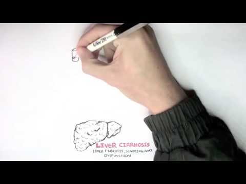
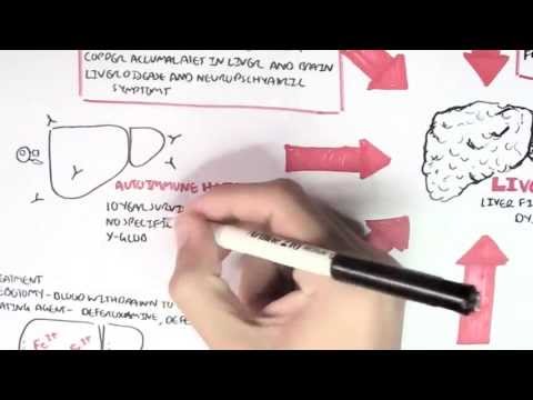
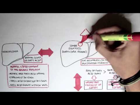
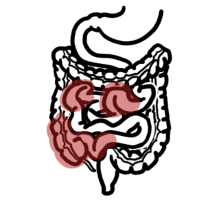
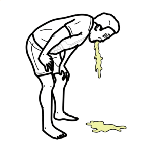
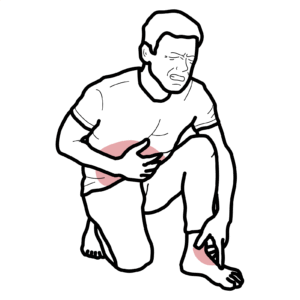
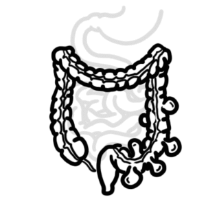
0:00 So, in this video, we're going to talk about liver cirrhosis. 0:09 I have a video that looks actually at the complications of cirrhosis, which 0:13 includes, you 0:13 know, the portal hypertension, ascites, hepatopalmonary syndrome, amongst many 0:18 other 0:18 things. 0:19 We will mainly look at, in this video, the signs and symptoms, the etiology, 0:24 the pathophysiology, 0:25 and the investigations and treatment for cirrhosis. 0:28 So, cirrhosis is a light stage progressive hepatic fibrosis characterized by 0:33 destruction 0:34 of the hepatic architecture and formation of regenerative nodules. 0:39 So, here we have a normal healthy liver, and draining into the liver is the 0:46 portal vein. 0:47 Now, the portal vein is what the GIT veins essentially drain into, so all those 0:54 nutrients 0:55 absorbed by the gut, by the GIT, it will drain or it will pass through the 1:00 liver through 1:01 the portal vein. 1:02 So, here is a person with liver cirrhosis. 1:07 A cirrhotic liver, we get fibrosis and nodule formation. 1:15 And the portal vein, which under normal conditions, actually, you know, drain 1:20 blood into the 1:21 liver, the draining is not effective because of the fibrosis of the liver, and 1:28 so there 1:29 is backflow due to the buildup of pressure here. 1:33 So, what causes this progression from a normal liver to a cirrhotic liver, 1:39 which is an irreversible 1:40 progression? 1:41 Well, common causes of liver cirrhosis include chronic viral hepatitis, 1:46 alcoholic liver disease, 1:49 and hematochromatosis. 1:51 Less common causes include autoimmune hepatitis, medications, wilson's disease, 1:56 and celiac 1:57 disease. 1:59 So, signs and symptoms of liver cirrhosis. 2:03 The neurological signs include extractsis, I think I pronounced that right, 2:08 which is the 2:09 hepatic flap, and then you have head and neck involvement, which is prodigy 2:13 gland swelling. 2:15 This usually involves alcohol. 2:19 Skin findings, you get spartanivi, which is also known as angiomata, as well as 2:24 jaundice 2:24 with bilirumin greater than two milligrams per deciliter. 2:29 Chest findings, you can find gynecomastia. 2:32 Abdominal findings, signs and symptoms include hepato megalae, spina megalae, 2:38 acides, caputmidusae, 2:40 as well as a grooving high lower bound gut and murmur, I hope I pronounced that 2:44 one right 2:44 too. 2:45 Urogenital involvement, men can have testicular atrophy. 2:49 The extremities, you can have pulmonary thema, you can nail changes like club 2:53 bing, you can 2:54 have hypertrophic osteoarthropathy, as well as dupitous contractures. 3:00 So those are the signs and symptoms associated with cirrhosis. 3:03 The complications of cirrhosis are a result mainly of portal hypertension, 3:08 which as we 3:09 mentioned briefly, before is basically due to the fibrosis of the liver, the 3:13 increase 3:14 in pressure results in the portal vein having a backflow. 3:19 And this will backflow into the gut associated, like the organs of the GIT. 3:25 So these complications include, and this in all these complications are not all 3:29 a result 3:30 of the portal hypertension, by the way, but it's mainly. 3:33 So these complications include acides, hepatic encellopathy, variceal hemorrh 3:38 age, bacterial 3:39 peritonitis, hepatopreneal syndrome, portal hypertension, gastropathy, hepatic 3:45 hydrothorax, 3:46 hepatopulmonary syndrome, porto, pulmonary hypertension, and serotic cardiomy 3:54 opathy. 3:55 So we looked at the signs and symptoms of liver cirrhosis, we looked at some 3:58 complications, 3:59 which if you want to learn more about, I have a video on that. 4:03 And next, I should look at the progression, I guess, the pathophysiology of how 4:08 liver 4:09 cells just become cirrhotic. 4:12 So here are your liver cells. 4:15 You have the blood vessel, and here is your liver, which is, I'm just 4:20 representing here 4:20 to be normal. 4:21 So we're looking at a normal liver. 4:24 Again here are your hepatocytes. 4:26 Now these are your sinusoidal cells, and so this is your hepatic sinusoid, 4:32 which is 4:32 essentially where the portal vein drains into, and the portal vein will then 4:37 drain, pass 4:38 through, and form the central vein, drain into the central vein. 4:42 So you have your stelate cells here, and your stelate cells are very important 4:45 in the pathogenesis 4:47 of liver cirrhosis, because these stelate cells, they're usually dormant and 4:51 they're 4:52 arrest, but in cirrhosis they are activated, and we'll talk about it. 4:59 You also have your cupful cells, which are essentially your macrophages of the 5:04 liver. 5:04 Now the causes of liver damage we looked at earlier, such as, you know, 5:09 autoimmune hepatitis, 5:10 Wilson's disease, medication, alcohol, and all these can lead to liver injury, 5:17 causing 5:18 hepatosellular necrosis and/or apoptosis. 5:23 As a result, we see these changes in the liver. 5:26 So we can say this is an advanced stage, which is cirrhosis already. 5:31 So you know, the dead cells with the dead liver cells release damage associated 5:38 molecular 5:39 patterns, dams, as well as reactive oxygen species. 5:43 Now this will stimulate the cupful cells and the stelate cells. 5:48 The stelate cells, which are dormant, are now activated. 5:53 The activated stelate cells do a few things, including further stimulating the 5:59 macrophages, 6:00 the cupful cells, to release cytokines, for example, amongst many other things. 6:07 Also the stelate cells, when they're activated, they secrete chemokines, such 6:11 as CCL2, which 6:12 attract more innate immune cells to the area via the CCL2 receptor. 6:20 As a result, this is what we see an accumulation of immune cells, activation of 6:25 stelate cells, 6:26 which release many more cytokines, mainly TNF-alpha, interleukin-6, interleukin 6:32 -1b, and 6:33 TGF. 6:35 Okay, let's focus on TGF now. 6:40 So TGF stands for transforming growth factor. 6:43 TGF, the transforming growth factor, is released primarily by stelate cells. 6:50 And it's released by stelate cells. 6:52 It actually further stimulates more other stelate cells to transform 6:57 essentially to become cells 6:59 known as myofibroblasts. 7:01 It causes myofibroblasts proliferation. 7:06 These myofibroblasts further release the transforming growth factor, which 7:10 stimulates stelate cells 7:12 to proliferate and become more myofibroblasts. 7:16 Now these fibroblasts are the ones that produce the matrix, the collagen, which 7:21 leads to the 7:21 fibrosis of liver, associated with liver cirrhosis. 7:28 So again, the fibroblasts release the matrix, which actually causes the fib 7:34 rosis of the liver. 7:37 Now going back to the cytokines, the TNF-alpha and the other cytokines attract 7:42 more neutrophils 7:42 and T cells into the area. 7:45 These T cells and neutrophils contribute to the inflammation early in liver 7:51 damage, which 7:52 will lead to necrosis and eventually the cirrhosis, the fibrosis. 8:01 Also the inflammation, it sort of stimulates the nodule formation because of 8:07 the liver 8:08 trying to repair itself. 8:14 So cirrhosis is the late stage of liver disease, aside from liver failure, but 8:19 when the liver 8:20 is really damaged and the complications are severe, liver transplantation can 8:27 be performed, 8:29 but this in itself is a long process and requires much regulation. 8:34 Next, let's look at investigations of a person suspected with liver disease. 8:43 So investigations, liver biopsy is the gold standard, taking a sample of the 8:51 liver. 8:52 Number two, you can perform a liver function test, this is ALT, AST, alanine, 9:00 bilirubin. 9:01 Three, you can perform an ultrasound, so here you're looking at liver 9:07 architecture. 9:09 Four, you can perform a metabolic breath test. 9:12 This is to assess functional reserve of the liver rather than the structure. 9:19 Five, you can perform a hepatic venous pressure gradient. 9:27 And finally, you can also perform a transient elastography. 9:34 And a transient elastography, it measures liver stiffness, which may reflect 9:39 the fibrosis 9:40 or edema and inflammation associated with the liver. 9:44 So here you can see the transducer measuring the liver stiffness. 9:49 Finally, let's look at the management for liver cirrhosis. 9:54 The main thing to do is trying to slow or reserve the progression of liver 10:02 disease. 10:04 And this actually leads to the second management, which is essentially 10:08 preventing the superimposed 10:10 insults to the liver. 10:11 So, you know, vaccinating to, in case of, liver infections, avoiding hepatotox 10:19 ins, which include 10:20 your alcohol, which is a main cause of liver cirrhosis, one of the main causes, 10:25 as well 10:26 as to adjust medications, as certain medications cause the liver to work harder 10:32 . 10:32 And third, medication adjustment and/or avoidance, as we talked about, is very 10:41 important. 10:42 And then four, we manage the symptoms and also the laboratory abnormalities 10:48 associated 10:48 with liver cirrhosis. 10:50 So we address the muscle cramps, umbilical hernias, hyponaturmia, thrombocytop 10:58 enia, and 10:59 the increase in IONR, which basically means your body's not clotting properly. 11:03 It's important to address all these factors associated with liver cirrhosis. 11:09 Management also includes preventing and identifying and treating the 11:13 complications of cirrhosis, 11:15 which we talked about earlier, including, you know, your peritonitis, your asc 11:20 ites, 11:21 hepatopolymer syndrome, et cetera. 11:23 And finally, the last management, of course, would be liver transplantation, if 11:29 all else, 11:30 and of course, if there's a suitable donor. 11:33 So thank you for watching. 11:34 Hope you enjoyed this video on liver cirrhosis.