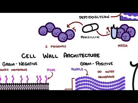
0:00 In this video we're going to talk about bacterias, and it's just going to look 0:20 at the structure 0:20 specifically. 0:22 So bacteria, bacteria are the largest group of microorganisms or medical 0:27 significance, 0:28 and so that's pretty important to know because they are literally everywhere. 0:34 Bacteria occur in three main shapes, coccus, a circular shape, spheria, a basil 0:39 is, a rod 0:40 shape and a spiral. 0:43 Now there's also other different shapes bacteria can take such as a coco basil 0:48 is which is in 0:49 between a coccus and a basilis, and there's also one, a bacteria can take shape 0:54 of what's 0:54 called a vibrio, and it's sort of spiral and basilis in between, it's sort of 0:58 going 0:59 to bend into it. 1:00 Now some bacteria are surrounded by polysaccharides containing capsule, so a 1:05 capsule containing 1:06 polysaccharides. 1:08 Now this capsule, also known as a slime layer, actually allows the bacteria to 1:14 bind to surfaces 1:15 of different cells in our body for example, and also to evade phagocytosis, to 1:20 avoid phagocytosis. 1:22 So it's very important essentially for the bacteria survival. 1:27 So what other parts does a bacteria have? 1:32 What other structures do a bacteria have? 1:35 Here I'm drawing a rod shape bacteria, it can be any type of rod shape bacteria 1:39 , and 1:39 now let's learn about part of its anatomy. 1:44 Some bacterias have what's known as a flagella, which are thin rigid filaments, 1:49 and these 1:49 allow for bacterial movement. 1:52 Flagella are essentially made up of proteins, and these proteins can move, can 1:55 propel the 1:56 bacteria forward, or move it around. 2:00 Bacteria can also possess hair-like appendages coming out of their surface. 2:05 There are two types of hair appendages, fimbriae, and pilae, and these are used 2:11 interchangeably. 2:13 A pilae is typically shorter than a fimbriae, but they essentially do the same 2:19 thing. 2:19 The fimbriae contributes to the bacteria's ability to cause a disease by 2:23 binding onto 2:24 a surface of a cell in our body for example, and this can also be done by the 2:29 pilae. 2:30 The pilae is another hair-like appendage, just like the fimbriae, and the pilae 2:34 help 2:34 attach the bacteria to mucosal cell surfaces, and the pilae are also involved 2:39 in what's 2:40 called bacterial conjugation. 2:43 Bacterial conjugation is essentially the transfer of the genetic material of a 2:48 bacteria from 2:50 one bacteria to another by forming a bridge using a pilae. 2:57 Now let's look at the different layers of bacteria has. 3:00 The bacteria's outermost layer is known as a capsule, and as we know it has 3:05 polysaccharide, 3:07 it enables it to bind to cell surfaces and also avoid phagocytosis, such as 3:13 avoiding 3:14 macrophages and also neutrophils. 3:17 The bacteria's innermost layer is known as a plasma membrane, and plasma 3:22 membranes are 3:23 also found in human cells, for example in eukaryotes, and the structure is very 3:28 similar 3:28 in both bacteria and humans. 3:30 The plasma membranes are semi-permeable membranes allowing transportation of 3:34 substances in and 3:35 out of the cells. 3:38 And plasma membranes, as I mentioned, in bacteria are similar to those in 3:42 humans in bacteria. 3:44 The plasma membrane is also a phospholipid bilayer and contains proteins as 3:50 well within 3:51 the membrane. 3:52 The phospholipid bilayer is made up of molecules known as phospholipids. 3:57 A phospholipid has a hydrophilic region, which means a region that loves water, 4:05 hydrophilic 4:06 as in love, and also has a hydrophobic region, hydrophobic and phobic as in 4:12 skin. 4:13 And this characteristic of a phospholipid basically enables it to form the 4:19 phospholipid 4:20 bilayer. 4:21 And so that's for the plasma membrane. 4:24 Now in between the capsule and plasma membrane, we have the cell wall. 4:29 And this is a very complex semi-rigid structure. 4:33 And the cell wall enables bacteria to be classified into two major groups, gram 4:39 -positive bacteria 4:41 or gram-negative bacteria. 4:43 And there's a difference. 4:47 First of all, they are grouped into gram-positive and gram-negative bacteria 4:52 because when you 4:53 dye them with different dyes, they have different colors. 4:56 And the different colors are caused by the different cell composition of both 5:01 these types 5:01 of bacteria. 5:03 So what is the difference in cell wall composition? 5:05 Well, the gram-positive bacteria has a cell membrane with a structure above it. 5:09 Let's just say here's the outside of the bacteria and here's the inside of the 5:13 bacteria. 5:14 So here we have the cell membrane. 5:16 And the structure above it is known as a peptidoglycan and it's composed of 5:20 amino acids and sugars. 5:22 The gram-negative bacteria also has a cell membrane, but it has a very thin 5:27 peptidoglycan 5:28 layer compared to the gram-positive. 5:31 So gram-negative has a thin peptidoglycan layer, the gram-positive has a thick 5:34 peptidoglycan layer. 5:36 In addition to that, the gram-negative has an extra cell membrane above this 5:40 thin peptidoglycan 5:41 layer. 5:43 The other notable difference between gram-positive and gram-negative bacteria 5:46 is that the gram-positive 5:47 bacteria has what's called a lipid-ticolic acid, which enables it to bind to 5:53 cell walls 5:54 or be recognized by macrophiles or agrocytes. 5:59 The gram-negative bacteria on the other hand has a lot of lipid-like structures 6:05 on the outer 6:06 cell membrane, such as lipoproteins and also what's called lipid A. Lipid A are 6:14 toxins 6:15 and they cause certain diseases and shock syndromes within the human body. 6:22 So now because of these different cell wall compositions, when we use dye, 6:26 these bacteria, 6:26 depending on what type of cell composition it has, will have a different color. 6:31 The dyes used to stain a particular bacteria in successive order is crystal 6:38 violet and then 6:39 we use iodine treatment and then we de-colorize it using alcohol and then we 6:45 use finally 6:46 saffronin and essentially, I'll make a video hopefully on how this process 6:51 works, but essentially 6:53 the bacteria with a thick peptidoglycan layer and one cell membrane will have 6:59 the crystal 7:00 violet color which is a bit purplish at the end and the gram and the bacteria 7:06 which has 7:06 two cell membranes and a thin will have a pinkish red color and so this is how 7:12 we classify 7:13 the bacteria. 7:14 If it has a purple color after being dyed, it's a gram positive. 7:18 If it has a pinkish color after being dyed, it's a gram-negative, hope this 7:24 makes sense. 7:25 So now that we know about the layers of the bacteria has, let's look at the cy 7:31 toplasm 7:32 of a bacterial. Cytoplasm contains water, enzymes, ribosomes and circular DNA 7:41 and this 7:42 is a fundamental difference between a bacteria and a eukaryote or human for 7:49 example. 7:50 So ribosomes are, as we know, are the site of protein synthesis and they could 7:54 be found 7:55 all around the cytoplasm here. 7:58 The DNA in bacteria is circular and not linear such as the DNA in humans, but 8:06 it should be 8:07 noted that humans also have circular DNA in the mitochondria, but anyway, the 8:12 main DNA 8:13 that make up the bacteria are in a circular form and they're all tangled up and 8:17 this is 8:18 the genetic material. 8:21 And we also have additional genetic information, genetic material known as pl 8:26 asmids, that are 8:27 in the cytoplasm and they're not part of the circular DNA. 8:31 They're like a small circular genetic information around the cytoplasm. 8:38 And plasmids, as I mentioned, have additional genetic information and they 8:42 could be incorporated 8:44 in the circular DNA, the big one, or they could be transferred between two 8:48 bacterias 8:49 through conjugation through the fimbriene or pili. 8:54 So that's all on the bacterial structure. 8:56 In the next video, look at the bacteria's growth, reproduction, and also class 9:02 ifications.