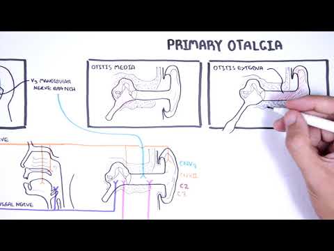
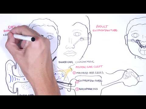
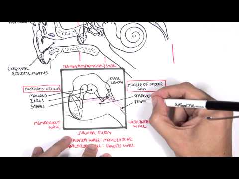
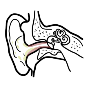
0:00 In this video we will learn about the external and middle ear. 0:10 So here is the ear. 0:12 We can see the external, middle and inner ear. 0:16 Let us learn about the innervation of the external ear first. 0:22 So here we have the oracle. 0:24 There are many nerves innervating the external ear, aspect of the ear. 0:28 And these are your auricular temporal branch of the mandibular nerve, V3, which 0:36 is a branch 0:37 of the trigeminal nerve, cranial nerve 5. 0:41 The other part are supplied by the lesser occipital nerve, which originates 0:48 from the C2 spinal cord. 0:52 And the greater auricular nerve, which is from the C2 and C3, spinal branches. 0:59 There are also auricular branches of the facial and vagus nerves. 1:06 Recapping on the external acoustic meatus, the lateral 2/3, so the outer 2/3 of 1:12 the external 1:13 acoustic meatus is mainly made up of cartilage or surrounded by cartilage, 1:18 whereas the medial 1:20 1/3 is bone. 1:23 The external acoustic meatus does not follow a straight line as well, so you 1:28 must pull 1:29 the ear a certain way in order to examine the external ear properly. 1:35 So looking at some clinical relevance here, swimmer's ear, also known as otitis 1:42 externa, 1:42 is an inflammation of the external acoustic meatus. 1:46 Clinical sign is pulling the tragus elicits pain. 1:52 The tragus, what I'm talking about, is this part of the oracle. 1:58 And another important anatomical part of the oracle is the helix here. 2:03 There is also another condition of the external ear, known as surfrazier, which 2:08 is where there 2:09 is a development of bony lumps in the external acoustic meatus. 2:14 It's not dangerous, but can potentially lead to hearing loss. 2:17 But surfrazier is common in the surfing population groups. 2:24 Okay, so that was for the external ear. 2:27 Let us now look at the eardrum. 2:29 This is what you see on an otoscopic view, where you are examining the external 2:37 ear. 2:37 So on the eardrum, we're going to look at some important parts of the 2:40 examination. 2:41 So here is what's known as the cone of light, the umpo, the lateral process of 2:47 the malleus, 2:48 the pause flaccida, and the posterior malleola folds. 2:55 So the malleus is important here because it is actually the first bony ossicle 3:01 that articulates 3:02 with the eardrum. 3:03 Anyway, the eardrum receives sound vibrations, which triggers a cascade of 3:10 vibrations within 3:11 the middle ear, which then send these vibrational signals to the inner ear. 3:18 Problems such as tympanic membrane perforation, which is a result of many 3:22 causes, mainly trauma 3:23 and infection, can be a problem in transmitting such processes. 3:30 But also it can be very painful. 3:34 Most tympanic membrane perforation heals spontaneously, but if it's large, the 3:42 perforation, it would 3:43 require surgery to fix. 3:46 Okay, now let us concentrate on the middle ear. 3:51 So again, here is the middle ear. 3:53 It contains the auditory ossicles, the smallest bones in the body. 3:58 And remember, the malleus, which is one of the auditory ossicles, the bones, 4:04 articulates 4:05 with the eardrum. 4:06 And that's why you can see some projections of the malleus on the otoscopic 4:16 view. 4:17 Anyway, the middle ears, the bony ossicles of the middle ear will send 4:23 vibrations to 4:24 the inner ear. 4:26 Yeah. 4:28 And here is your pharyngeal tympanic tube, which will connect to the nasopharyn 4:33 x. 4:34 And the pharyngeal tympanic tube is important because it sort of equalizes 4:40 pressure within 4:41 the middle ear and the outside world. 4:46 An important part of the pharyngeal tympanic tube is that in children is more 4:50 horizontal 4:50 and shorter. 4:51 Most children are more susceptible to ear infections, or middle ear infections, 4:57 should 4:57 I say, that arise from the nose or the mouth. 5:00 And it can travel up to the middle ear. 5:04 Anyway, inferior to the middle ear, you can find the internal jugular vein. 5:12 And this will lead us to the next topic, which are the boundaries of the middle 5:16 ear. 5:17 So the middle ear, I'm talking about, you know, where the bony ossicles are. 5:21 So the boundaries that make up the middle ear. 5:24 And we're going to look at the right middle ear. 5:30 The inferior wall of the middle ear is also known as a jugular wall or jugular 5:34 floor because 5:35 this is where you can find the internal jugular vein right at its course. 5:40 We have the tegmental wall on the top, which is your frontal bone. 5:44 So it's made up of your frontal bone. 5:48 The lateral wall, the one most towards the outside, is the membranous wall 5:54 because this 5:55 is basically your eardrum. 5:58 The medial aspect of the wall is the labyrinth. 6:01 And this is where you can find the inner ear. 6:05 There are important structures that make up the labyrinth wall. 6:09 And these are the oval window and the round window. 6:13 Also the prominence of the facial canal and the lateral semicircular canal can 6:19 be found 6:19 here. 6:21 The posterior wall is known as the mastoid wall because here is where we find 6:26 the mastoid 6:27 bone. 6:29 On this wall we can find the mastoid anterum. 6:32 Now the mastoid anterum is important because it is relevant to mastoiditis. 6:38 The mastoid anterum is a cavity continuous with a collection of air filled 6:45 spaces known 6:46 as the mastoid cells. 6:49 Now infection of the middle ear can spread to these mastoid cells and cause 6:53 mastoiditis, 6:55 which is very painful. 6:59 The anterior wall of the middle ear is known as a crowded wall. 7:02 And here we can find the pharyngeal tympanic tube and tensor tympanic muscle 7:07 run its course. 7:10 So that was it I guess on the middle ear and the external ear. 7:16 If you haven't watched the other video on the anatomy of the ear overview, 7:20 please watch 7:20 that. 7:21 Thank you.