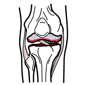
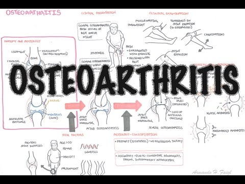
Transient osteoporosis of the hip – pathophysiology & diagnosis
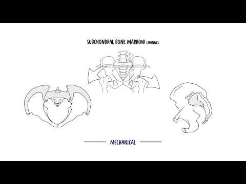
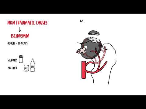
Transient osteoporosis of the hip causes sudden, severe groin or buttock pain and reversible bone marrow edema—learn how to spot, diagnose, and manage it. In this clear, clinically focused video you’ll discover the defining features of transient osteoporosis of the hip, why it’s distinct from general osteoporosis and avascular necrosis, and how MRI identifies diffuse femoral head and neck edema without collapse. Learn the suspected mechanisms—transient vascular disturbance, microvascular spasm, venous outflow obstruction, neurogenic and hormonal factors—and how these produce pain through increased intraosseous pressure and inflammation.
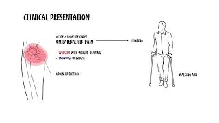
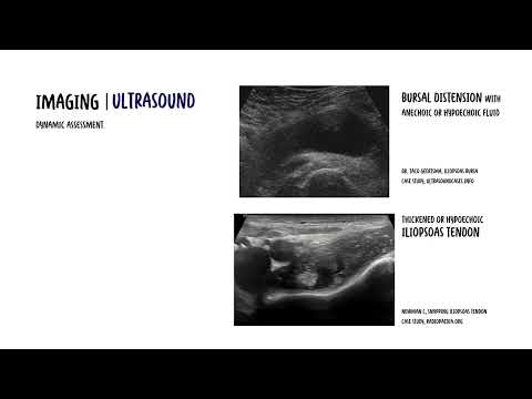
You’ll understand the typical presentation in healthy middle-aged men and pregnant women in the third trimester: acute unilateral hip pain that worsens with weight bearing, minimal systemic signs, and normal inflammatory labs. The video explains imaging clues (early normal X-rays, later proximal femoral osteopenia, MRI edema), key differentials (avascular necrosis, stress fracture, infection, inflammatory arthritis, malignancy), and why recognizing this self-limiting condition prevents unnecessary surgery. Practical management guidance covers protected weight bearing, analgesia, physiotherapy, and follow-up imaging—most patients recover fully within 6–12 months. Recurrence is rare but possible, sometimes affecting the opposite hip.
If you want a concise, evidence-informed primer on diagnosis, pathophysiology, and conservative treatment of transient osteoporosis of the hip, this video equips clinicians and patients with the signs to watch for and the steps to take—watch now to learn how timely recognition can avoid overtreatment and support full recovery.

Notes Articles