Overview
DiGeorge syndrome (22q11.2 deletion syndrome) is a congenital immunodeficiency and multisystem disorder caused by a microdeletion on chromosome 22. It leads to defective development of the third and fourth pharyngeal pouches, resulting in thymic hypoplasia/aplasia, hypocalcaemia due to parathyroid hypoplasia, and congenital heart defects. Prevalence is estimated at 1 in 4000 live births. Clinical presentation varies from severe neonatal disease with immunodeficiency and cardiac defects to milder phenotypes presenting later in life.
Definition
Microdeletion: A chromosomal deletion too small to be detected on standard karyotype; detected by FISH/array.
Pharyngeal pouch: Failure in embryologic development of the pharyngeal pouches, which is driven by TBX1, leads to absence or hypoplasia of the thymus and parathyroid glands.
Hypocalcaemia: Low serum calcium, often symptomatic with tetany or seizures.
Conotruncal anomalies: A congenital heart defect, abnormalities of the great arterial connections and arrangement. They include Tetralogy of Fallot, Pulmonary atresia with ventricular septal defect, Double outlet ventricles, Truncus arteriosus, Transposition of the great arteries and Interrupted aortic arch with ventricular septal defect.
Anatomy & Physiology
- Thymus: Site of T-cell maturation (positive/negative selection).
- Parathyroid glands: Regulate calcium via parathyroid hormone (PTH).
- 3rd and 4th pharyngeal pouches: Embryological precursors to thymus, parathyroids, parts of cardiac outflow tract.
- Defect in pouch development → thymic aplasia, parathyroid hypoplasia, cardiac outflow tract malformations.
“CATCH-22” → hallmark features of DiGeorge (Cardiac defects, Abnormal facies, Thymic hypoplasia, Cleft palate, Hypocalcaemia; Chromosome 22q11.2).
Aetiology and Risk Factors
- Cause: 22q11.2 microdeletion (most due to de novo deletion; ~10% inherited).
- Family history of 22q11.2 deletion.
- Parental balanced translocation (rare).
Pathophysiology
- 22q11.2 deletion → abnormal neural crest migration and pharyngeal pouch development.
- Thymic hypoplasia → impaired T-cell maturation → variable immunodeficiency.
- Parathyroid hypoplasia → ↓ PTH → hypocalcaemia.
- Abnormal aortic arch / conotruncal development → congenital heart disease.
- Craniofacial anomalies due to neural crest migration defects.
Severity depends on extent of thymic dysfunction → partial DiGeorge (mild T-cell dysfunction) vs complete (SCID-like).
Clinical Manifestations
- Immunodeficiency: Recurrent viral, fungal, opportunistic infections (T-cell defect).
- Hypocalcaemia: Neonatal tetany, seizures, muscle cramps.
- Congenital heart disease: Conotruncal anomalies (Tetralogy of Fallot, truncus arteriosus, interrupted aortic arch, VSD).
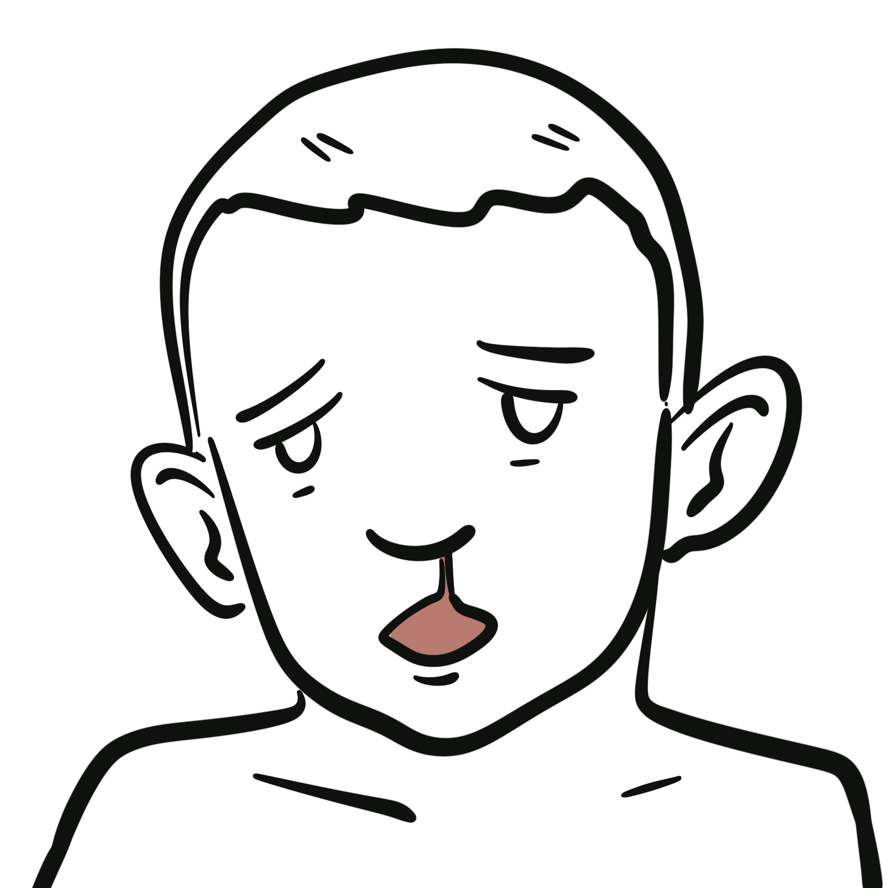
- Craniofacial anomalies: Low-set ears, micrognathia, cleft palate, hypertelorism.
- Neurodevelopmental/psychiatric: Learning difficulties, increased risk of schizophrenia.
Tetany in neonate + congenital heart disease + recurrent infections = DiGeorge until proven otherwise.
Diagnosis
- Genetic testing: FISH or chromosomal microarray for 22q11.2 deletion.
- Labs: Low/absent T-cells, reduced PHA (phytohaemagglutinin) response, hypocalcaemia (↓ PTH).
- Imaging: CXR → absent thymic shadow.
- Differentials:
- SCID (but usually earlier severe infections, absent both T & B function).
- Hypoparathyroidism (isolated, without thymic/heart defects).
- Other syndromic congenital heart diseases.
Treatment
- Cardiac surgery: Repair congenital heart defects.
- Calcium & vitamin D supplementation: For hypocalcaemia.
- Infection management: Prophylactic antibiotics, antifungals as needed.
- Immune restoration: Thymic transplant or HSCT in complete DiGeorge.
- Avoid live vaccines if severe T-cell deficiency.
- Speech therapy, psychological support for craniofacial/learning issues.
Tailor treatment to severity — complete DiGeorge may require thymic or stem cell transplant, partial often managed supportively.
Complications & Prognosis
- Severe infections in infancy if untreated.
- Congenital heart disease → morbidity and mortality.
- Chronic hypocalcaemia → seizures, neuromuscular irritability.
- Increased risk of autoimmune disease and psychiatric disorders later in life.
- Prognosis variable: partial form survivable into adulthood; complete form fatal without intervention.
References
- McDonald-McGinn DM, Sullivan KE, Marino B, et al. 22q11.2 deletion syndrome. Nat Rev Dis Primers. 2015;1:15071.
- Markert ML, Devlin BH, McCarthy EA. Thymus transplantation. Clin Immunol. 2010;135(2):236–46.
- Jawad AF, McDonald-McGinn DM, Zackai EH, Sullivan KE. Immunologic features of chromosome 22q11.2 deletion syndrome. J Pediatr. 2001;139(5):715–23.
- Ryan AK, Goodship JA, Wilson DI, et al. Spectrum of clinical features associated with interstitial chromosome 22q11 deletions. J Med Genet. 1997;34(10):798–804.
- Sullivan KE. Chromosome 22q11.2 deletion syndrome: DiGeorge syndrome/velocardiofacial syndrome. Immunol Allergy Clin North Am. 2008;28(2):353–66.


Discussion