

Head and Brain Trauma
Overview
Head and Brain Trauma encompasses injury to the scalp, skull, or brain tissue (Traumatic Brain Injury, TBI) resulting from external mechanical forces.
Classification:
- Primary Injury: Damage occurring at the moment of impact (e.g., contusions, lacerations, diffuse axonal injury, intracranial hematomas).
- Secondary Injury: Downstream cellular and physiological cascade (hypoxia, hypotension, cerebral edema, elevated intracranial pressure [ICP], ischemia) progressing over hours-to-days.
- Severity (by GCS)
- Severe (GCS 3–8)
- Moderate (GCS 9–12)
- Mild (GCS 13–15).
The primary goal in acute TBI management is the prevention of secondary brain injury. The two most critical and preventable drivers of secondary brain damage are hypoxia (SpO2 < 90%) and hypotension (Systolic BP < 100 mmHg for ages 50–69, or < 110 mmHg for ages 15–49 and >= 70).
Approach
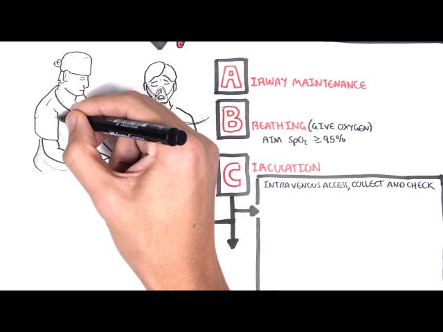
Step 1: Primary Survey & Immediate Stabilization (ATLS ABCDE)
- Airway with Cervical Spine Motion Restriction: Secure airway if GCS <= 8 or patient unable to protect airway.
- Breathing: Maintain normocapnia (PaCO2 35–45 mmHg; ETCO2 35–40 mmHg). Avoid routine hyperventilation.
- Circulation: Resuscitate aggressively with isotonic crystalloids (0.9% Normal Saline or Plasmalyte; avoid hypotonic fluids like 0.45% NaCl or D5W which worsen cerebral edema). Target SBP >= 100–110 mmHg.
- Disability: Assess GCS, pupil size and reactivity, and focal neurological signs.
- Exposure: Log roll to examine scalp/spine; maintain normothermia.
Step 2: Recognize Signs of Impending Brain Herniation / Severe Elevated ICP
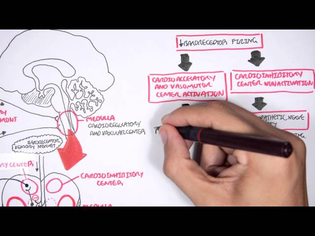
- Cushing’s Triad: Severe Hypertension (widened pulse pressure) + Bradycardia + Irregular Respirations (Cheyne-Stokes).
- Pupillary Signs: Anisocoria (unilaterally dilated, sluggish/fixed pupil) $\rightarrow$ Uncal herniation compressing ipsilateral CN III.
- Posturing: Decorticate (flexor) or Decerebrate (extensor) posturing.
Step 3: Branching Logic & Imaging Decisions (Canadian CT Head Rule for Mild TBI GCS 13–15)
- High Risk for Neurosurgical Intervention (CT Mandatory):
- GCS < 15 at 2 hours post-injury.
- Suspected open or depressed skull fracture.
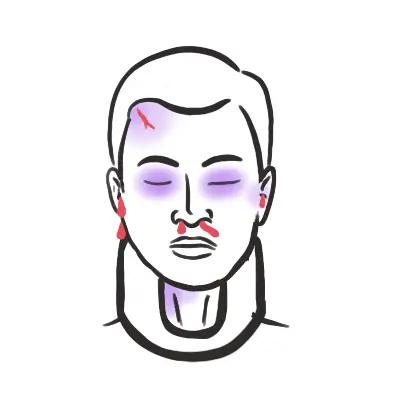
- Any sign of basal skull fracture (hemotympanum, “racoon eyes”, Battle’s sign, CSF otorrhea/rhinorrhea).
- = 2 episodes of vomiting.
- Age >= 65 years.
- Medium Risk (CT Recommended): Amnesia before impact > 30 minutes, dangerous mechanism (pedestrian struck, ejected occupant, fall > 3 feet or 5 stairs).
Unilateral fixed and dilated pupil + contralateral hemiparesis = Uncal Herniation until proven otherwise. Immediately administer hyperosmolar therapy (3% Hypertonic Saline or Mannitol) and obtain urgent neurosurgical consultation.
Differential Diagnosis
Extra-Axial Intracranial Hemorrhages:
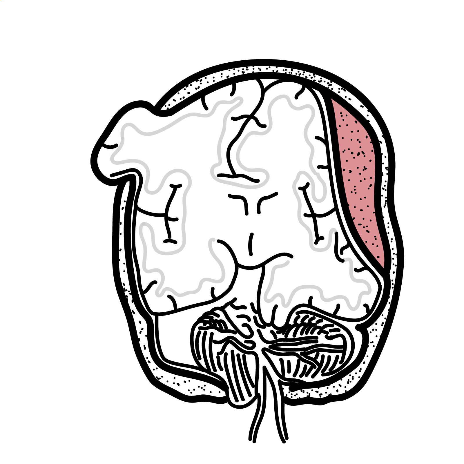
- Epidural Hematoma (EDH):
- Mechanism: Tear of Middle Meningeal Artery associated with temporal bone fracture.
- Distinguishing features: Classic “Lucid Interval” (brief loss of consciousness $\rightarrow$ transient clarity $\rightarrow$ rapid neurological decline); Biconvex / Lens-shaped hyperdensity on CT that does NOT cross suture lines.
- Subdural Hematoma (SDH):
- Mechanism: Tear of Bridging Veins (common in elderly, alcoholics, or anticoagulated patients).
- Distinguishing features: Crescent-shaped hyperdensity on CT that crosses suture lines; can be acute (hyperdense), subacute (isodense), or chronic (hypodense).
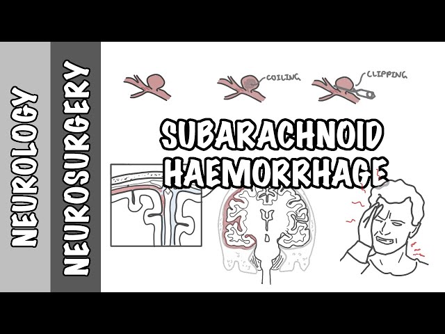
- Subarachnoid Hemorrhage (Traumatic SAH):
- Mechanism: Disruption of superficial cortical vessels; most common intracranial lesion in TBI.
- Distinguishing features: Blood tracking within sulci and basal cisterns; thunderclap headache, photophobia, meningismus.
Intra-Axial Injuries:
- Cerebral Contusion & Intraparenchymal Hemorrhage (IPH):
- Mechanism: Coup (site of impact) or contrecoup (opposite site) impact against bony skull protuberances (frontal/temporal poles).
- Distinguishing features: “Punctate / Salt and Pepper” hyperdensities on CT; high risk of delayed expansion within 24–48 hours.
- Diffuse Axonal Injury (DAI):
- Mechanism: High-speed deceleration / rotational shearing forces (e.g., high-speed motor vehicle collision).
- Distinguishing features: Severe profound coma out of proportion to CT findings; microscopic punctate hemorrhages at grey-white matter junction, corpus callosum, and brainstem (best seen on MRI DWI/FLAIR).
Skull Fractures:
- Basal Skull Fracture:
- Distinguishing features: Battle’s sign (mastoid ecchymosis), Raccoon eyes (periorbital ecchymosis), CSF rhinorrhea/otorrhea, CN VII/VIII palsy. STRICTLY CONTRAINDICATED: Nasogastric tube insertion (risk of intracranial entry; use orogastric tube instead).
Epidural hematomas do NOT cross suture lines (limited by dural attachments), whereas Subdural hematomas DO cross suture lines but are limited by dural reflections (falx cerebri and tentorium).
Investigations
First-Line / Bedside Diagnostics:
- Serial Glasgow Coma Scale (GCS) & Pupillary Exam: Repeat every 15–30 minutes in acute stages.
- Point-of-Care Blood Glucose & ABG/VBG: Exclude hypoglycemia/hypoxia/hypercapnia as primary or contributing causes of altered mental status.
- Coagulation Profile (INR, APPT, Fibrinogen, Platelets): Critical in patients on anticoagulants/antiplatelets (e.g., Warfarin, DOACs).
- Serum Electrolytes (Na+, K+): Baseline before hyperosmolar therapy; check for SIADH or CSW.
Targeted / Diagnostic Imaging Strategy:
- Non-Contrast CT Head (Gold Standard): Immediate modality of choice for acute head trauma. Evaluates hemorrhage, mass effect, midline shift (> 5 mm indicates significant risk), ventricular effacement, and skull fractures.
- CT Angiography (CTA) Head & Neck: Indicated if blunt cerebrovascular injury (BCVI) is suspected (e.g., neck trauma, transverse process fractures, basilar skull fractures).
- MRI Brain (Non-emergent / Secondary): Superior for identifying Diffuse Axonal Injury (DAI), brainstem lesions, and unexplained persistent neurological deficits when CT is normal.
Never delay an emergent non-contrast CT head in a patient with a deteriorating GCS or focal neurological deficits. If the patient is on anticoagulants with an acute intracranial bleed, reverse anticoagulation immediately while awaiting CT confirmation.
Critical Management
Emergency Management of Elevated ICP / Impending Herniation:
- Positioning: Elevate head of bed 30 degrees; keep head/neck in midline position (prevents jugular venous compression).
- Hyperosmolar Therapy:
- 3% Hypertonic Saline: 250 mL IV bolus over 15–20 minutes (or 2 mL/kg). Target serum Na+ 145–155 mmol/L.
- Mannitol 20%: 0.5–1.0 g/kg IV bolus over 15–20 minutes (avoid if SBP < 90 mmHg / hypotensive).
- Controlled Hyperventilation (Emergency Bridging ONLY): Target PaCO2 30–35 mmHg only during active herniation crises; stop once hyperosmolar therapy takes effect.
Hemodynamic & Physiological Targets (Brain Trauma Foundation Guidelines):
- Blood Pressure: Target SBP >= 100 mmHg (ages 50–69) or >= 110 mmHg (ages 15–49 or >= 70).
- Intracranial Pressure (ICP): Keep ICP < 22 mmHg.
- Cerebral Perfusion Pressure (CPP = MAP – ICP): Target CPP between 60–70 mmHg.
- Seizure Prophylaxis: Levetiracetam (or Phenytoin) for 7 days post-injury to prevent early post-traumatic seizures in severe/moderate TBI.
Anticoagulation Reversal:
- Warfarin: Prothrombin Complex Concentrate (PCC) + IV Vitamin K 10 mg.
- DOACs (Dabigatran / Factor Xa inhibitors): Idarucizumab (for Dabigatran) or Andexanet Alfa / PCC (for Rivaroxaban/Apixaban).
Prophylactic hyperventilation is HARMFUL—it causes profound cerebral vasoconstriction and worsens cerebral ischemia. Hyperventilation should only be used as a temporary emergency rescue measure during active brain herniation.
References
- National Institute for Health and Care Excellence. Head injury: assessment and early management (NG232) [Internet]. 2023 [cited 2026 Aug 20]. Available from: https://www.nice.org.uk/guidance/ng232
- Brain Trauma Foundation. Guidelines for the management of severe traumatic brain injury, 4th edition [Internet]. 2016 [updated 2020; cited 2026 Aug 20]. Available from: https://braintrauma.org/coma/guidelines/current

Members only discussions coming soon…