

Catastrophic Antiphospholipid Syndrome (CAPS)
Overview
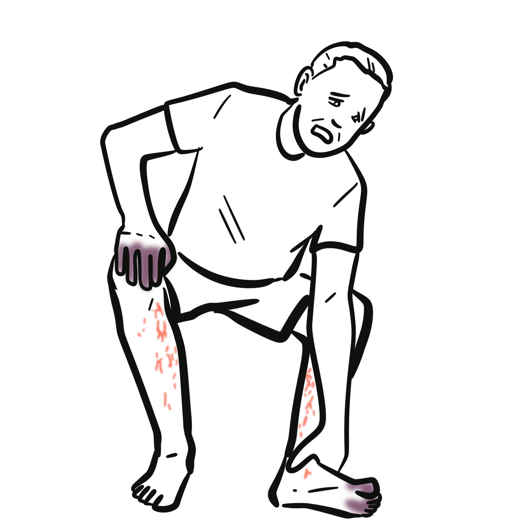
Catastrophic antiphospholipid syndrome (CAPS) is a rare, life-threatening form of antiphospholipid syndrome (APS) characterised by rapidly developing thrombosis affecting multiple organs, typically over days. Small-vessel thrombosis and thrombotic microangiopathy are prominent, although large-vessel arterial or venous thromboses may occur simultaneously.1
CAPS is a medical emergency. The combination of widespread thrombosis, systemic inflammation and progressive organ dysfunction can resemble severe sepsis, disseminated intravascular coagulation (DIC) or another thrombotic microangiopathy. Treatment should begin when clinical suspicion is high rather than waiting for every classification criterion to be fulfilled.2
Most patients have an identifiable precipitating event, particularly infection. CAPS may occur in patients with known primary APS, APS associated with systemic lupus erythematosus (SLE), or previously unrecognised APS.
Definition
- Catastrophic antiphospholipid syndrome (CAPS)
- A fulminant form of APS in which rapidly developing, widespread thrombosis causes dysfunction of multiple organs, usually within one week.
- Antiphospholipid antibodies (aPL)
- Autoantibodies associated with thrombosis and pregnancy morbidity. The routinely tested antibodies are lupus anticoagulant, anticardiolipin antibodies and anti-β2-glycoprotein I antibodies.
- Thrombotic microangiopathy
- Small-vessel thrombosis associated with tissue ischaemia and organ injury; thrombocytopenia and microangiopathic haemolytic anaemia may accompany it.
Aetiology & Risk Factors
Aetiology
CAPS develops when pathogenic antiphospholipid antibodies interact with an additional inflammatory or prothrombotic stimulus. Activation of endothelial cells, monocytes, platelets, coagulation pathways and complement can produce an escalating cycle of inflammation and thrombosis.2
CAPS may be associated with primary APS, SLE-associated APS or, less commonly, another autoimmune disease, malignancy or systemic inflammatory disorder. A previous diagnosis of APS is not required.
Risk Factors
- Infection, including respiratory, urinary, gastrointestinal and soft-tissue infection
- Surgery or significant trauma
- Interruption of anticoagulation or subtherapeutic anticoagulation
- Pregnancy, the postpartum period and obstetric complications
- Active SLE or another inflammatory disease
- Malignancy
- Certain medications, particularly oestrogen-containing therapies
- Prolonged immobilisation
Search actively for a precipitating event. Treating infection, restoring interrupted anticoagulation or controlling an SLE flare is part of treating CAPS—not a separate issue to address later.
Pathophysiology
Antiphospholipid antibodies bind phospholipid-associated proteins, particularly β2-glycoprotein I, and promote endothelial and monocyte tissue-factor expression, platelet activation, complement activation, impaired physiological anticoagulant and fibrinolytic activity, and small- and large-vessel thrombosis.
Tissue ischaemia causes further endothelial injury and inflammation, creating a thrombo-inflammatory amplification loop. The pathological hallmark is a predominantly non-inflammatory thrombotic vasculopathy, helping distinguish CAPS from primary vasculitis.
Why can CAPS deteriorate so quickly? Complement activation, coagulation and endothelial injury reinforce one another across multiple vascular beds. New organ injury may therefore appear over hours or days rather than as isolated events separated by months or years.
Clinical Manifestations
Consider CAPS when dysfunction of several organs develops rapidly, particularly in a patient with APS, SLE, previous thrombosis or a positive aPL history.
Renal
- Acute kidney injury, oliguria or renal thrombotic microangiopathy
- New or worsening hypertension
- Proteinuria or haematuria
Respiratory
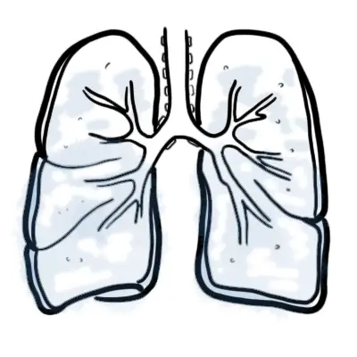
- Hypoxaemia, respiratory failure or acute respiratory distress syndrome
- Pulmonary embolism or pulmonary microvascular thrombosis
- Diffuse alveolar haemorrhage
- Pulmonary hypertension
Neurological
- Encephalopathy or altered consciousness
- Ischaemic stroke, focal deficits or cerebral venous sinus thrombosis
- Seizures or headache
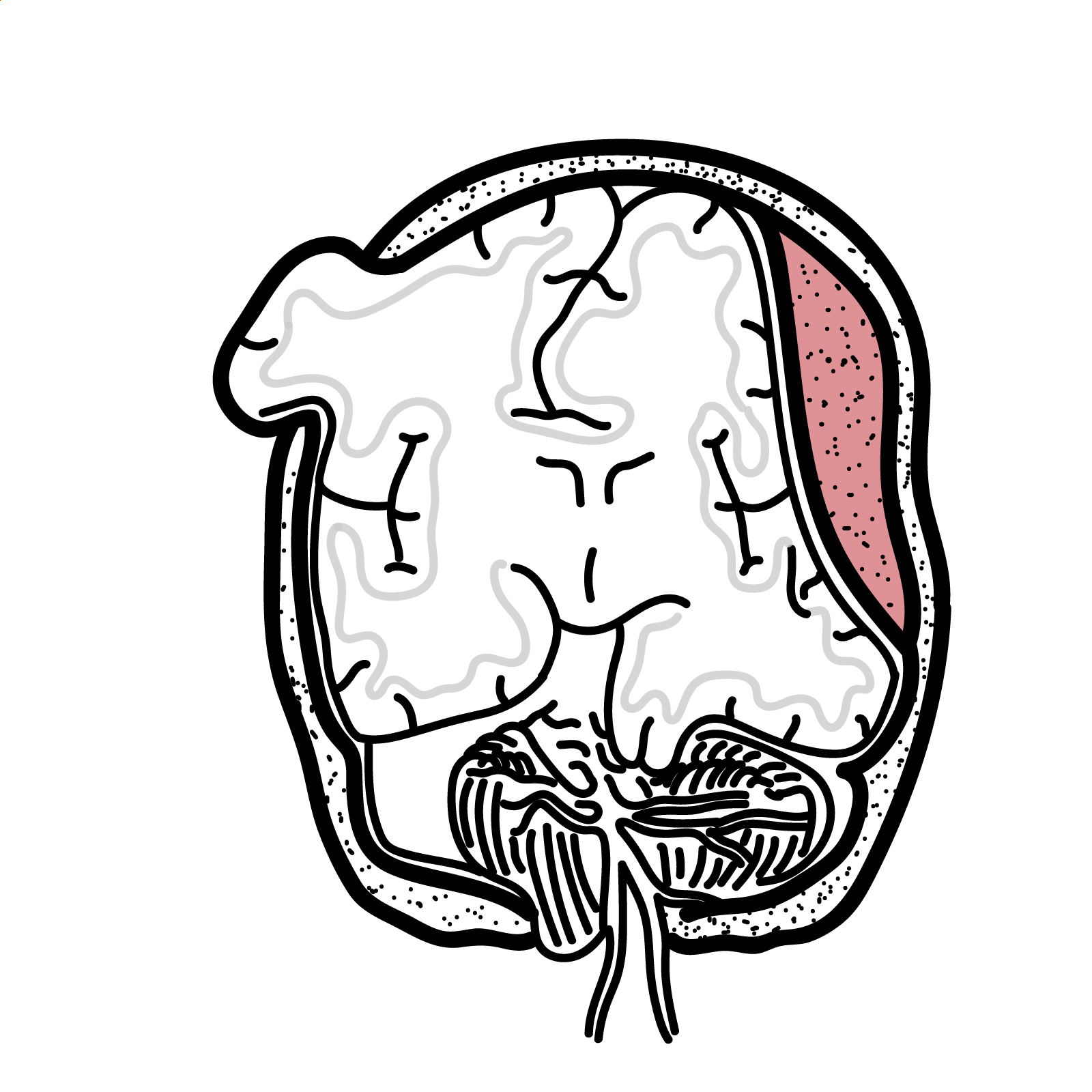
- Intracranial haemorrhage
Cardiovascular
- Myocardial ischaemia or infarction
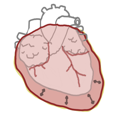
- Heart failure, valvular lesions or intracardiac thrombus
- Microvascular myocardial injury or shock
Cutaneous
- Livedo reticularis or livedo racemosa
- Retiform purpura, painful digital ischaemia or ulceration
- Skin necrosis or gangrene
Gastrointestinal and Abdominal
- Abdominal pain, mesenteric ischaemia or bowel infarction
- Hepatic, splenic or pancreatic injury
- Adrenal infarction or haemorrhage
Haematological
- Thrombocytopenia
- Microangiopathic haemolytic anaemia, elevated lactate dehydrogenase and schistocytes
- Coagulation abnormalities when CAPS overlaps with DIC or severe systemic illness
CAPS does not always produce the complete laboratory pattern of a thrombotic microangiopathy. A normal platelet count or absence of obvious schistocytes should not override compelling evidence of rapidly evolving, multisystem thrombosis.
Diagnosis & Investigations
CAPS is a clinicopathological diagnosis. Investigations should occur in parallel with stabilisation and treatment. They should establish thrombosis or thrombotic microangiopathy, identify aPL, map threatened organs and test whether a close mimic better explains the presentation.
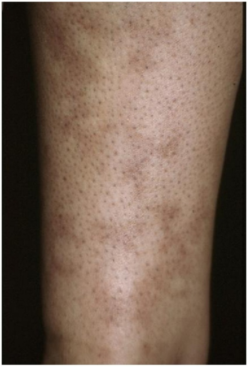
Livedo reticularis produces a violaceous, net-like cutaneous pattern. It may indicate impaired cutaneous blood flow but is not specific to CAPS. Image: Uva L/Wikimedia Commons, CC BY 3.0.
What to notice
Detailed image description
Source: https://commons.wikimedia.org/wiki/File:Livedo_reticularis_of_an_arm.png Creator: Uva L Licence: Creative Commons Attribution 3.0 Unported (CC BY 3.0) Licence URL: https://creativecommons.org/licenses/by/3.0/ Modifications: None.
Initial Laboratory Assessment
- Full blood count and peripheral blood film: thrombocytopenia, anaemia and schistocytes
- Haemolysis screen: lactate dehydrogenase, bilirubin, haptoglobin and reticulocyte count
- Coagulation profile: PT/INR, aPTT, fibrinogen and D-dimer
- Renal assessment: creatinine, electrolytes, urinalysis and urine protein quantification
- Liver function tests
- Lactate and blood gas: tissue hypoperfusion and severity
- Troponin and ECG: when myocardial injury is suspected
- Cultures: targeted to a possible infectious precipitant
Serial trends may be more informative than isolated results: falling platelets, rising creatinine, increasing lactate or new haemolysis may reveal ongoing disease.
Antiphospholipid Antibody Testing
- Lupus anticoagulant
- Anticardiolipin IgG and IgM
- Anti-β2-glycoprotein I IgG and IgM
Persistent positivity on repeat testing at least 12 weeks later supports underlying APS, but repeat confirmation must not delay emergency treatment. Anticoagulants, acute inflammation and coagulation abnormalities may interfere with lupus anticoagulant assays; record anticoagulant exposure and discuss uncertain results with the laboratory.
An isolated prolonged aPTT does not mean that the patient is protected from thrombosis. Lupus anticoagulant may prolong phospholipid-dependent clotting assays in vitro while being strongly associated with thrombosis in vivo.
Targeted Organ Assessment
- CT pulmonary angiography for suspected pulmonary embolism
- CT or MRI brain for focal signs, seizures or unexplained encephalopathy
- CT angiography of the abdomen for suspected mesenteric or solid-organ ischaemia
- Echocardiography for cardiac dysfunction, valvular disease or intracardiac thrombus
- Vascular ultrasound for suspected limb deep-vein thrombosis
- Chest imaging for pulmonary oedema, haemorrhage, infection or acute respiratory distress syndrome
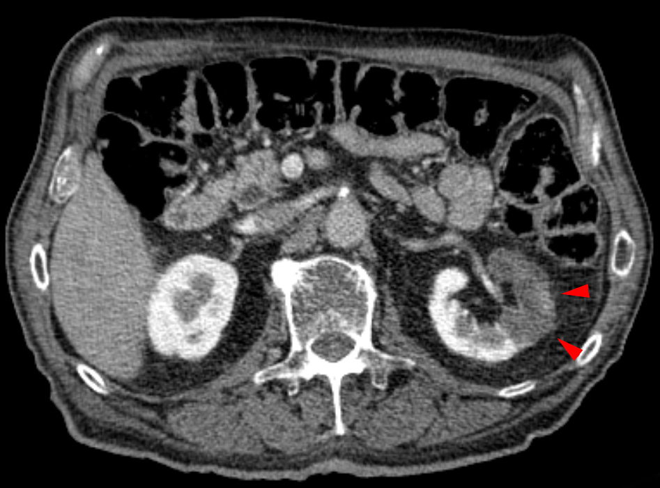
Contrast-enhanced CT showing a partial left renal infarction. Infarcted tissue is relatively hypoenhancing compared with adjacent viable renal parenchyma. Image: Hellerhoff/Wikimedia Commons, CC BY-SA 3.0.
What to notice
Detailed image description
Source: https://commons.wikimedia.org/wiki/File:Niereninfarkt_in_der_Computertomographie.jpg Creator: Hellerhoff Licence: Creative Commons Attribution-ShareAlike 3.0 Unported (CC BY-SA 3.0) Licence URL: https://creativecommons.org/licenses/by-sa/3.0/ Modifications: None.
Histopathology
Biopsy of safely accessible affected tissue may show small-vessel thrombosis with relatively little vessel-wall inflammation and support definite CAPS classification. It should only be performed when it will influence management, bleeding risk is acceptable and treatment will not be delayed.
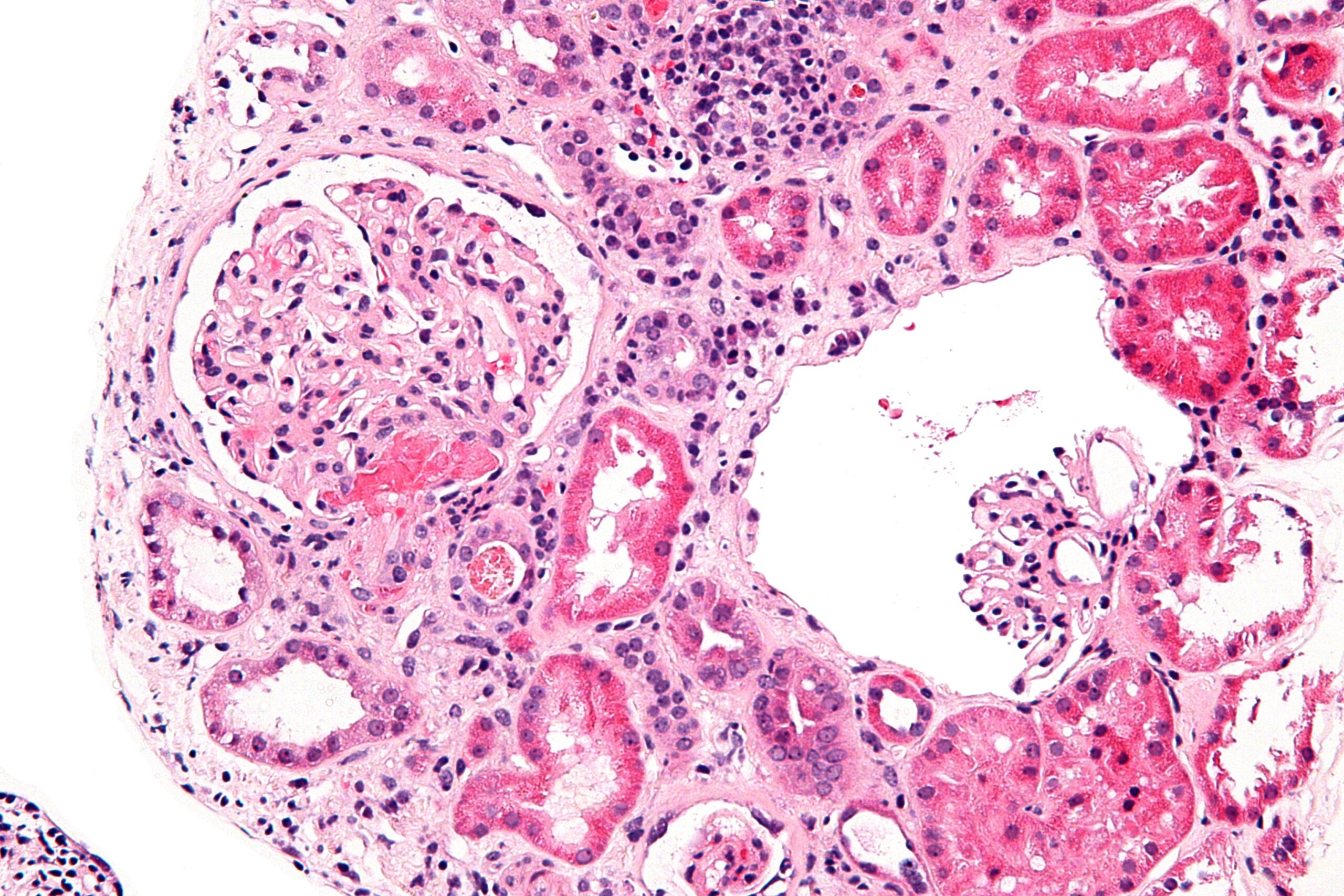
High-magnification H&E-stained kidney biopsy showing acute thrombotic microangiopathy. This pattern supports a thrombotic vasculopathy but is not specific to CAPS. Image: Nephron/Wikimedia Commons, CC BY-SA 3.0.
What to notice
Detailed image description
Source: https://commons.wikimedia.org/wiki/File:Acute_thrombotic_microangiopathy_-_high_mag.jpg Creator: Nephron Licence: Creative Commons Attribution-ShareAlike 3.0 Unported (CC BY-SA 3.0) Licence URL: https://creativecommons.org/licenses/by-sa/3.0/ Modifications: None.
Important Differentials
| Differential | Features favouring the alternative diagnosis |
|---|---|
| Sepsis with DIC | Clear infectious syndrome, consumption coagulopathy with reduced fibrinogen, prolonged PT and diffuse bleeding; sepsis may trigger or coexist with CAPS. |
| Thrombotic thrombocytopenic purpura | Prominent microangiopathic haemolytic anaemia and severe thrombocytopenia with severe ADAMTS13 deficiency. |
| Complement-mediated haemolytic uraemic syndrome | Renal-dominant thrombotic microangiopathy without the typical aPL-associated multisystem thrombotic pattern. |
| Heparin-induced thrombocytopenia | Compatible heparin exposure followed by platelet fall and thrombosis; assess with the 4Ts score and appropriate testing. |
| HELLP syndrome | Pregnancy-associated haemolysis, elevated liver enzymes and low platelets, usually with hypertension or pre-eclampsia. |
| Systemic vasculitis | Inflammatory vessel-wall injury with compatible systemic features and supportive serology or histology. |
| Malignant hypertension | Severe hypertension with retinal, cardiac and renal injury; it may itself produce thrombotic microangiopathy. |
| Cholesterol embolisation | Recent vascular procedure, livedo or blue toes, eosinophilia and cholesterol clefts on histology. |
Sepsis and CAPS are not mutually exclusive. Infection is a common CAPS trigger, while CAPS itself can produce fever, elevated inflammatory markers, shock and DIC-like abnormalities. Ask whether infection alone explains the number, distribution and speed of thrombotic organ injuries.
Classification
The preliminary international consensus criteria remain the commonly used CAPS classification framework.1
- Involvement of three or more organs, systems or tissues
- Development of manifestations simultaneously or within less than one week
- Histological confirmation of small-vessel thrombosis in at least one organ or tissue
- Laboratory confirmation of antiphospholipid antibodies
Definite CAPS requires all four criteria. Probable CAPS includes closely related presentations in which one element is incomplete—for example, only two organs are involved, histology or repeat aPL confirmation is unavailable, or a third event occurs outside the one-week interval despite anticoagulation.1
These are classification criteria for consistent research groups, not rules that replace clinical judgement. The 2023 ACR/EULAR APS classification criteria were not designed as CAPS diagnostic criteria.4
Do not wait for a third organ, a biopsy or a 12-week repeat antibody result before treating a rapidly progressive CAPS-compatible presentation.
Treatment
CAPS requires urgent multidisciplinary management, usually involving intensive care, haematology and rheumatology. Management simultaneously suppresses thrombosis, controls thrombo-inflammation, removes or neutralises circulating factors and treats the precipitant.
Immediate Supportive Care
- Stabilise airway, breathing and circulation
- Provide ventilation, renal replacement therapy or circulatory support when required
- Treat suspected infection promptly after appropriate cultures
- Correct triggers such as interrupted anticoagulation
- Avoid unnecessary invasive procedures and additional thrombotic risks
First-Line “Triple Therapy”
Recommended initial treatment is therapeutic anticoagulation, high-dose glucocorticoids, and plasma exchange and/or intravenous immunoglobulin (IVIG).2 Treatment should generally begin when CAPS is strongly suspected unless a specific contraindication is present.
Anticoagulation
Intravenous unfractionated heparin is often preferred initially because it acts rapidly, can be titrated or stopped quickly, and is practical in unstable patients, severe renal impairment or those requiring procedures. Anti-factor Xa monitoring may be required when lupus anticoagulant prolongs the baseline aPTT.
Thrombocytopenia alone does not automatically contraindicate anticoagulation; decisions should integrate platelet count, active bleeding, threatened organs and overall thrombotic burden.
Glucocorticoids
High-dose intravenous glucocorticoids suppress the intense inflammatory response and are commonly followed by an oral regimen once the patient stabilises. Dose and duration are individualised because controlled trial evidence is limited.
Plasma Exchange and IVIG
Plasma exchange may remove aPL, inflammatory mediators and complement components. IVIG has immunomodulatory effects and may be used instead of, or with, plasma exchange. When both are used, IVIG is generally administered after plasma exchange so that it is not immediately removed.
Treat the Underlying Trigger
- Antimicrobial therapy and source control for infection
- Re-establishment of therapeutic anticoagulation
- Treatment of an SLE flare
- Management of an obstetric complication
- Investigation and treatment of malignancy
SLE-Associated CAPS
Cyclophosphamide should be considered with active SLE, particularly significant inflammatory organ disease. Its role is less clear in primary APS without SLE activity.3
Refractory or Recurrent CAPS
If thrombosis or organ injury progresses despite conventional treatment, specialist teams may consider
- Rituximab
- Eculizumab
CAPS treatment is time-critical: anticoagulation + high-dose glucocorticoids + plasma exchange and/or IVIG, together with immediate treatment of the precipitating event.
Complications & Prognosis
Complications
- Multiorgan failure
- Acute kidney injury and chronic kidney disease
- Respiratory failure or acute respiratory distress syndrome
- Stroke, seizures and persistent neurological disability
- Myocardial infarction and heart failure
- Mesenteric ischaemia and bowel infarction
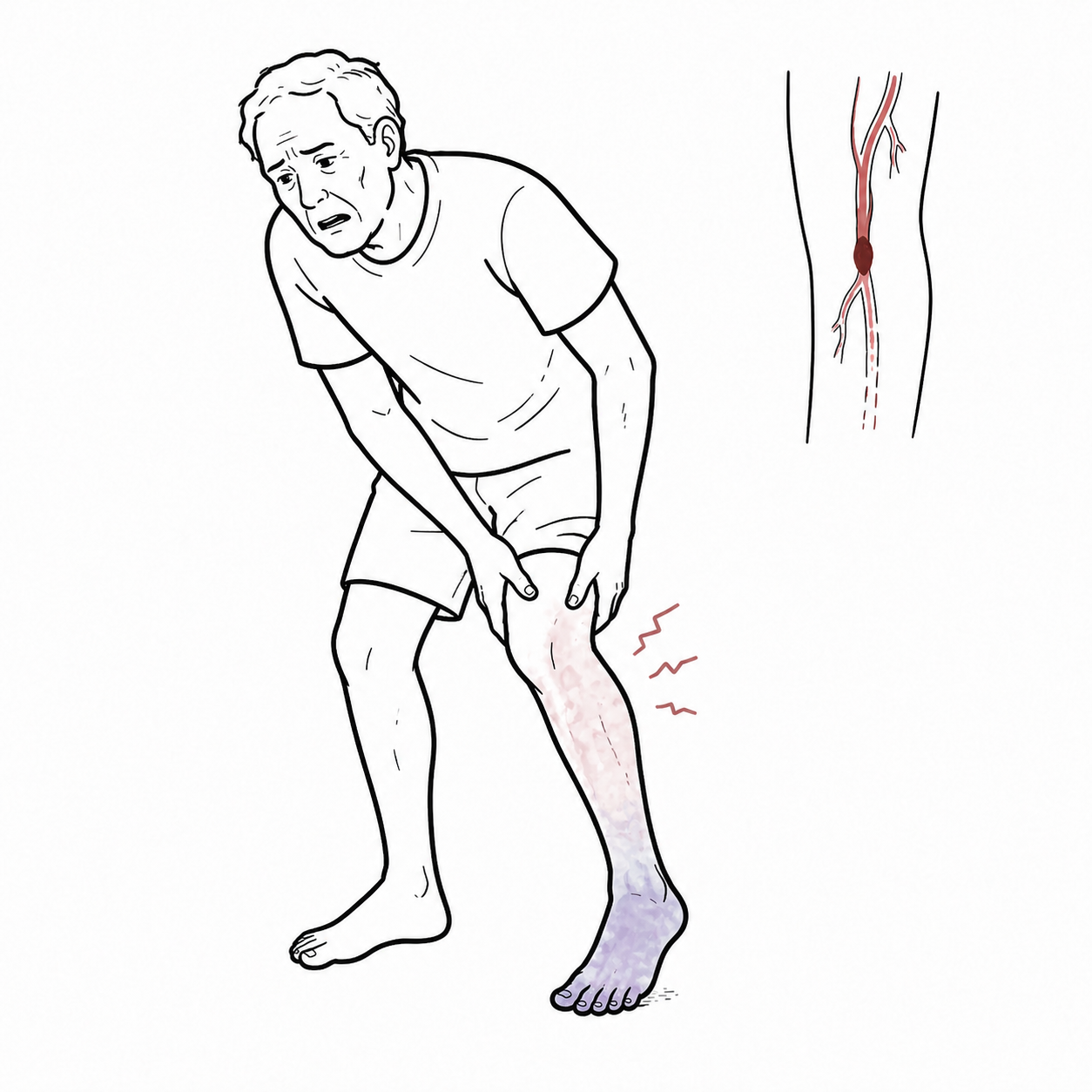
- Limb or digital ischaemia requiring amputation
- Adrenal infarction or haemorrhage
- Haemorrhage related to thrombocytopenia, DIC, tissue injury or anticoagulation
- Secondary infection and recurrent thrombosis
Prognosis
CAPS continues to have substantial mortality despite treatment. Poorer prognosis is associated with extensive organ involvement, severe renal, pulmonary, cerebral or cardiac injury, treatment delay, infection or severe SLE activity, inability to deliver anticoagulation and poor early treatment response.
Survivors may have permanent renal, neurological, cardiac or peripheral vascular impairment. CAPS recurrence is uncommon, but patients remain at risk of further APS-related thrombosis and usually require lifelong specialist follow-up.
Early recognition matters. Rapidly evolving multisystem thrombosis with thrombocytopenia or haemolysis should prompt immediate consideration of CAPS—even when infection, DIC or another thrombotic microangiopathy initially appears more familiar.
References
- Asherson RA, Cervera R, de Groot PG, Erkan D, Boffa MC, Piette JC, et al. Catastrophic antiphospholipid syndrome: international consensus statement on classification criteria and treatment guidelines. Lupus. 2003;12(7):530–534. doi:10.1191/0961203303lu394oa.
- Tektonidou MG, Andreoli L, Limper M, Amoura Z, Cervera R, Costedoat-Chalumeau N, et al. EULAR recommendations for the management of antiphospholipid syndrome in adults. Ann Rheum Dis. 2019;78(10):1296–1304. doi:10.1136/annrheumdis-2019-215213.
- Kazzaz NM, McCune WJ, Knight JS. Treatment of catastrophic antiphospholipid syndrome. Curr Opin Rheumatol. 2016;28(3):218–227. doi:10.1097/BOR.0000000000000269.
- Salter BM, Crowther MA. Catastrophic antiphospholipid syndrome: a CAPS-tivating hematologic disease. Hematology Am Soc Hematol Educ Program. 2024;2024(1):214–222. doi:10.1182/hematology.2024000544.
- Rodríguez-Pintó I, Espinosa G, Erkan D, Shoenfeld Y, Cervera R; CAPS Registry Project Group. The effect of triple therapy on the mortality of catastrophic anti-phospholipid syndrome patients. Rheumatology (Oxford). 2018;57(7):1264–1270. doi:10.1093/rheumatology/key082.









Members only discussions coming soon…