

Orthostatic Hypotension
OVERVIEW
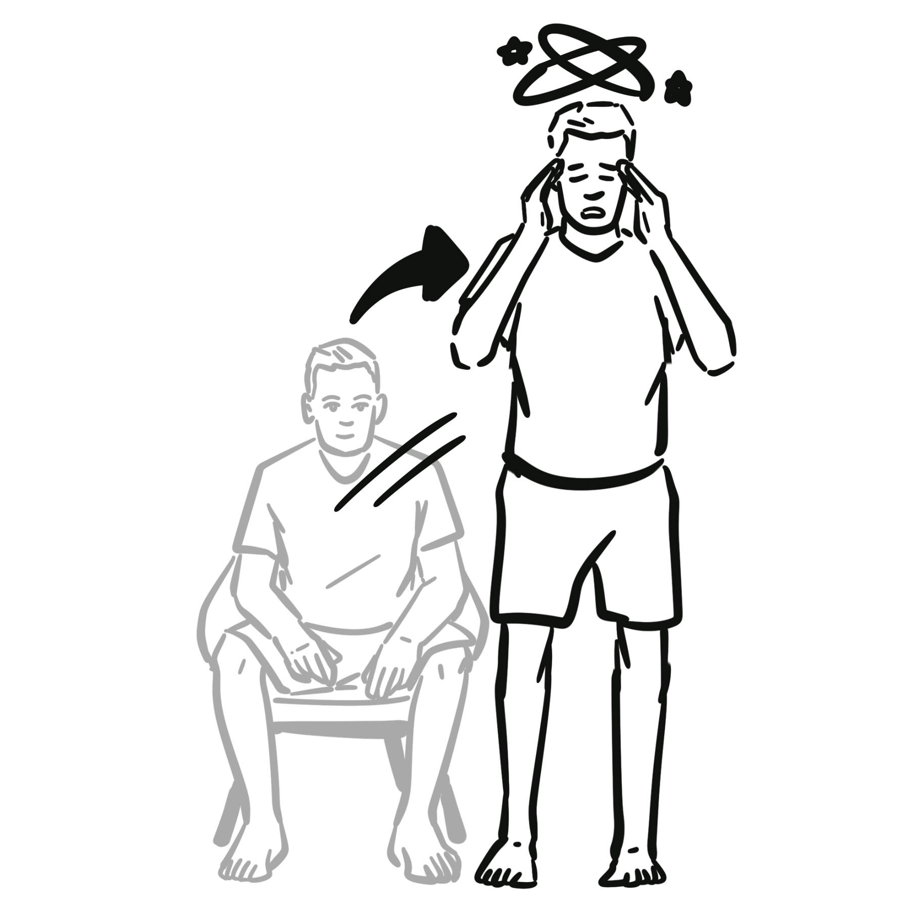
Orthostatic Hypotension (OH) is defined as a sustained drop in blood pressure occurring within 3 minutes of standing or head-up tilt:
- Systolic BP: Decrease of >= 20 mmHg AND/OR
- Diastolic BP: Decrease of >= 10 mmHg
(In patients with supine hypertension >= 150/90 mmHg, a drop of >= 30 mmHg SBP is required).
Normal Physiological Response to Orthostasis (standing up)
- Standing up from a recline or sitting position results in pooling of 500-1000mL of blood in the lower extremities and splanchnic circulation.
- The physiological response
- ↓venous return to the heart → ↓ventricular filling → ↓cardiac output → baroreceptor reflex
- Baroreceptor reflex
- → stimulation of the vasomotor center → peripheral vasoconstriction → ↑BP → ↑venous return + ↑cardiac output
- → stimulation of the cardiovascular center → ↑HR → ↑cardiac output +↑venous return
- Overall clinically there is a small reduction in systolic BP (<10mmHg), increase in diastolic BP and an increase in HR
Always measure blood pressure after at least 5 minutes of quiet rest in the supine position before performing an active stand test to establish a true baseline.
APPROACH
Step 1: Diagnostic Confirmation (Active Stand Test / Bedside Orthostatics)
- Measurement Protocol: Measure HR and BP after 5 minutes resting supine, then at 1 minute and 3 minutes post-standing.
Step 2: Emergency Triage & Red Flags
- Syncope with Trauma or Head Injury: Rule out intracranial hemorrhage/cervical spine injury.
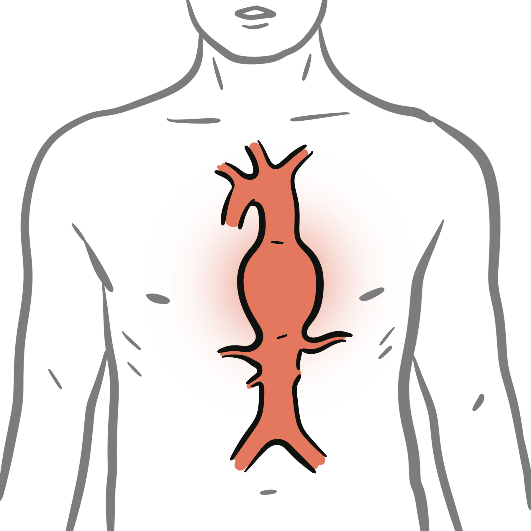
- Acute Volume Loss / Hemorrhage: Tachycardia + severe orthostasis -> GI bleed, ruptured ectopic pregnancy, ruptured AAA.
- Acute Cardiac Signs: Chest pain, dyspnea, irregular pulse -> Arrhythmia, ACS, aortic stenosis, PE.
- Neurological Red Flags: Rapidly progressive parkinsonism, ataxia, early autonomic failure, vertical gaze palsy -> Central neurodegenerative etiologies (e.g., Multiple System Atrophy).
Step 3: Branching Logic (Heart Rate Response Analysis)
- Neurogenic Orthostatic Hypotension (nOH): Blunted HR increase (< 15 bpm upon standing despite significant BP drop). Indicates baroreflex arc failure or sympathetic denervation.
- Non-Neurogenic Orthostatic Hypotension: Compensatory reflex tachycardia (> 15 bpm HR increase upon standing). Indicates hypovolemia, vasodilation, or drug-induced etiology.
- POTS Differential: HR increase >= 30 bpm (or >= 40 bpm if age 12–19) within 10 minutes of standing WITHOUT a drop in SBP >= 20 mmHg.
A blunted heart rate response (< 15 bpm increase despite a > 20 mmHg drop in SBP) strongly points toward a neurogenic etiology (autonomic failure), whereas prominent compensatory tachycardia indicates volume depletion or medication-induced vasodilation.
DIFFERENTIAL DIAGNOSIS
Non-Neurogenic Causes (Intact Baroreflex, Compensatory Tachycardia):
- Volume Depletion / Hypovolemia:
- Distinguishing features: Dehydration, vomiting/diarrhea, overt GI bleeding, excessive diuresis, elevated BUN/creatinine ratio (> 20:1), dry mucous membranes.
- Medication-Induced (Most common overall cause in elderly):
- Distinguishing features: Antihypertensives (alpha-blockers e.g., tamsulosin/doxazosin, vasodilators, diuretics), psychotropics (TCAs, antipsychotics), SGLT2 inhibitors, nitrates.
- Cardiovascular Deconditioning / Prolonged Bedrest:
- Distinguishing features: Recent prolonged hospitalization, frailty, muscle wasting; intact autonomic reflexes.
- Adrenal Insufficiency (Addison’s Disease):
- Distinguishing features: Hyperpigmentation, hyponatremia, hyperkalemia, weight loss, fatigue, hypoglycemia.
Neurogenic Causes (nOH – Impaired Baroreflex, Blunted HR Response):
- Central Synucleinopathies (Primary Autonomic Failure):
- Parkinson’s Disease (PD): Resting tremor, rigidity, bradykinesia + orthostasis.
- Multiple System Atrophy (MSA): Orthostatic hypotension + prominent ataxia (MSA-C) or early severe autonomic failure with erectile dysfunction and urinary retention (MSA-P/Shy-Drager).
- Dementia with Lewy Bodies (DLB): Visual hallucinations, fluctuating cognition, parkinsonism + early orthostasis.
- Pure Autonomic Failure (PAF): Isolated autonomic failure without central neurological signs.
- Peripheral Neuropathies (Secondary Autonomic Failure):
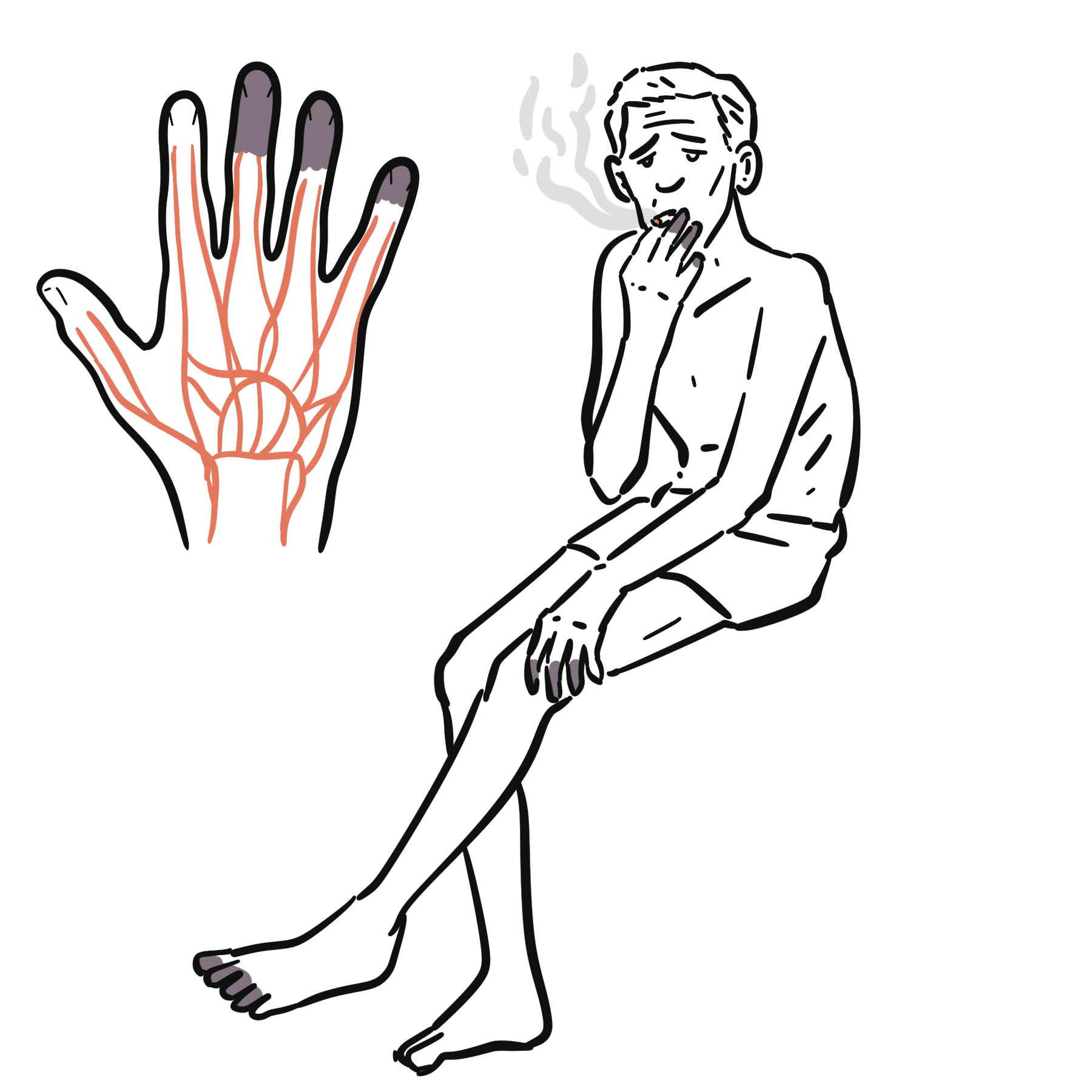
- Diabetic Autonomic Neuropathy: Longstanding diabetes, stocking-glove sensory loss, gastroparesis, resting tachycardia.
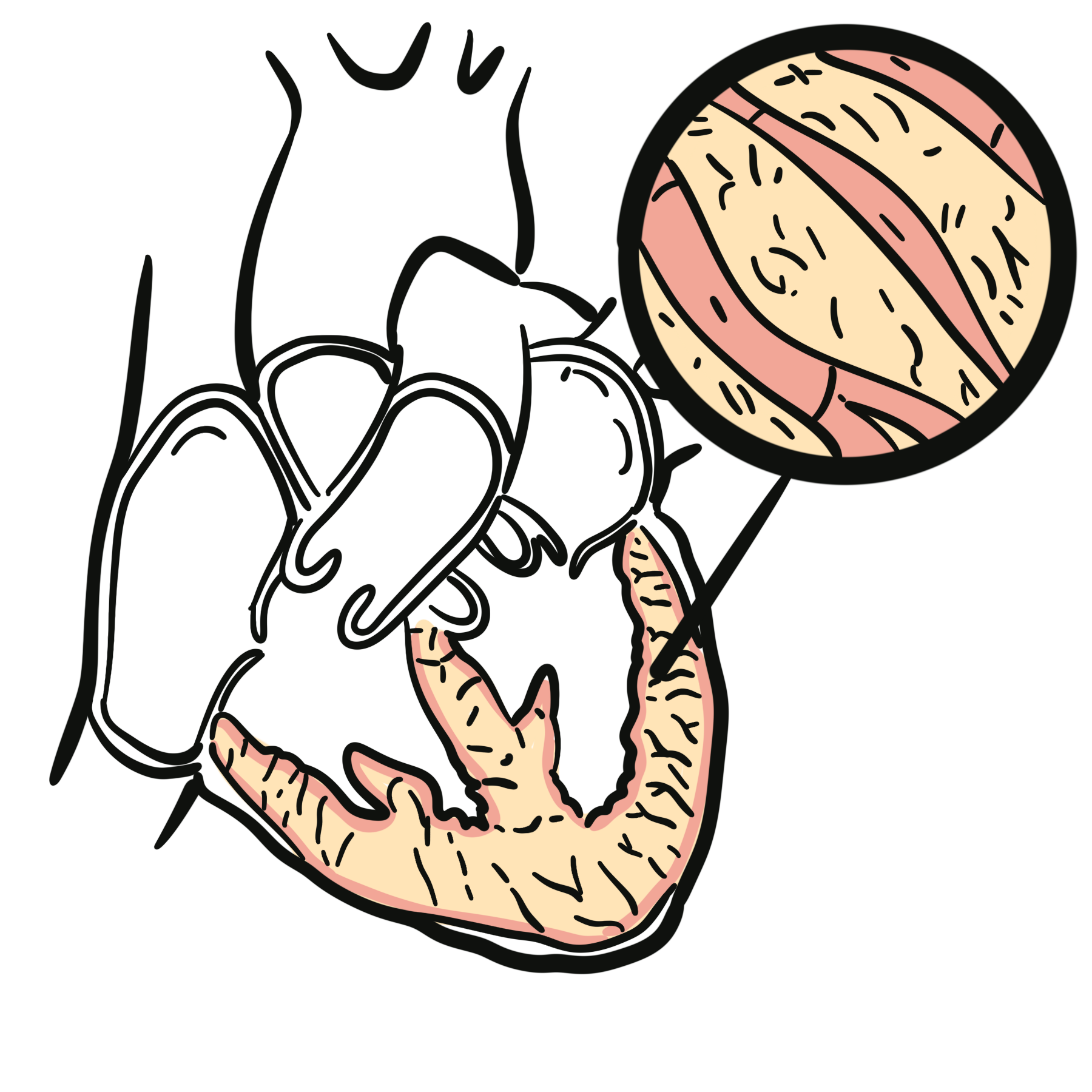
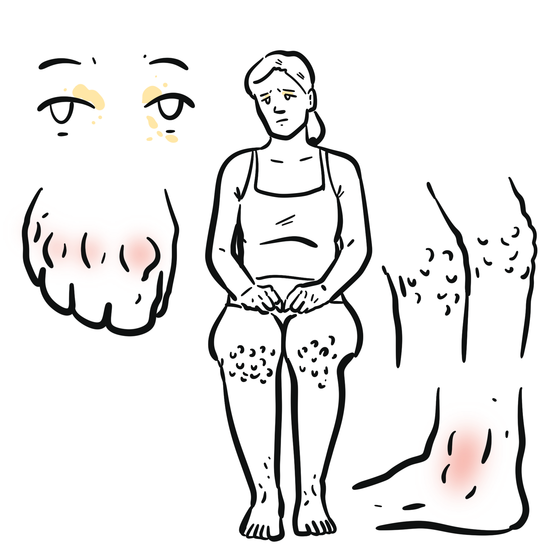
- AL Amyloidosis: Nephrotic syndrome, macroglossia, periorbital purpura (“raccoon eyes”), sensorimotor neuropathy + severe orthostasis.
- Autoimmune Autonomic Ganglionopathy (AAG): Acute/subacute severe pan-autonomic failure (fixed pupils, dry mouth, severe orthostasis; anti-gAChR antibodies).
- Other Neuropathies: Sjögren’s syndrome, HIV, Vitamin B12 deficiency, Guillain-Barré / Miller Fisher variant.
Tamsulosin and other alpha-1 blockers used for BPH are notorious causes of drug-induced orthostatic hypotension in older men. Always review the medication list before initiating an extensive neurological workup.
INVESTIGATIONS
First-Line / Bedside & Basic Diagnostic Tests:
- Active Stand Test (Bedside Orthostatics): Standard 3-minute test quantifying SBP, DBP, and HR changes.
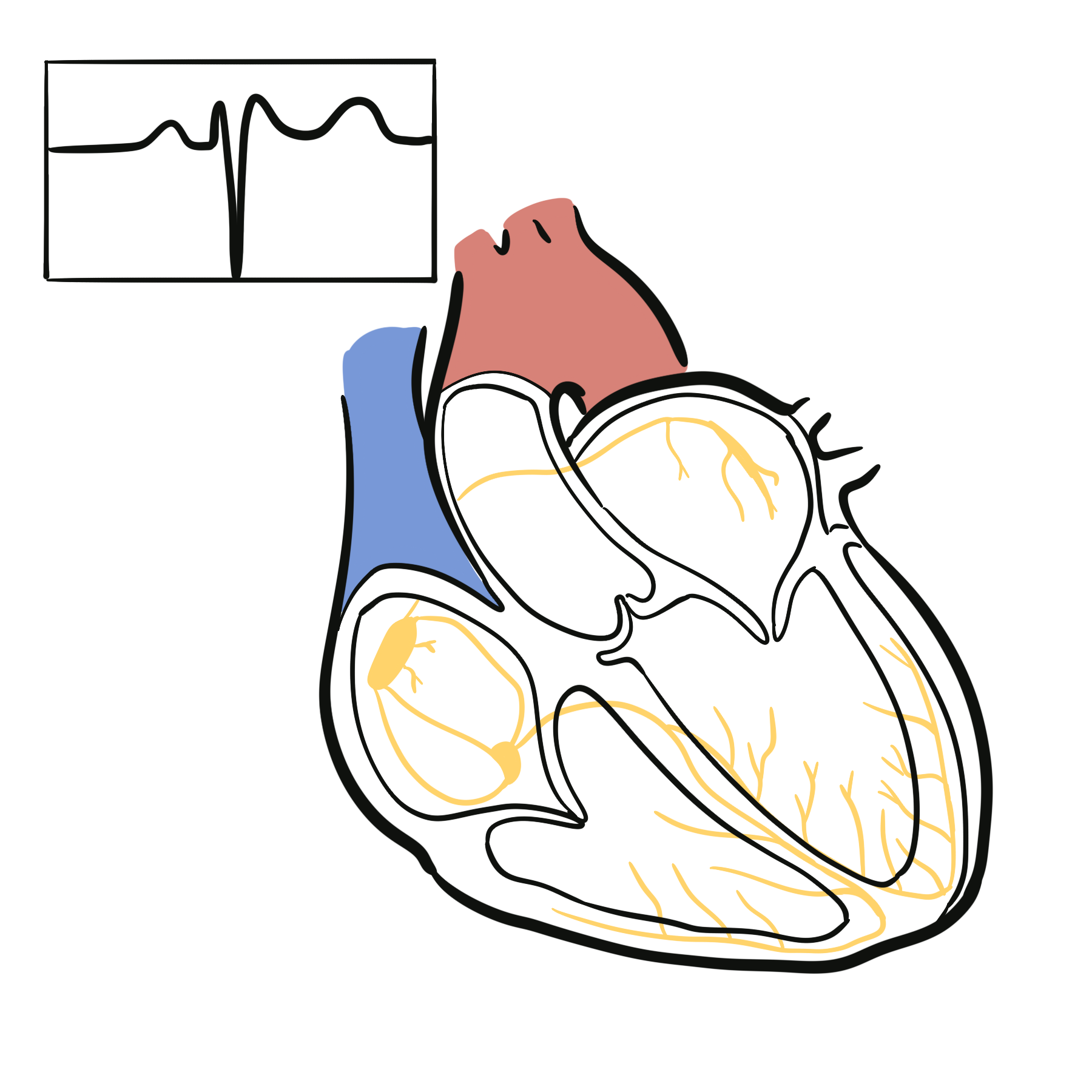
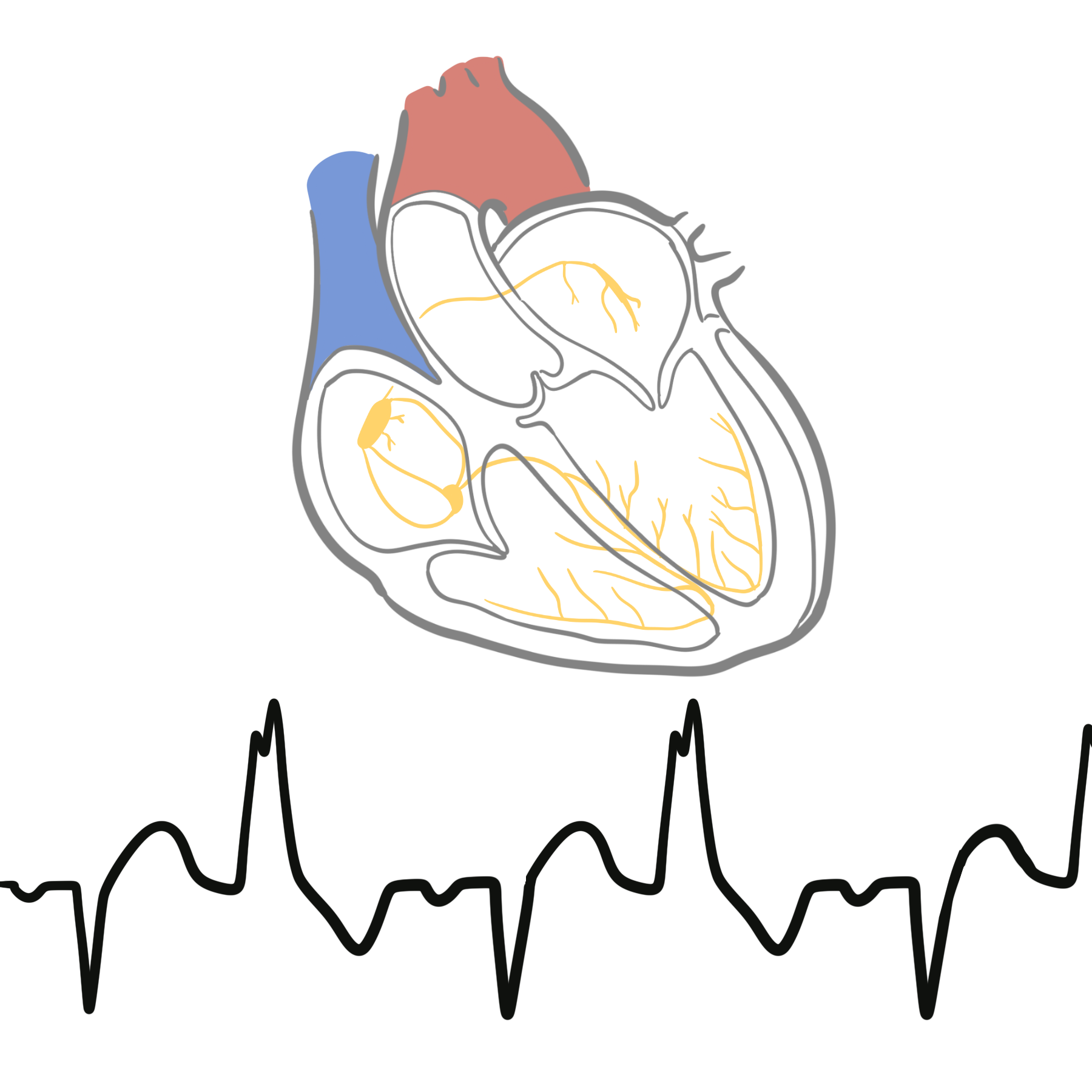
- 12-Lead ECG: Assess for arrhythmia, ischemic changes, conduction delay, or prolonged QTc.
- Full Blood Count (FBC): Screen for anemia (occult bleeding/hemolysis).
- Urea, Electrolytes, Creatinine: Screen for dehydration (elevated BUN/Cr), renal dysfunction, or Addisonian electrolyte shifts (low Na+, high K+).
- Fasting Glucose & HbA1c: Rule out diabetic neuropathy / metabolic causes.
- Morning Cortisol (8 AM): Screen for adrenal insufficiency.
Targeted / Specialized Autonomic Investigations:
- Head-Up Tilt Table Testing (HUTT): Indicated if active stand test is inconclusive, or to differentiate nOH from POTS or vasovagal syncope.
- Autonomic Function Tests:
- Ewing Battery: Heart rate variability with deep breathing (30:15 ratio), Valsalva maneuver phase IV overshoot, cold pressor test.
- Serum Free Light Chains, SPEP/UPEP: Screen for AL Amyloidosis / Plasma cell dyscrasia.
- Serum Anti-Neuronal Ganglionic Acetylcholine Receptor (gAChR) Antibodies: Confirm Autoimmune Autonomic Ganglionopathy.
- Supine and Standing Plasma Norepinephrine Levels:
- Normal: Norepinephrine doubles upon standing.
- nOH (Central/PAF): Low supine norepinephrine (< 100 pg/mL) with minimal rise upon standing (< 100% increase).
In normal individuals, standing causes plasma norepinephrine levels to double. A failure of norepinephrine to rise upon standing confirms neurogenic autonomic failure.
CRITICAL MANAGEMENT
Immediate Stabilization (Symptomatic Acute / Severe Drops):
- Positioning: Place patient supine with legs elevated (Trendelenburg position) to optimize venous return and cerebral perfusion.
- Acute Fluid Bolus: If hypovolemic or acutely symptomatic without heart failure -> Rapid infusion of 500 mL normal saline IV (or rapid ingestion of 500 mL cold water orally, which triggers a sympathetically mediated pressor response lasting 30–60 min).
- Drug Re-evaluation: Hold or depressurize non-essential antihypertensives, diuretics, and alpha-blockers immediately.
First-Line Non-Pharmacological Measures (Core Long-Term Strategy):
- Physical Counterpressure Maneuvers (PCM): Leg crossing, muscle squeezing, squatting, or toe-raising at onset of symptoms.
- Dietary Sodium & Fluid Intake: 2–3 L fluid/day + 6–10 g salt/day (unless contraindicated by severe heart failure).
- Abdominal Binder & Waist-High Compression Stockings: Abdominal compression (15–20 mmHg) is more effective than lower leg compression alone due to splanchnic venous pooling.
The splanchnic circulation holds up to 20–30% of total blood volume. An abdominal binder is often far more effective at preventing orthostatic drops than thigh-high compression stockings alone.


Members only discussions coming soon…