Chronic Exertional Compartment Syndrome (Exercise-induced Compartment Syndrome)

Overview

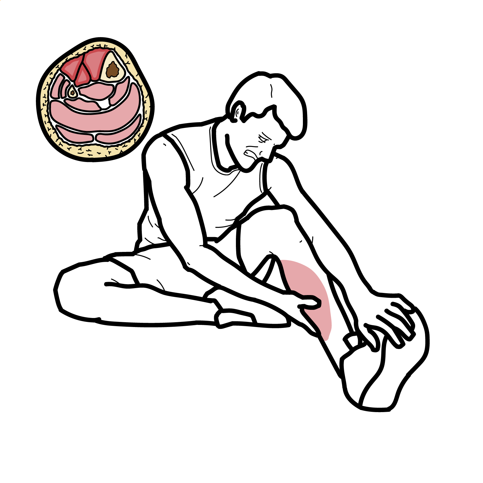
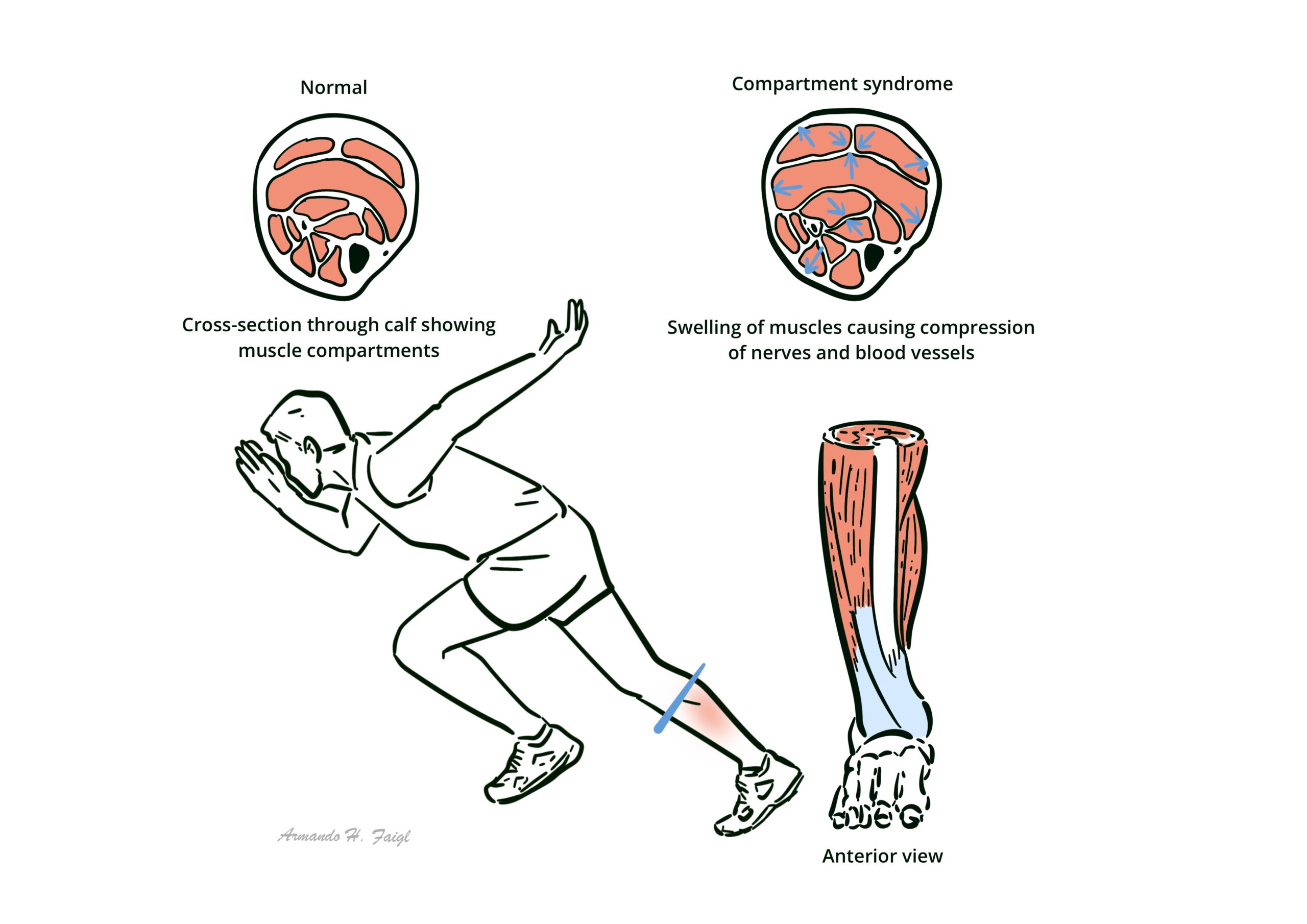
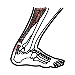
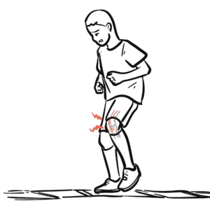
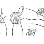
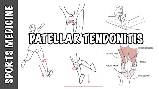
Chronic compartment syndrome (also known as chronic exertional compartment syndrome or exercise induced compartment syndrome) is an exertional condition in which repetitive activity causes transiently elevated pressure within a closed myofascial compartment, leading to ischemic pain, tightness, and sometimes neurologic symptoms that predictably begin with exercise and abate with rest. It most often affects young, physically active people (runners, team-sport athletes, military recruits) and may account for a substantial proportion of exercise-related lower-leg pain; upper-limb cases occur in rowers, climbers, weightlifters, and motorcycle racers. 1,2,3,4 Risk increases with high training volumes, running gait mechanics that increase anterior compartment demand, and fascial stiffness; outcomes range from successful return to sport with gait retraining or fasciotomy to persistent or recurrent symptoms if diagnosis is delayed. 1,5,6

Definition

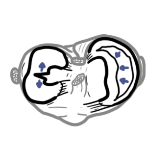
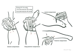
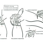
Compartment: A limb space bounded by rigid fascia containing muscle groups, nerves, and vessels.
Intracompartmental pressure (ICP): Pressure within a compartment; measured with a needle/catheter manometer at rest and after exercise.
Chronic exertional compartment syndrome (CECS): exertional condition in which repetitive activity causes transiently elevated pressure within a closed myofascial compartment, leading to ischemic pain, tightness, and sometimes neurologic symptoms that predictably begin with exercise and abate with rest.
Fasciotomy: Surgical decompression of a compartment by incising the fascia.
Ischemia: Inadequate perfusion causing exertional pain and paresthesia in CECS.

Anatomy & Physiology 

  • Lower leg compartments: Anterior (dorsiflexors, deep peroneal nerve), lateral (everters, superficial peroneal nerve), deep posterior (tibialis posterior, toe flexors, tibial nerve), superficial posterior (gastrocnemius/soleus, sural nerve).
  • Fascial mechanics: Inelastic fascia limits radial expansion; muscle volume acutely increases with hyperemia during exercise; venous outflow depends on muscle pump and low extravascular pressure.
  • Neurovascular layout: Segmental nerves traverse fixed fascial tunnels; rising tissue pressure first impairs low-pressure venous/lymphatic drainage → capillary perfusion

Exertional symptoms with a normal exam at rest are a clue to CECS rather than injury/strain.

Aetiology & Risk Factors

Aetiology

  • Repetitive exercise causing transient compartment pressure elevations exceeding perfusion pressure during activity. 1,2
  • Multifactorial contributors: fascial stiffness, muscle hypertrophy, altered biomechanics (e.g., over-stride, excessive dorsiflexor demand), and abnormal neuromuscular control patterns. 2,5

Risk Factors

  • High-volume running/marching, rapid training escalation, military basic training, young age/high fitness demands
  • Specific sports
    • Runners (especially anterior)
    • Rowers/climbers/weightlifters/motorcycle racers (forearm)
  • Gait mechanics increasing anterior compartment load (heel-strike/over-stride) and boot-induced marching mechanics; previous exertional leg pain

Bilateral lower-leg involvement is common; anterior and deep posterior compartments are most often affected. 1,2

Pathophysiology

  1. Exercise onset: Muscle hyperemia → acute volume expansion within a non-compliant fascial compartment.
  2. Pressure rise: ICP rises; venous outflow and lymphatic drainage are impeded first.
  3. Perfusion compromise: Tissue perfusion pressure (arterial minus ICP) falls → ischemic pain, cramping, tightness, exertional paresthesia/weakness.
  4. Symptom relief: Stopping exercise reduces metabolic demand; ICP gradually normalizes and symptoms resolve.
  5. Chronicity: Recurrent cycles reinforce pain behavior, can lead to performance limitation and, rarely, neurologic deficits if continued unchecked

Clinical Manifestations

  • Exertional pain/tightness/cramping starting after a reproducible time or distance
    • Resolves with rest within minutes
    • Often bilateral lower-leg; forearm symptoms with gripping/handlebar load
  • Paresthesia or numbness 
  • Transient weakness (drop-foot, loss of grip) during activity

Examination

  • Usually normal at rest
  • Immediately post-exercise—tense, tender compartment(s), pain with passive stretch of involved muscles, sometimes visible muscle herniation
  • Pulses are typically normal

Triad Predictable exertional pain + compartment tightness/firmness + rapid symptom relief with rest.

Normal resting exam does not exclude CECS; examine immediately after symptom-provoking activity.

Diagnosis

Investigations

  • Intracompartmental pressure (ICP) testing—still the most used test; performed at rest and serially after symptom-provoking exercise
  • Updated thresholds (supporting evidence): Traditional Pedowitz criteria (rest ≥15 mmHg, 1-min post ≥30 mmHg, 5-min post ≥20 mmHg) remain common
  • MRI after exercise: T2/STIR signal increase in symptomatic compartments correlates with CECS; helpful when ICP equivocal
  • Near-infrared spectroscopy (NIRS): Noninvasive muscle oxygenation patterns differ in CECS, but evidence is insufficient to fully replace ICP

Diagnostic criteria: Commonly applied thresholds based on ICP testing of affected muscle

  • Resting ICP ≥15 mmHg
  • 1-min post-exercise ≥30 mmHg
  • 5-min post-exercise ≥20 mmHg

Differential diagnoses

ConditionDistinguishing featuresKey tests
Medial tibial stress syndrome (MTSS)Diffuse posteromedial tibial border pain; worse after activity too; no neurologic symptomsClinical ± MRI/bone scan
Tibial/fibular stress fractureFocal bony tenderness; night painMRI
Popliteal artery entrapment / endofibrosisExertional claudication, diminished pulses with provocative ankle/knee positionsDoppler/CTA
Nerve entrapments (superficial peroneal, saphenous)Paresthesia independent of exertional pressure pattern; Tinel’sNerve conduction ± US
Exertional rhabdomyolysisSystemic symptoms, dark urine, CK↑Labs
Lumbar radiculopathyDermatomal pain/weakness; not strictly distance-linkedMRI spine

Measure the symptomatic compartments and side(s); test immediately post-provocation for maximal yield.

Treatment

  • Conservative (first-line, especially early or in military/runner cohorts):
    • Gait retraining (reduce over-stride, increase cadence, transition away from excessive dorsiflexor demand; marching technique modifications); can reduce surgeries and enable return to duty/sport.
    • Load management, footwear/orthoses as needed, strength & neuromuscular control, calf/ankle mobility.
    • Emerging options: Botulinum toxin A injections may normalize ICP and relieve symptoms in small series/case reports; evidence low-quality; transient strength loss reported.
  • Surgical (for refractory, pressure-positive CECS or failed conservative care):
    • Open or endoscopic fasciotomy of involved compartments; both approaches show similar return-to-sport and satisfaction rates in comparative series.
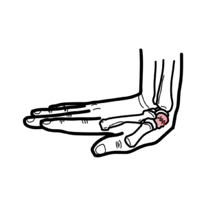
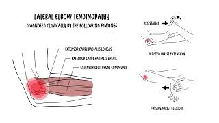
    • Forearm CECS: endoscopic/mini-open fasciotomy yields high return to sport with low recurrence in small series.
    • Post-op rehab protocols vary; progressive running/return-to-play typically staged over weeks to months.

Address biomechanics first; even pressure-positive anterior CECS may improve sufficiently with structured gait retraining to avoid surgery.

Complications & Prognosis

Complications

  • From disease: Persistent exertional pain limiting activity; rare neurologic deficits if continuing to train through symptoms.
  • From fasciotomy: Wound issues, hematoma, infection, nerve injury (e.g., superficial peroneal), DVT, need for revision; cosmetic scarring.

Prognosis

  • Return to sport/work: After fasciotomy, return-to-sport ranges widely (~26–100%) with satisfaction ~42–94%; open vs endoscopic shows similar outcomes overall. 12,16,17
  • Conservative success: Gait retraining programs in military/runner cohorts show meaningful symptom resolution and reduced surgery rates in many patients. 5
  • Poor prognostic indicators: Deep posterior involvement, multiple compartments, military occupation, and need for revision fasciotomy have been associated with lower satisfaction/RTS in some series. 12,15,17

References

  1. Vogels S, Ritchie ED, van der Burg BLSB, et al. Clinical Consensus on Diagnosis and Treatment of Patients with Chronic Exertional Compartment Syndrome of the Leg: A Delphi Analysis. Sports Med. 2022;52(12):3055-3068. (SpringerLink)
  2. Roscoe D, Roberts AJ, Hulse D. Intramuscular Compartment Pressure Measurement in Chronic Exertional Compartment Syndrome: New and Improved Diagnostic Criteria. Am J Sports Med. 2015;43(2):392-398. (upload.orthobullets.com)
  3. Maksymiak R, Ritchie E, Zimmermann W, et al. Historic cohort: outcome of CECS-suspected patients. BMJ Mil Health. 2021;167(6):387-393. (militaryhealth.bmj.com)
  4. Freedman BA, et al. Return to Sport After Fasciotomy for Forearm CECS: Systematic Review. Am J Sports Med. 2024. (CoLab)
  5. Zimmermann WO, Hutchinson MR, van den Berg R, et al. Conservative treatment of anterior CECS in the military with mid-term follow-up. BMJ Open Sport Exerc Med. 2019;5:e000532. (BMJ Open Sports & Exercise Medicine)
  6. Hartman J, Simpson S. Current Diagnosis and Management of CECS. Curr Phys Med Rehabil Rep. 2018;6:136-141. (SpringerLink)
  7. Tønning LU, et al. Muscle strength, oxygen saturation and activity in CECS vs controls. Int J Sports Phys Ther. 2023;18(2):348-357. (International Journal of Sports Therapy)
  8. Ringler MD, et al. MRI accurately detects CECS: validation study. Skeletal Radiol. 2012;42:385-392 (supporting modality use in modern reviews). (SpringerLink)
  9. OJSM Review: Reliable & Valid Noninvasive Tools for CECS Diagnosis (NIRS & others). Orthop J Sports Med. 2023;11:23259671221145151. (SAGE Journals)
  10. Baria MR, Sellon JL. Botulinum toxin for CECS: case report with 14-month follow-up. Clin J Sport Med. 2016. (SpringerLink)
  11. MIRROR project summary; case series data on BoNT-A for CECS. 2022. (mirrorusuhs.org)
  12. Hurley ET, et al. Return to sport & outcomes after open vs endoscopic release for CECS. Arthrosc Sports Med Rehabil. 2022;4(6):e1913-e1920. (ScienceDirect)
  13. Beldame J, Prado G. Endoscopic fasciotomy for forearm CECS—outcomes. World J Orthop. 2021;12(5):320-331. (WJGNET)
  14. Hsu RY, et al. Rehabilitation and return-to-activity criteria after CECS surgery: systematic review. Physician Sportsmed. 2023;52(2):125-133. (Europe PMC)
  15. Campano D, et al. Systematic review of fasciotomy in CECS (complications & revisions). J Vasc Surg. 2020;71:1541-1552. (jvascsurg.org)
  16. JISAKOS summary review 2023: satisfaction 42–94%, RTS 26–100% after fasciotomy; conservative RTS 25–35%. J ISAKOS. 2023. (jisakos.com)
  17. Waterman BR, et al. Risk factors & outcomes in military populations. Clin Sports Med. 2014;33:693-705; plus related analyses. (Context for prognosis/recurrence in military). (sportsmed.theclinics.com)

Related Content

Discussion

0 Comments
Most Voted
Newest Oldest
Inline Feedbacks
View all comments

Table Of Contents

AH Community Platform is coming
Get access to member features
Early access launches soon
Armando hasudungan brain logo
Armando Hasudungan
By Visualising Medicine
© 2025 Visualising Medicine. All rights reserved.
Become a member to access note taking
Orangise your medical learning
This is just one of the many AH community member perks
Become a member to access quizzes
Strengthen your medical knowledge
This is just one of the many AH community member perks
0
Would love your thoughts, please comment.x
()
x