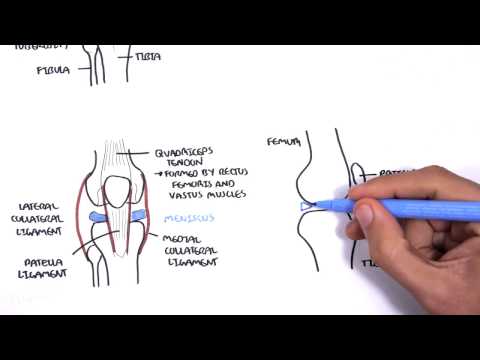
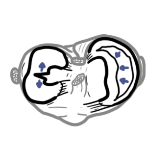
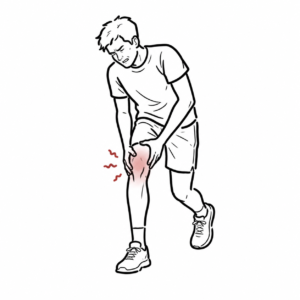
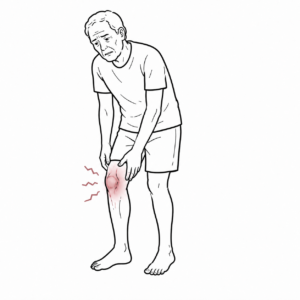
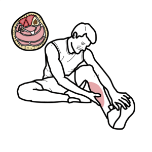
0:00 Hello, in this video we're going to talk about the meniscus, specifically the 0:08 meniscae 0:08 found in the knee joints. 0:12 So we'll first look at the right knee from an anterior view. 0:16 The knee is made up of three bones, the femur, the tibia and the patella. 0:23 On the surface of the ends of the bones we can find articular college, which 0:28 are shiny 0:29 colleges that protects the bone ends from damage. 0:34 You have many ligaments supporting the bone as well, the joint. 0:37 But also here in blue we have the lateral and medial meniscae. 0:43 The ligaments on the lateral side is the lateral collateral ligament and the 0:47 ligament 0:47 on the medial side is the medial collateral ligament. 0:52 Here is the transverse ligament which joins the two anterior parts of the men 0:56 iscae with 0:57 each other and supposedly prevents the meniscus from moving forward. 1:03 Let us now focus on the meniscae and look at the meniscus of the right knee 1:08 joint from 1:09 a superior view, so from the top. 1:13 Remember that the meniscus sits on the tibial bone. 1:17 To orientate ourselves here, here is the medial meniscus, it is C shaped. 1:25 Lateral meniscus is more of an O shaped. 1:28 Here is the fibula bone at the posterior lateral part of the tibia. 1:34 And again here is your transverse ligament connecting the anterior horns or 1:39 anterior 1:40 parts of the meniscus. 1:42 There are two other important ligaments inside the knee joint that help 1:47 stabilize the knee. 1:48 They are named according to where they attach on the tibial bone. 1:52 The ACL ligament or the anterior cruciate ligament joins to the anterior part 1:57 of the tibia and 1:59 the PCL the posterior cruciate ligament joins at the posterior part of the t 2:05 ibia. 2:06 So let us look at again the superior view of the tibial bone. 2:10 So looking from the top. 2:12 Now to orientate ourselves here again you have your lateral meniscus, then you 2:17 have 2:17 your head of the fibula here, then you have your medial meniscus, again your 2:23 inner cruciate 2:24 ligaments, your ACL connecting to the anterior part of the tibia and then you 2:30 have your PCL, 2:31 your posterior cruciate ligament attaching to the posterior part of the tibia. 2:36 Now in some people there is presence of another ligament at the back coming off 2:41 the posterior 2:42 aspect of the lateral meniscus known as the posterior meniscophimoral ligament. 2:47 And you either have an anterior or posterior but these ligaments essentially 2:53 help the posterior 2:54 cruciate ligament. 2:55 It is known to help it. 2:58 Then again here is the front of course and here is where you have the patella 3:02 ligament which 3:03 will eventually attach to the tibial tuberosity. 3:08 The medial clateral ligament is quite important because it has a deep and 3:13 superficial layer. 3:14 The deep part of the medial clateral ligament joins actually with the medial 3:21 meniscus. 3:23 This means that injury to the medial clateral ligament can cause medial menisc 3:28 al injury 3:29 as well. 3:31 The lateral clateral ligament here joins to the head of the fibula. 3:37 Finally, another important fascia that runs down and joins to the anterior 3:43 lateral part 3:45 of the tibia is the ileotibial band. 3:51 Now let us talk about the function of the meniscus and there are four main 3:55 functions. 3:56 One, it absorbs shock so it is a shock absorber, two, it allows for increased 4:01 congruency 4:02 between joint surfaces, three, it enhances joint stability and four, it aids in 4:08 the distribution 4:09 of the synovial fluid. 4:13 But remember two things if you can. 4:15 One is that the meniscus is a shock absorber and two, it helps in joint 4:21 stability. 4:23 Echosignificance, there is something called the unhappy triad or Donahue triad 4:28 and this 4:28 is a serious injury coming from the lateral part of the knee joint with the 4:34 knee twisting 4:35 and this can cause what is known as the unhappy triad. 4:39 The unhappy triad consists of a medial clateral ligament here which will then 4:44 tear the medial 4:44 meniscus and will also, the injury will also result in an anterior crucial lig 4:50 ament tear. 4:51 This is an indication for surgery. 4:55 Finally, again, drawing the same image looking at the right tibia from the 5:00 superior view, 5:02 we can see the meniscus again. 5:04 The meniscus can be divided into three sections. 5:08 The anterior part are the anterior horns. 5:13 The anterior horns are connected by the transverse ligament. 5:19 But this is only seen sorry in some people, not all. 5:24 Then you have the posterior horns at the back. 5:29 It's also important to remember that compared to the lateral meniscus, the 5:33 medial meniscus 5:34 is firmly attached to the tibia and it's not very mobile, thus it is easily 5:41 torn. 5:42 The lateral meniscus on the other hand is slightly more mobile because the 5:47 posterior 5:47 horns of the lateral meniscus does not attach firmly onto the tibia. 5:52 Finally, we can further divide the lateral and medial meniscus into the 5:58 peripheral third. 6:00 So, the outside edge is the peripheral third. 6:05 Then we have the middle third and we have the inner third. 6:08 The peripheral third is also known as the red red zone. 6:14 It is called the red red zone because it has blood supply, branches of the gen 6:19 iculate 6:19 arteries. 6:20 Thus, damage to the peripheral meniscus is repairable. 6:26 The inner third is the white white zone. 6:29 This is an avascular zone. 6:31 There is no blood supply and so it is unrepairable if damaged. 6:37 Let's now look at the meniscus from a different view. 6:39 Let's cut a slice of the meniscus from the side and look at it from that angle. 6:44 Here again is your peripheral third, the red red zone. 6:48 The middle third is called the red white zone and the inner third is the white 6:53 white zone. 6:55 The red white zone and the white white zone is classified as avascular. 7:01 Damage to this part of the meniscus means that repairing it is meaningless and 7:06 so removal 7:07 would be the best option if patients are symptomatic. 7:12 What I mean by this is if the damage causes pain. 7:18 So clinical relevance of the meniscus, well, meniscal tears are very common. 7:24 It can occur in healthy young active adolescents or in adults and the elderly. 7:31 So we can say meniscal tears can either be acute or degenerative. 7:36 Red meniscal tears are usually bucket handle tears from longitudinal tears. 7:41 Degenerative meniscal tears are from old age and is due to wearing tear with 7:47 accompanying 7:48 osteoarthritis. 7:49 It is a complex type of tear which may involve other things. 7:55 These injuries can present with a sensation of catching or the knee's locking. 8:01 Knee locking is essentially where the torn meniscus, if it is big enough, it 8:06 can get caught 8:07 between the tibia and the femur bone essentially and this is known as the 8:12 locking sensation 8:13 or the knee's catching. 8:15 I hope you enjoyed this video on the clinical anatomy of the Neimanisca. 8:19 Thank you.