

Acute Respiratory Distress Syndrome
Overview
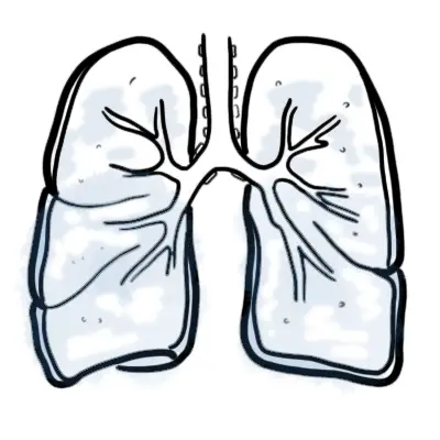
ARDS is an acute, diffuse, inflammatory lung injury that leads to increased pulmonary vascular permeability, increased lung weight, and a loss of aerated tissue. Clinical hallmarks of ARDS are hypoxemia and bilateral radiographic opacities, while the pathological hallmark is diffuse alveolar damage.
Overview
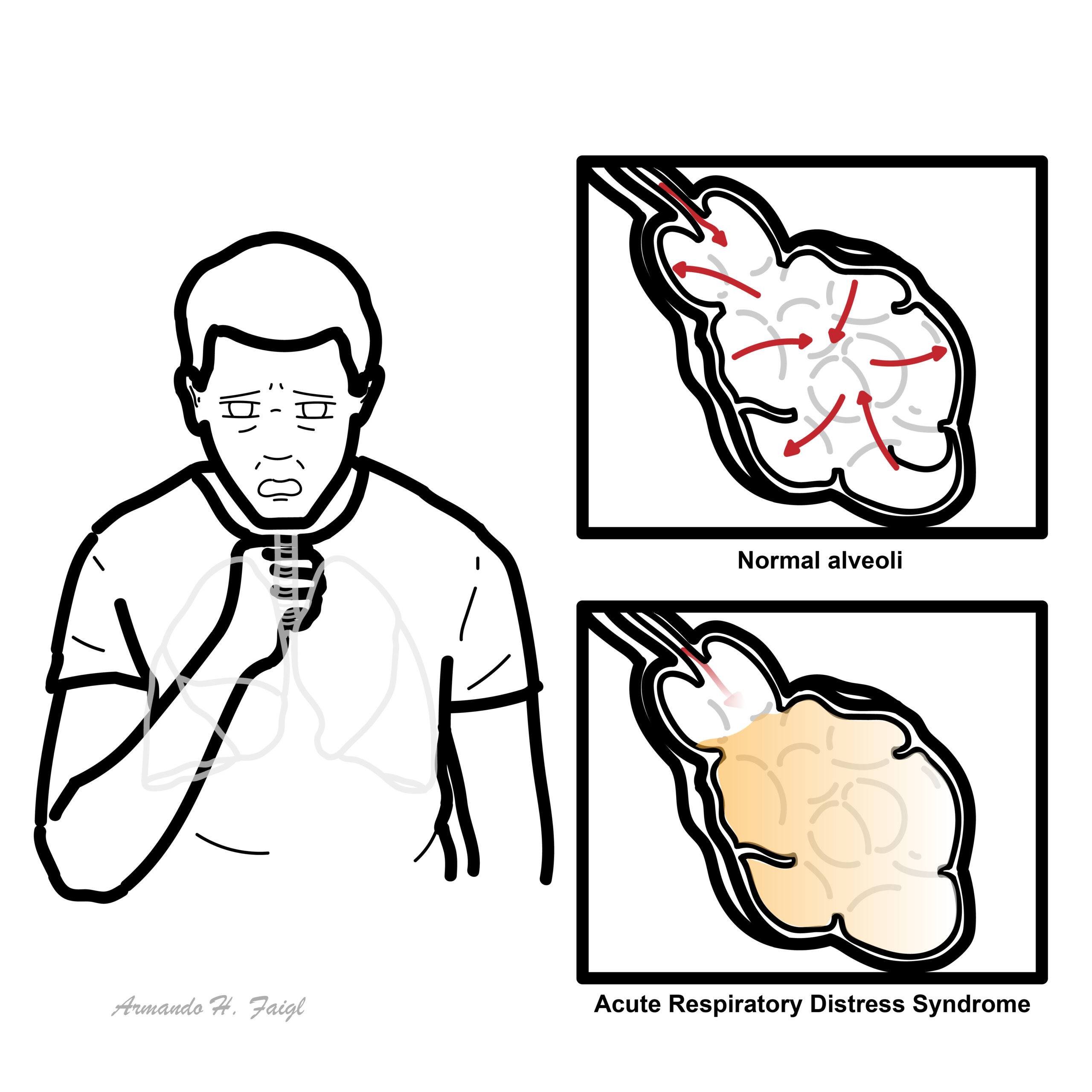
Acute Respiratory Distress Syndrome (ARDS) is a life-threatening form of acute, diffuse inflammatory lung injury caused by direct or indirect pulmonary insults. The hallmark of the condition is a rapid increase in alveolar-capillary membrane permeability, resulting in protein-rich pulmonary edema, loss of aerated lung tissue, severe ventilation-perfusion (V/Q) mismatching, and profound, refractory hypoxemia.
As the inflammatory cascade progresses, damage to alveolar epithelial and capillary endothelial cells leads to surfactant inactivation, widespread microatelectasis, and decreased pulmonary compliance (“stiff lungs”). ARDS is not a primary disease itself, but rather a catastrophic syndrome triggered by various critical illnesses—most commonly sepsis. Despite advances in supportive care and mechanical ventilation strategies, ARDS carries high morbidity and mortality, with surviving patients frequently facing long-term physical and cognitive impairments.
Definition
PaO2/FiO2 (P/F) Ratio: The ratio of arterial oxygen partial pressure (in mmHg) to fractional inspired oxygen concentration (expressed as a decimal, e.g., 21% = 0.21); the primary index used to grade ARDS severity.
Berlin Definition: The standardized clinical diagnostic criteria for ARDS requiring acute onset, bilateral opacities on imaging, non-cardiogenic origin of edema, and a P/F ratio <= 300 mmHg on PEEP >= 5 cmH2O.
Shunt Physiology (Intrapulmonary Shunt): Perfusion of non-ventilated, collapsed alveoli (V/Q = 0), resulting in venous admixture that is characteristically refractory to supplemental oxygen administration.
Baby Lung Concept: The physiological phenomenon in ARDS where the functional, aerated portion of the lung is severely reduced to the size of a healthy young child’s lung, making it highly vulnerable to volutrauma and barotrauma.
Driving Pressure (Delta P): Calculated as Plateau Pressure minus PEEP (Pplat – PEEP); reflects the stress applied to the aerated lung during mechanical ventilation and strongly correlates with survival.
Classification
Graded based on the degree of hypoxemia evaluated with a minimum PEEP or CPAP >= 5 cmH2O:
| Severity Grade | PaO2 / FiO2 Ratio | Associated Mortality |
| Mild ARDS | 201 – 300 mmHg | ~ 27% |
| Moderate ARDS | 101 – 200 mmHg | ~ 32% |
| Severe ARDS | <= 100 mmHg | ~ 45% |
Aetiology & Risk Factors
Aetiologies are divided into direct (pulmonary) and indirect (systemic) insults to the lung microvasculature and epithelium.
Direct Lung Injury (Epithelial Insult)
- Pneumonia: Bacterial, viral (e.g., Influenza, COVID-19), fungal, or atypical pneumonia (most common direct cause).
- Aspiration of Gastric Contents: Chemical pneumonitis secondary to acidic stomach contents damaging the alveolar epithelium (e.g., Mendelson syndrome).
- Pulmonary Contusion: Blunt thoracic trauma.
- Inhalation Injury: Toxic gas, smoke inhalation, or chemical vapors.
- Near-Drowning: Fresh or saltwater aspiration disrupting surfactant function.
Indirect Lung Injury (Endothelial Insult)
- Sepsis: Non-pulmonary severe infection/septic shock (most common overall cause of ARDS).
- Severe Trauma & Shock: Non-thoracic major trauma, hypovolemic shock, or crush injuries.
- Acute Pancreatitis: Release of circulating pancreatic enzymes (phospholipase A2) causing systemic endothelial damage.
- Transfusion-Related Acute Lung Injury (TRALI): Immune-mediated capillary leak within 6 hours of blood product transfusion.
- Drug Overdose: Opioids, salicylates, tricyclic antidepressants, or chemotherapy agents.
Sepsis is the single most common cause of ARDS overall, whereas pneumonia is the most common cause of direct lung injury leading to ARDS.
Pathophysiology
Aetiology
- Indirect lung injury: sepsis, shock, trauma, pancreatitis, drug OD, intracranial haemorrhage, blood-transfusion related lung injury
- Direct lung injury: aspiration, inhalational injury, embolism
- Insult → release of inflammatory mediators promoting neutrophil accumulation in microcirculation oflung
- Neutrophils damage vascular endothelium + alveolar epithelium
- Leads to pulmonary oedema, hyaline membrane formation & difficult gas exchange
Clinical Manifestation
- Respiratory Manifestations
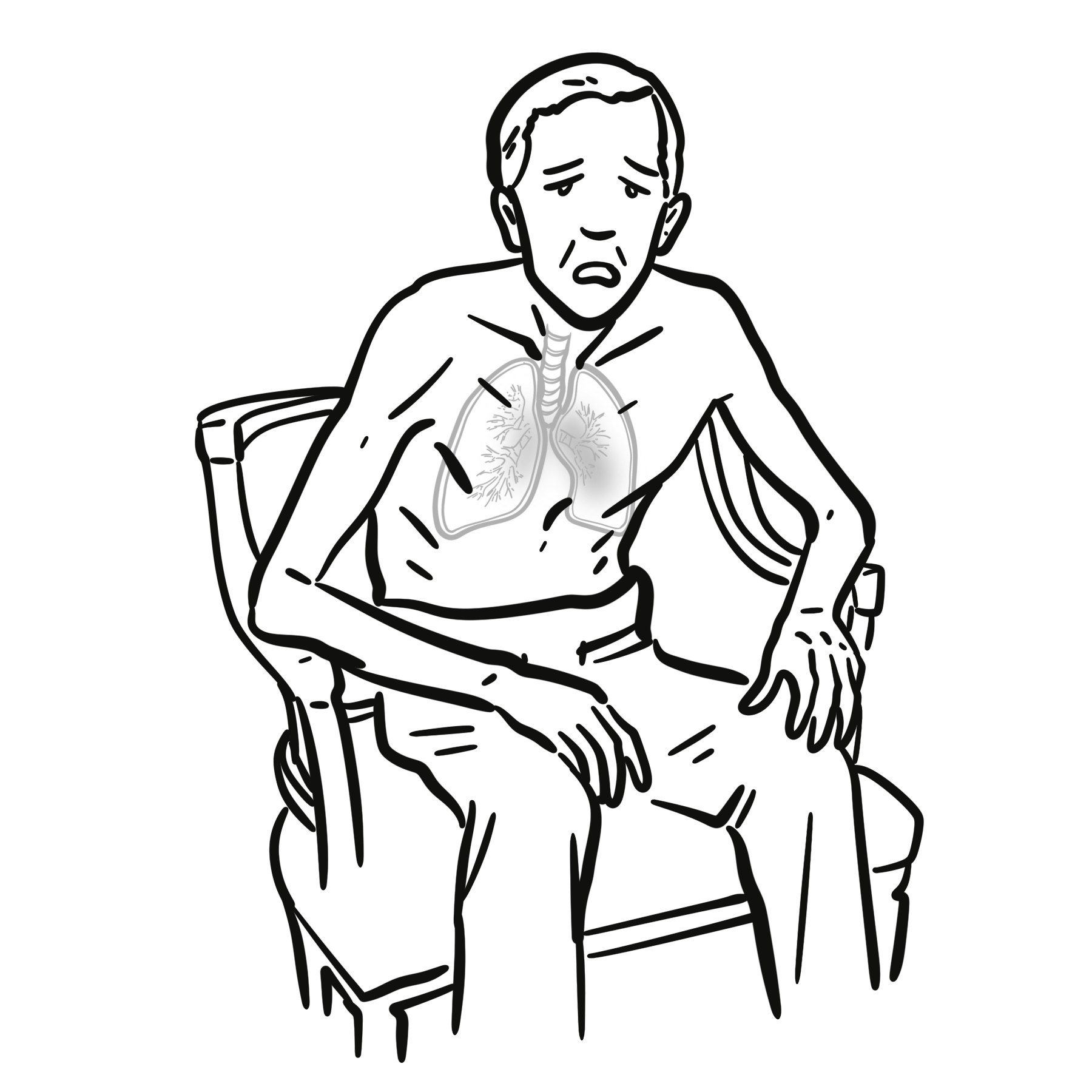
- Acute Severe Dyspnoea & Tachypnoea: Develops rapidly within 24–72 hours of the triggering insult.
- Refractory Hypoxemia: Severe arterial hypoxemia that fails to respond significantly to increasing concentrations of supplemental oxygen (due to shunt physiology).
- Auscultatory Findings: Diffuse, bilateral end-inspiratory crackles/rales and bronchial breath sounds throughout all lung fields.
- Work of Breathing: Intercostal retractions, accessory muscle use, nasal flaring, and tachypnea (> 30 breaths/min).
- Systemic & Hemodynamic Manifestations
- Tachycardia & Diaphoresis: Secondary to sympathetic surge driven by severe hypoxia and hypercapnia.
- Cyanosis: Central cyanosis resistant to oxygen delivery.
- Neurological Impairment: Agitation, anxiety, confusion, and lethargy due to cerebral hypoxia and hypercapnia.
- SIRS / Septic Features: Fever or hypothermia, hypotension, leukocytosis, and signs of primary underlying disease (e.g., severe abdominal pain in pancreatitis).
Hypoxemia in ARDS is refractory to supplemental oxygen because blood is passing through non-ventilated, collapsed, fluid-filled alveoli (intrapulmonary shunt). Opening these alveoli requires positive pressure (PEEP), not just higher FiO2
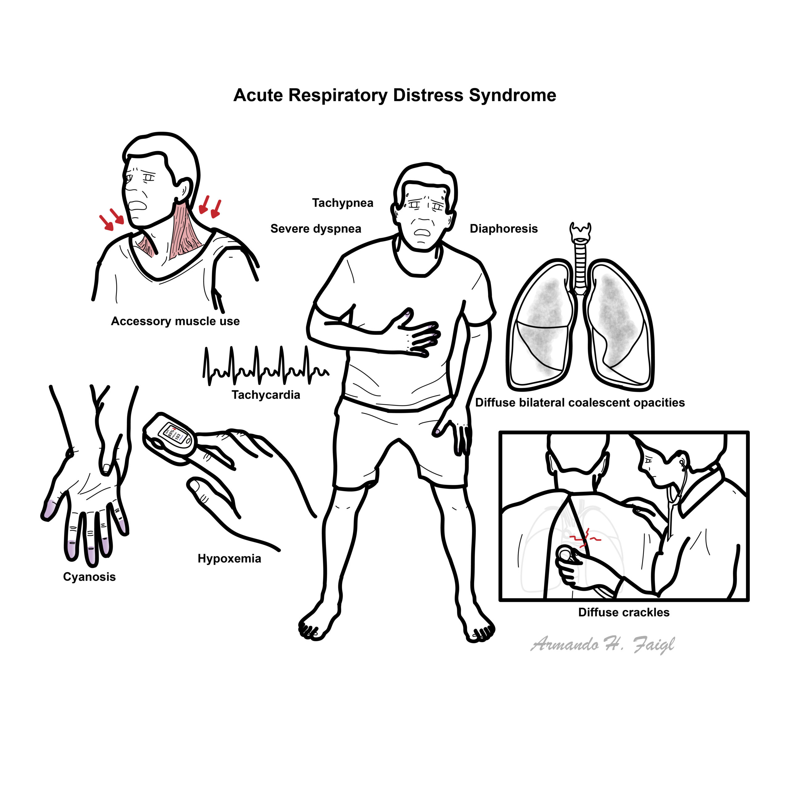
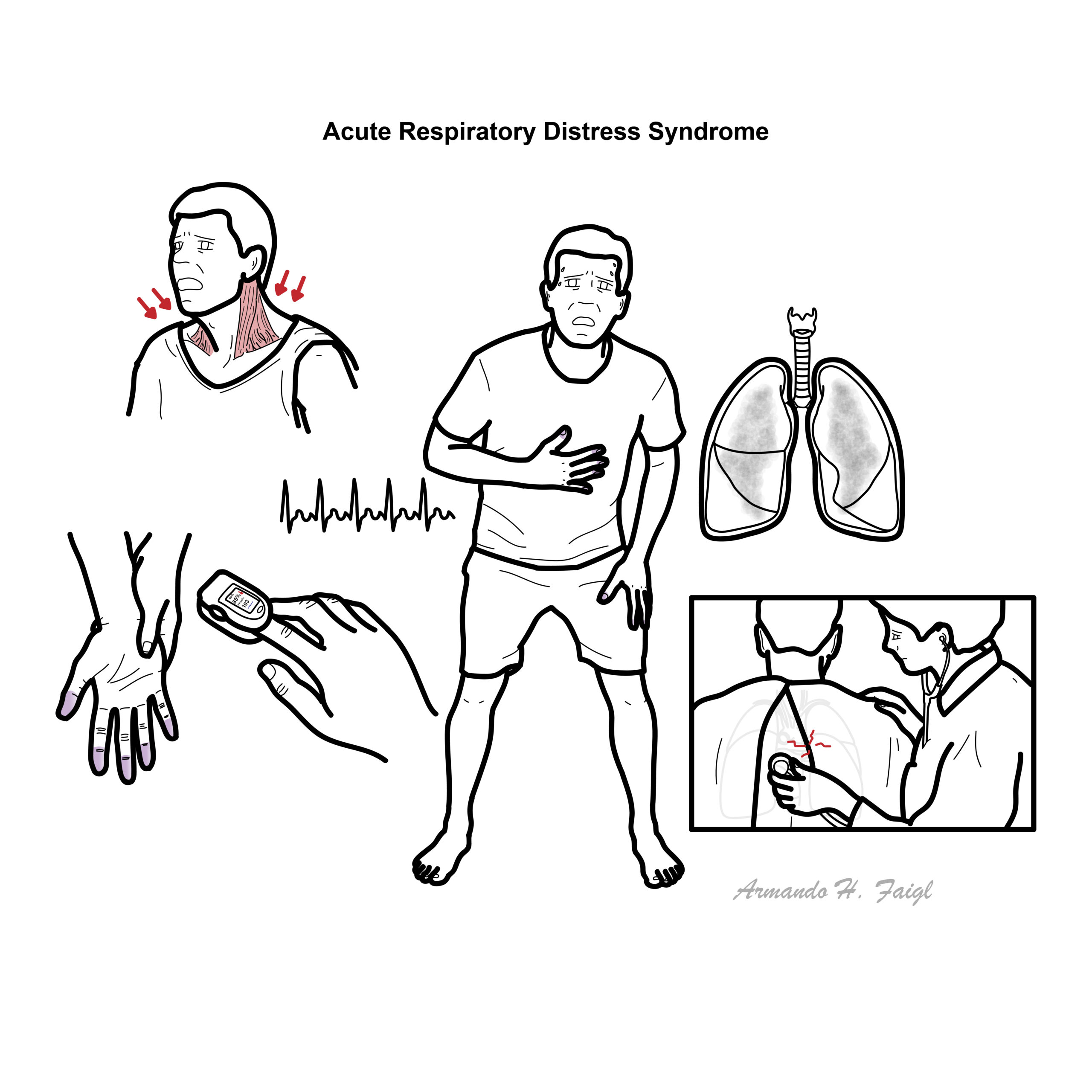
Clinical hallmarks of ARDS are hypoxemia and bilateral radiographic opacities, while the pathological hallmark is diffuse alveolar damage.
Clinical Course
- The first several days of ARDS are characterized by hypoxemia requiring a moderate to high concentration of inspired oxygen
- Most patients who survive this initial course begin to exhibit better oxygenation and decreasing alveolar infiltrates over the next several days.
- Some patients, however, have persistent, severe hypoxemia and remain ventilator-dependent. Pulmonary proliferative changes and fibrosis may progressively replace the pathological findings of diffuse alveolar damage as early as ten days after the onset of the respiratory failure.
Diagnosis
Diagnostic Criteria – Berlin Definition
- Acute onset of symptoms of a known clinical insult (<1 week)
- Bilateral opacities consistent with pulmonary edema on Chest X-ray or CT. Other causes ruled out:
- Pulmonary nodules
- Lung collapse
- Pleural effusion
- Cardiac failure and fluid overload ruled out
- Decreased (moderate to severe) oxygenation must be present
- Mild
- Moderate
- Severe
Diagnostic work up
- Arterial Blood Gas (ABG):
- Early: Respiratory alkalosis with severe hypoxemia (low PaO2, low PaCO2, elevated pH) due to compensatory hyperventilation.
- Late: Mixed respiratory and metabolic acidosis with hypercapnia (high PaCO2, low pH) as respiratory muscles fatigue.
- Chest X-Ray / CT Scan:
- CXR: Bilateral, patchy alveolar opacities (“white-out” lung in severe cases); characteristically lacks cardiomegaly, pleural effusions, or pulmonary vascular redistribution.
- Chest CT: Heterogeneous ground-glass opacities with dense consolidation in dependent lung zones.
- Echocardiography: Essential to rule out left ventricular dysfunction, mitral valve disease, or fluid overload (cardiogenic pulmonary edema). Normal LV ejection fraction and normal left atrial pressure confirm non-cardiogenic etiology.
- Laboratory Investigations: Complete blood count, blood cultures, inflammatory markers (CRP/procalcitonin), amylase/lipase, and renal/liver panels to identify the underlying cause and assess multi-organ dysfunction.
On CXR, ARDS classically presents with bilateral fluffy infiltrates without cardiomegaly or pleural effusions. Echocardiogram is the quickest way to rule out cardiogenic pulmonary edema (e.g., acute heart failure).
Treatment
- Mechanical ventilation using high PEEP & low tidal volumes to prevent ventilator-associated injury
- O2 therapy: aim PaO2>60mmHg or SpO2>90% with permissive hypercapnia
- Treat underlying disorder/precipitant
- Conservative fluid Mx strategy: aim to minimize or eliminate positive fluid balance that can → pulmonary oedema
- Supportive care
- Haemodynamic support to maintain MAP>60mmHg
- Enteral nutritional support
- DVT & stress ulcer prevention
Mortality is 30-40% and mostly from extra-pulmonary complications.
Complications & Prognosis
Complications
- Ventilator-Induced Lung Injury (VILI): Includes barotrauma (pneumothorax, pneumomediastinum, subcutaneous emphysema), volutrauma, and atelectotrauma.
- Pulmonary Complications: Ventilator-Associated Pneumonia (VAP), pulmonary fibrosis, and chronic pulmonary hypertension.
- Systemic Complications: Multiple Organ Dysfunction Syndrome (MODS) (acute kidney injury, DIC, stress ulcers), ICU-acquired weakness, and deep vein thrombosis (DVT).
- Post-Intensive Care Syndrome (PICS): Long-term cognitive impairment, depression, anxiety, PTSD, and physical deconditioning in survivors.
- Overall Mortality: Remains high at approximately 35–45% (correlates with Berlin severity grade).
- Primary Cause of Death: Most ARDS patients die from sepsis and Multiple Organ Dysfunction Syndrome (MODS), not isolated hypoxemia.
- Predictors of Poor Outcome: Advanced age, underlying liver disease/cirrhosis, non-pulmonary organ failures, persistent high driving pressure, and delayed diagnosis.
The primary cause of death in patients with ARDS is not isolated hypoxemia, but rather sepsis and Multiple Organ Dysfunction Syndrome (MODS).
References
Matthay MA, Zemans RL, Zimmerman GA, et al. Acute respiratory distress syndrome. Nat Rev Dis Primers. 2019;5(1):18. doi:10.1038/s41572-019-0069-0
Force AD, Ranieri VM, Rubenfeld GD, et al. Acute respiratory distress syndrome: the Berlin Definition. JAMA. 2012;307(23):2526-2533. doi:10.1001/jama.2012.5669
Thompson BT, Chambers RC, Liu KD. Acute Respiratory Distress Syndrome. N Engl J Med. 2017;377(6):562-572. doi:10.1056/NEJMra1608077






Members only discussions coming soon…