

Chronic Intestinal Ischaemia
Overview
Chronic intestinal ischaemia, more commonly called chronic mesenteric ischaemia, results from an inadequate increase in intestinal blood flow after eating. It is usually caused by atherosclerotic narrowing of one or more mesenteric arteries.
Patients classically develop recurrent postprandial abdominal pain, leading to reduced food intake, fear of eating and progressive weight loss. Diagnosis is frequently delayed because symptoms overlap with more common gastrointestinal disorders.1–3
Untreated disease may cause severe malnutrition or progress to acute-on-chronic mesenteric ischaemia, bowel infarction and death. Symptomatic disease therefore requires prompt specialist assessment and consideration of mesenteric revascularisation.1–3
Definition
- Chronic mesenteric ischaemia
- Inadequate intestinal blood flow, particularly after eating, caused by a persistent limitation of the mesenteric circulation.
- Intestinal angina
- Recurrent postprandial abdominal pain caused by insufficient mesenteric blood flow to meet the increased metabolic demand of digestion.
- Food fear
- Avoidance or restriction of eating because meals repeatedly trigger abdominal pain.
- Acute-on-chronic mesenteric ischaemia
- Sudden deterioration in a patient with chronic mesenteric arterial disease, potentially resulting in bowel infarction.
- Mesenteric revascularisation
- Restoration of blood flow through an obstructed mesenteric artery using an endovascular or open surgical procedure.
Anatomy & Physiology
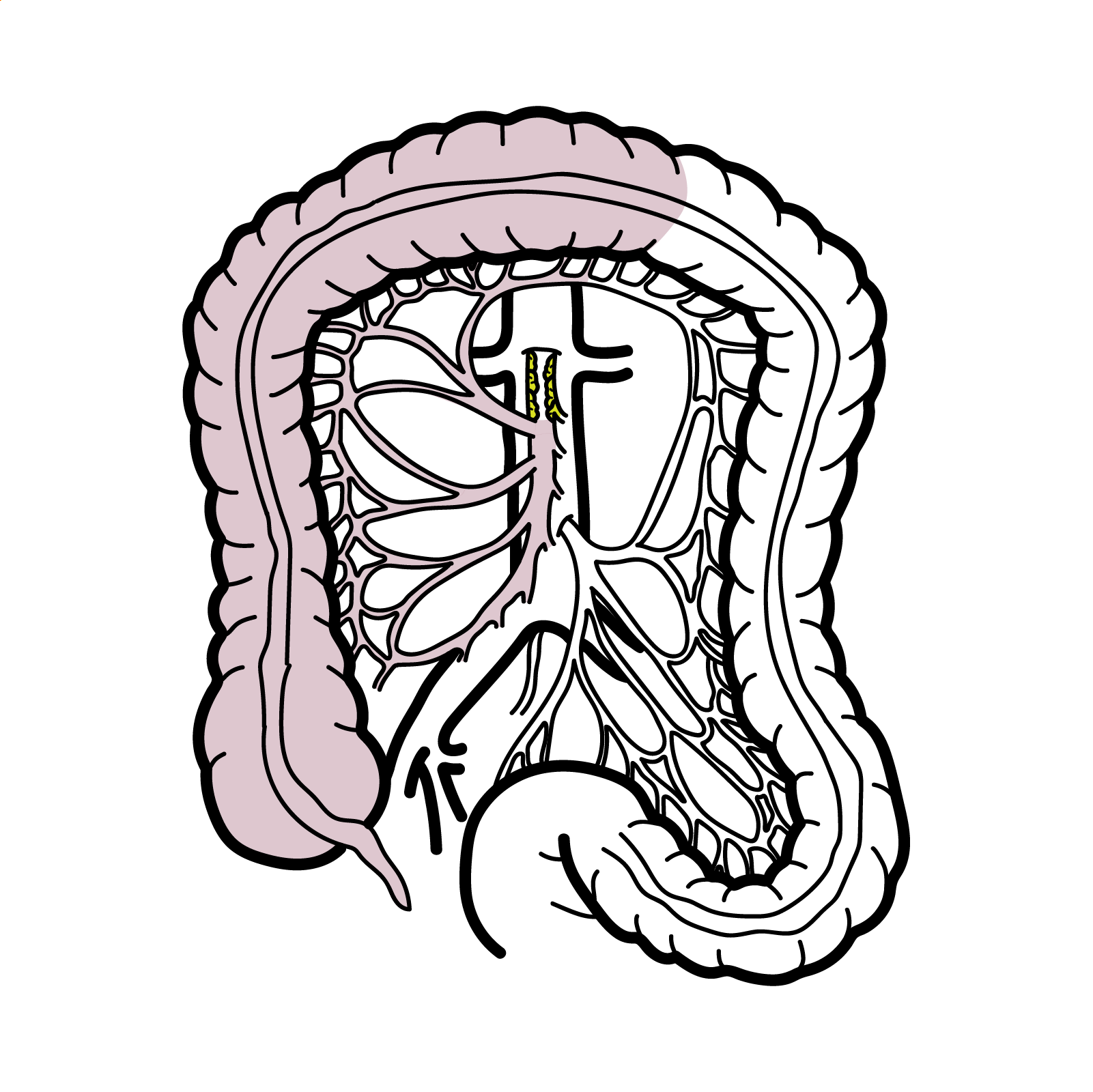
The gastrointestinal tract receives arterial blood from three major branches of the abdominal aorta:
- Coeliac artery: supplies the foregut, including the stomach, proximal duodenum, liver, spleen and pancreas.
- Superior mesenteric artery (SMA): supplies the midgut, including the distal duodenum, small intestine and proximal two-thirds of the transverse colon.
- Inferior mesenteric artery (IMA): supplies the hindgut, including the distal third of the transverse colon, descending colon, sigmoid colon and upper rectum.
Extensive collateral connections normally allow one vessel to compensate for gradual obstruction of another. Consequently, symptomatic atherosclerotic disease often involves more than one mesenteric artery, although clinically important single-vessel disease can occur, particularly when the SMA is affected.1–3
Intestinal blood flow increases substantially after a meal. A severe arterial stenosis may provide adequate resting perfusion but fail to meet this postprandial demand, producing intestinal angina.
Aetiology & Risk Factors
Aetiology
Atherosclerotic mesenteric arterial disease (most common cause).
Median arcuate ligament syndrome: The median arcuate ligament compresses the proximal coeliac artery.
Other uncommon causes include:
- Mesenteric artery dissection.
- Fibromuscular dysplasia.
- Large- or medium-vessel vasculitis.
- Radiation-associated arterial injury.
- Thromboangiitis obliterans.
- Chronic non-occlusive mesenteric ischaemia associated with severe cardiac, pulmonary or small-vessel disease.
- Previous abdominal vascular surgery or aortic intervention affecting the mesenteric circulation.
Risk Factors
Risk factors for atherosclerotic chronic mesenteric ischaemia are similar to those for atherosclerosis elsewhere:
- Increasing age.
- Cigarette smoking.
- Hypertension.
- Dyslipidaemia.
- Diabetes mellitus.
- Chronic kidney disease.
- Established coronary, cerebrovascular or peripheral arterial disease.
- Family history of premature cardiovascular disease.
Women are diagnosed more frequently than men, although the reason for this association is not fully understood.
Pathophysiology
Arterial narrowing reduces the ability of the mesenteric circulation to increase blood flow during digestion. Ischaemia develops when postprandial metabolic demand exceeds the available blood supply.
Pain commonly begins after eating and may last for one to several hours. Patients learn that smaller meals, skipped meals or fasting reduce their symptoms. Progressive dietary restriction then contributes to weight loss, protein-energy malnutrition, vitamin and mineral deficiencies, frailty and impaired wound healing.
Severe stenosis may eventually progress to thrombosis or critically reduced resting perfusion. This can produce acute-on-chronic mesenteric ischaemia, characterised by a sudden change in the nature or severity of pain and a risk of irreversible bowel necrosis.
The intestine may receive sufficient blood at rest but become ischaemic after a meal, when its metabolic and perfusion requirements rise substantially.
Clinical Manifestations
Typical Presentation
- Recurrent, dull or cramping abdominal pain after eating.
- Pain usually begins within approximately 30 minutes of a meal.
- Symptoms may persist for one to several hours.
- Food avoidance or consumption of progressively smaller meals.
- Unintentional weight loss.
- Early satiety, nausea or vomiting.
- Diarrhoea, constipation or abdominal bloating.
- Reduced physical reserve, muscle wasting or other evidence of malnutrition.
The traditional triad of postprandial pain, weight loss and an abdominal bruit is not present in every patient. Its absence does not exclude chronic mesenteric ischaemia.1
Clinical Examination
- Low body weight or visible muscle wasting.
- Signs of dehydration or malnutrition.
- Abdominal bruit.
- Evidence of generalised atherosclerosis, such as reduced peripheral pulses or carotid bruits.
- Usually a soft abdomen without peritonism in uncomplicated chronic disease.
Consider chronic mesenteric ischaemia in an older patient with atherosclerotic risk factors, otherwise unexplained postprandial abdominal pain and progressive weight loss—even when the abdominal examination is unremarkable.
Diagnosis & Investigations
Diagnosis requires correlation between a compatible clinical presentation and significant mesenteric arterial disease. An incidental stenosis on imaging does not, by itself, establish symptomatic chronic mesenteric ischaemia.
No blood test reliably confirms or excludes chronic mesenteric ischaemia. Investigations may include:
- Serum lactate may be elevated.
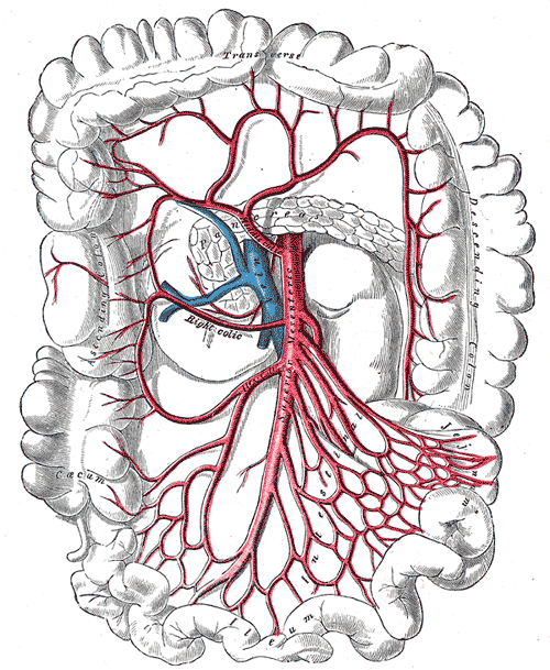
The superior mesenteric artery and its intestinal branches. Gray’s Anatomy, 1918; public domain.
What to notice
Detailed image description
Source: https://commons.wikimedia.org/wiki/File:Superior_mesenteric_a.gifCreator/source work: Gray’s Anatomy, 20th US edition, 1918.Licence: Public Domain Mark 1.0 — https://creativecommons.org/publicdomain/mark/1.0/Modifications: None.
CT Angiography
CT angiography of the abdomen and pelvis is the principal diagnostic imaging test. It demonstrates the site and severity of mesenteric arterial stenosis or occlusion, the aorta and collateral vessels, and possible alternative intra-abdominal pathology.1–4
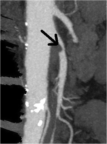
CT angiogram demonstrating stenosis of the superior mesenteric artery. Ryan Spangler, CC BY 4.0.
What to notice
Detailed image description
Source: https://commons.wikimedia.org/wiki/File:Superior_mesenteric_artery_stenosis.jpgCreator: Ryan Spangler.Licence: CC BY 4.0 — https://creativecommons.org/licenses/by/4.0/Modifications: None.Originating publication: Spangler R, Van Pham T, Khoujah D, Martinez JP. Abdominal emergencies in the geriatric patient. Int J Emerg Med. 2014;7:43. doi:10.1186/s12245-014-0043-2.
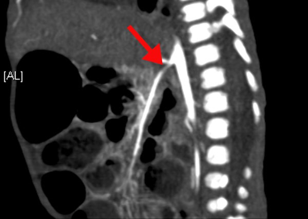
CT angiogram showing superior mesenteric artery stenosis indicated by the arrow. Omar Oda, CC BY 2.0.
What to notice
Detailed image description
Source: https://commons.wikimedia.org/wiki/File:Stenosis_of_the_superior_mesenteric_artery.jpgCreator: Omar Oda.Licence: CC BY 2.0 — https://creativecommons.org/licenses/by/2.0/Modifications: Commons version cropped before import; no further modification by Armandoh.Originating publication: Oda O, Zamakhshary M, Al Namshan M, Al Jadaan S, Al Shalaan H. Bowel ischemia in a baby with unspecified renovascular hypertension: a case report. J Med Case Rep. 2011;5:569. doi:10.1186/1752-1947-5-569.
Alternative Vascular Imaging
- Contrast-enhanced MR angiography
- Duplex ultrasound: accuracy can be limited by body habitus, bowel gas and operator dependence.
- Catheter angiography: generally reserved for patients undergoing endovascular treatment or when non-invasive imaging remains inconclusive.
The diagnosis is clinical and radiological: compatible symptoms plus significant mesenteric arterial disease. Mesenteric stenosis without compatible symptoms may be incidental.
Treatment
Immediate Management
Patients with symptomatic chronic mesenteric ischaemia should receive expedited referral to a vascular or multidisciplinary mesenteric-ischaemia service. The aims are to relieve symptoms, restore normal eating, reverse malnutrition and prevent bowel infarction.
Analgesia and careful correction of dehydration or electrolyte abnormalities may be required. Nutritional status should be assessed, but attempts to rapidly increase oral or enteral feeding before restoring adequate blood flow may worsen postprandial ischaemia. Revascularisation should not be unnecessarily delayed for prolonged nutritional treatment.
Mesenteric Revascularisation
Revascularisation is indicated for most patients with symptomatic atherosclerotic chronic mesenteric ischaemia.
Endovascular revascularisation: This is usually the initial approach when the arterial anatomy is suitable:
- Balloon angioplasty followed by stent placement across the stenosis.
- Lower early morbidity and shorter recovery than open surgery.
- Particularly suitable for older or medically comorbid patients.
Restenosis is more frequent after endovascular treatment than after open reconstruction, so clinical and imaging surveillance is important.
Open surgical revascularisation
Medical and Cardiovascular Management
Medical treatment alone does not restore adequate intestinal perfusion in symptomatic occlusive disease. It remains essential as secondary prevention before and after revascularisation:
- Stop smoking.
- Commence antiplatelet therapy where appropriate.
- Use lipid-lowering therapy, usually a statin.
- Optimise blood pressure and diabetes management.
- Encourage appropriate physical activity once symptoms and nutritional status permit.
- Treat associated coronary, cerebrovascular and peripheral arterial disease.
Following mesenteric stenting, antiplatelet treatment is prescribed according to the procedural protocol and the patient’s bleeding and thrombotic risks.
Median Arcuate Ligament Syndrome
Coeliac artery stenting without first releasing the external compression is generally inappropriate.
Asymptomatic Mesenteric Arterial Stenosis
Incidental asymptomatic single-vessel stenosis usually does not require revascularisation. Management focuses on cardiovascular risk reduction and monitoring.
Complications & Prognosis
Complications
- Progressive weight loss and protein-energy malnutrition.
- Vitamin and mineral deficiencies.
- Frailty, muscle wasting and reduced quality of life.
- Acute-on-chronic mesenteric ischaemia.
- Bowel infarction, perforation, peritonitis and sepsis.
- Multiorgan failure and death.
- Restenosis or occlusion after revascularisation.
- Access-site bleeding, arterial dissection, embolisation or contrast-associated complications following endovascular treatment.
- Cardiac, respiratory, wound or graft complications following open surgery.
Prognosis
Successful revascularisation usually produces substantial improvement or resolution of postprandial pain, permits restoration of normal eating and supports weight recovery.
Delayed diagnosis increases the risk of severe malnutrition and acute bowel infarction.
A sudden change from episodic postprandial pain to severe, continuous pain may indicate acute-on-chronic mesenteric ischaemia and requires emergency assessment.
References
- Terlouw LG, Moelker A, Abrahamsen J, et al. European guidelines on chronic mesenteric ischaemia: joint United European Gastroenterology and multidisciplinary clinical guidelines on the diagnosis and treatment of patients with chronic mesenteric ischaemia. United European Gastroenterol J. 2020;8(4):371–395. doi:10.1177/2050640620916681. Available from: https://pmc.ncbi.nlm.nih.gov/articles/PMC7226699/
- Huber TS, Björck M, Chandra A, et al. Chronic mesenteric ischemia: clinical practice guidelines from the Society for Vascular Surgery. J Vasc Surg. 2021;73(1 Suppl):87S–115S. doi:10.1016/j.jvs.2020.10.029. Available from: https://pubmed.ncbi.nlm.nih.gov/33171195/
- Koelemay MJ, Geelkerken RH, Kärkkäinen J, et al. Editor’s Choice—European Society for Vascular Surgery 2025 clinical practice guidelines on the management of diseases of the mesenteric and renal arteries and veins. Eur J Vasc Endovasc Surg. 2025;70(2):153–274. doi:10.1016/j.ejvs.2025.06.010. Available from: https://pubmed.ncbi.nlm.nih.gov/40513642/
- Ginsburg M, Obara P, Lambert DL, et al. ACR Appropriateness Criteria imaging of mesenteric ischemia. J Am Coll Radiol. 2018;15(11S):S332–S340. doi:10.1016/j.jacr.2018.09.018. Available from: https://acsearch.acr.org/docs/70909/narrative/
- Spangler R, Van Pham T, Khoujah D, Martinez JP. Abdominal emergencies in the geriatric patient. Int J Emerg Med. 2014;7:43. doi:10.1186/s12245-014-0043-2. Available from: https://pmc.ncbi.nlm.nih.gov/articles/PMC4306086/
- Oda O, Zamakhshary M, Al Namshan M, Al Jadaan S, Al Shalaan H. Bowel ischemia in a baby with unspecified renovascular hypertension: a case report. J Med Case Rep. 2011;5:569. doi:10.1186/1752-1947-5-569. Available from: https://pmc.ncbi.nlm.nih.gov/articles/PMC3285044/
Members only discussions coming soon…