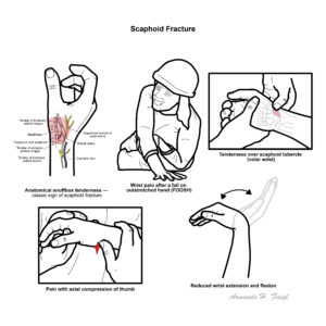
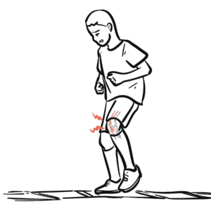
Scaphoid fracture is the most common carpal bone fracture, accounting for ~60–70% of all carpal fractures. It occurs most frequently in young adults (15–40 years) following a fall on the outstretched hand (FOOSH). The scaphoid’s tenuous blood supply predisposes to non-union and avascular necrosis, especially in proximal fractures. Early recognition is essential as delayed or missed diagnosis is common and may lead to long-term disability.
Definition
FOOSH: “Fall On Outstretched Hand,” common mechanism of wrist injuries. Anatomical snuffbox: Depression between tendons of extensor pollicis longus and brevis; tenderness here is pathognomonic for scaphoid fracture. Avascular necrosis (AVN): Bone death due to disruption of blood supply. A common site for avascular necrosis is to the scaphoid bone as well as the hip. Non-union: Failure of bone fragments to heal within expected timeframe.
Anatomy & Physiology
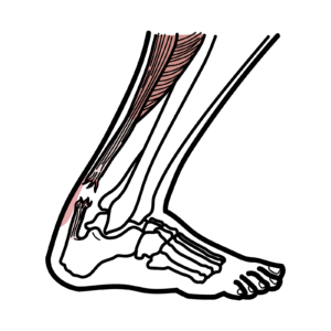
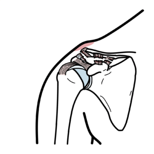
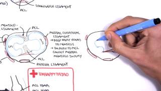
Scaphoid: Boat-shaped carpal bone on radial side of wrist; articulates with radius, trapezium, trapezoid, capitate, lunate.
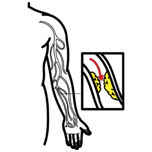
Blood supply: Retrograde from dorsal carpal branch of radial artery → enters distally → supplies proximal pole last.
Clinical relevance: Proximal fractures at highest risk of AVN due to poor vascularity.
Proximal pole = poor healing → high risk AVN.
Aetiology and Risk Factors
Aetiology
Direct trauma: FOOSH (most common).
Less common: Punching injuries, direct blow to wrist.
Risk Factors
Young, active adults (sports injuries, falls).
Males > females.
High-energy trauma (e.g., motor vehicle accidents).
Pathophysiology
FOOSH → axial load transmitted through radial styloid.
Scaphoid bone compressed against distal radius.
Fracture occurs, most often at the waist (70%), less at proximal pole (20%) or distal pole (10%).
Retrograde blood supply disrupted → risk of delayed healing or AVN (especially proximal).
Mechanism + vascular anatomy explains why scaphoid fracture is high risk despite being a “small bone.”
Clinical Manifestations
Wrist pain after FOOSH.
Clinical examination
Anatomical snuffbox tenderness (classic).
Tenderness over scaphoid tubercle (volar wrist).
Pain with axial compression of thumb.
↓ wrist motion, swelling may be minimal.
Often subtle → patients may continue to use wrist.
Snuffbox tenderness = scaphoid fracture until proven otherwise.
Diagnosis
Clinical suspicion: FOOSH + snuffbox tenderness.
X-ray (wrist, scaphoid views): May be normal initially (up to 30% missed).
Repeat imaging: X-ray after 10–14 days if initial negative.
MRI (gold standard): Detects occult fractures within 24 hours.
CT scan: Best for assessing union and fracture displacement.
Differential Diagnosis
Condition
Differentiating Features
Distal radius fracture
More swelling/deformity, visible on plain films
Wrist sprain
No bony tenderness, normal MRI
Scapholunate ligament injury
Pain on Watson’s test, widening on imaging
Always immobilise if suspicion is high, even with negative initial X-ray.
Classification
Distal pole (~10%) – good healing, low risk AVN.
Waist (~70%) – most common.
Proximal pole (~20%) – poor healing, high risk AVN.
Treatment
Non-displaced distal/waist fractures: Immobilisation in thumb spica cast for 6–12 weeks.
Proximal or displaced fractures: Surgical fixation (percutaneous screw or open reduction).
Follow-up imaging: CT/MRI for union assessment.
Rehabilitation: Gradual mobilisation once union confirmed.
Ibrahim T, Qureshi A, Sutton AJ, et al. Surgical vs non-surgical treatment of acute scaphoid fractures: systematic review and meta-analysis. Cochrane Database Syst Rev. 2008;(4):CD006770.
Buijze GA, Ochtman L, Ring D. Management of scaphoid fractures in adults: what’s new? Injury. 2010;41(11):1120–8.
Discussion