Armando Hasudungan site title and tagline

X-linked Agammaglobulinaemia (XLA / Bruton’s Disease)

Overview

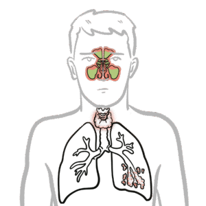
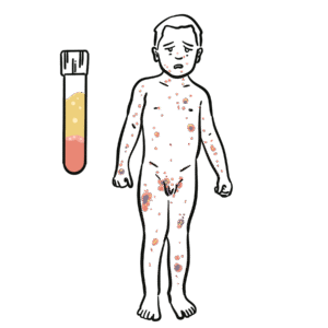
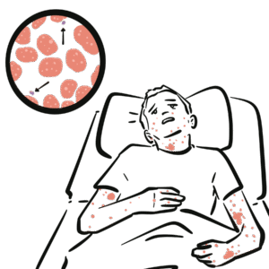
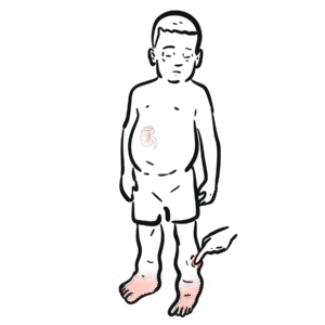
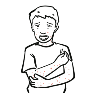
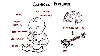
X-linked agammaglobulinaemia (XLA) is a rare primary immunodeficiency caused by mutations in the Bruton’s tyrosine kinase (BTK) gene, leading to failure of B-cell maturation. It results in profound hypogammaglobulinaemia and susceptibility to recurrent bacterial infections, particularly with encapsulated organisms. It usually presents after 6 months of age (once maternal IgG wanes). Prevalence is ~1 in 200,000 live male births.

Triad “Bruton’s = Boys, B-cells, Bacteria.”

Definition

BTK (Bruton’s tyrosine kinase): Enzyme critical for B-cell maturation.
Hypogammaglobulinaemia: Low levels of immunoglobulins (IgG, IgA, IgM).
Encapsulated bacteria: Bacteria such as S. pneumoniae, H. influenzae that require opsonising antibodies for clearance.
Opsonisation: Process where antibodies coat bacteria to promote phagocytosis.

Anatomy & Physiology 

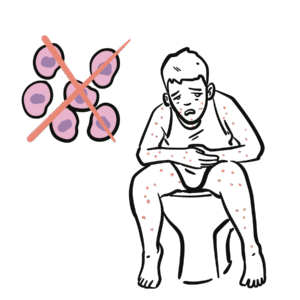
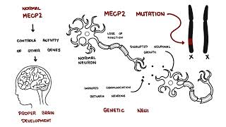
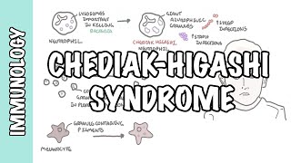
  • Normal B-cell development: Pro-B cell → pre-B cell (BTK required) → immature B cell → plasma cell producing antibodies.
  • In XLA, mutation in BTK halts differentiation at the pre-B stage, leading to:
    • Absent circulating B cells.
    • Absent plasma cells.
  • Absent immunoglobulins (IgG, IgA, IgM, IgE).

“XLA = no B cells, no antibodies.”

Aetiology & Risk Factors

Aetiology

  • Genetics: Mutations in BTK gene (Xq21.3–Xq22).
  • Inheritance: X-linked recessive → almost exclusively affects boys.

Risk Factors

  • Family history of affected males.
  • Carrier mothers (asymptomatic).

Pathophysiology

  1. BTK mutation → defective pre-B to mature B-cell transition.
  2. Absent mature B cells and plasma cells.
  3. Profound hypogammaglobulinaemia.
  4. No antibody-mediated opsonisation → impaired clearance of encapsulated bacteria.
  5. Recurrent bacterial infections from ~6 months of age (loss of maternal IgG).

Timing is key – healthy until 6 months, then recurrent infections.

Clinical Manifestations

  • Onset after 6 months of age.
  • Recurrent bacterial infections:
    • Otitis media, sinusitis, pneumonia.
    • Septicaemia, meningitis.
    • Organisms: Streptococcus pneumoniae, Haemophilus influenzae, Enteroviruses (esp. polio, echovirus).
  • GI infections: Chronic diarrhoea from Giardia lamblia.
  • Absent lymphoid tissue: Very small or absent tonsils, adenoids, lymph nodes.
  • Chronic lung disease: Bronchiectasis if untreated.

“Boy with recurrent bacterial infections + absent tonsils” = XLA.

Diagnosis

  • CBC: Normal lymphocyte count.
  • Immunoglobulins: Profound ↓ IgG, IgA, IgM.
  • Flow cytometry: Absent/very low CD19+ B cells.
  • Genetic testing: BTK mutation.
  • Physical exam: Absent tonsils/lymph nodes.

Differential Diagnosis

ConditionDifferentiating Feature
CVIDLater onset, some B cells present, not complete absence
Selective IgA deficiencyOnly IgA absent, often asymptomatic
SCIDSevere infections earlier (<3 months), T- and B-cell defects
Secondary hypogammaglobulinaemiaDrug-induced (rituximab, steroids), protein loss

Treatment

  • Lifelong IVIG or SCIG replacement therapy.
  • Prompt antibiotic therapy for infections.
  • Prophylactic antibiotics may be used.
  • Avoid live vaccines (esp. oral polio)
  • Aggressive management of chronic lung disease (bronchiectasis).

Treatment is supportive (antibodies supplied), no curative therapy currently.

Complications & Prognosis

  • Recurrent severe infections → bronchiectasis, chronic lung disease.
  • Enteroviral meningoencephalitis.
  • Arthritis (from Mycoplasma or Ureaplasma).
  • Prognosis: excellent with immunoglobulin replacement and infection prevention.

Without treatment, most die in childhood; with Ig replacement, can live near-normal lifespan.

References

  1. Bruton OC. Agammaglobulinemia. Pediatrics. 1952;9(6):722–728.
  2. Conley ME, Dobbs AK, Quintana AM, et al. Genetic basis of X-linked agammaglobulinemia. N Engl J Med. 2009;360(7):676–81.
  3. Winkelstein JA, Marino MC, Lederman HM, et al. X-linked agammaglobulinemia: report on a United States registry of 201 patients. Medicine (Baltimore). 2006;85(4):193–202.
  4. Ochs HD, Smith CIE. X-linked agammaglobulinemia: a model primary immunodeficiency. J Allergy Clin Immunol. 1996;98(4):687–99.
  5. Picard C, Al-Herz W, Bousfiha A, et al. Primary immunodeficiency diseases: 2015 IUIS classification. J Clin Immunol. 2015;35(8):696–726.

Related Content

Discussion

0 Comments
Most Voted
Newest Oldest
Inline Feedbacks
View all comments

Table Of Contents

Become a member
Ready to take your education seriously?
Armando hasudungan brain logo
Armando Hasudungan
By Visualising Medicine
© 2026 Visualising Medicine. All rights reserved.
Become a member to access note taking
Orangise your medical learning
This is just one of the many AH community member perks
Become a member to access quizzes
Strengthen your medical knowledge
This is just one of the many AH community member perks
0
Would love your thoughts, please comment.x
()
x