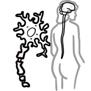
0:00 Hello, in this video, we're going to look at the light reflex. 0:09 So essentially, the mechanism as to how the pupils constrict when you shine a 0:15 light to 0:15 the eyeball. 0:17 So in order to do so, let's just recap the object nerve pathway, so the visual 0:22 pathway. 0:23 So the eyeball here, the optic nerve, optic chiasm and the optic tract, the occ 0:28 ipital lobe. 0:29 But here I'm drawing the midbrain in the center because it is important to know 0:32 the light reflex. 0:35 The midbrain contains many structures, anatomical structures and nuclei that 0:38 are important to 0:39 the light reflex. 0:41 From the back here, the back of the midbrain, we have the superior colicum, and 0:49 then we 0:49 have the pretectal nucleus, in front of the pretectal nucleus, you have the 0:54 nucleus of 0:55 Erenge Westphile. 0:57 Now the nucleus of Erenge Westphile is important because it is where the oculom 1:03 otor nerve, cranial 1:04 nerve number three, it's where the oculomotor nerve originates. 1:09 Oculomotor nerve will travel along this pathway here, so oculomotor nerve here. 1:14 Now it's also important to know that the oculomotor nerve contains the parasymp 1:19 athetic 1:19 nerve fibers. 1:20 So the parasympathetic nerve fibers, some of them travel along with the oculom 1:25 otor nerve. 1:26 And the oculomotor nerve will travel towards the ciliary ganglion where it will 1:29 synapse 1:30 with another neuron and this other neuron is then going to send this whatever 1:35 signal to 1:36 the pupil. 1:39 Alright, so let's zoom into the eyeball here and look at some important 1:45 anatomical structures. 1:46 Let's cut a cross section of the right eyeball here. 1:50 Here we have the ciliary muscles. 1:51 Now the ciliary muscles is important because it essentially changes the shape 1:56 of our lens 1:57 and thus how much light is coming into our eye. 2:00 The iris is also very important. 2:03 Now the iris is the color in our eye and the color that makes up our eye and 2:09 the iris is 2:09 important because it contains muscles and it's actually the iris that is 2:14 responsible for how 2:14 big or how small our pupils are. 2:18 The iris contains circular or radial muscles and now depending on what muscles 2:22 are stimulated 2:23 if it's circular or radial this will determine if the pupils are constricting 2:28 or dilating. 2:29 So here in this case I'm drawing a pupil, I'm drawing the eyeball and the 2:33 pupils are constricting 2:35 because the circular muscles here in red are constricting. 2:40 So let's just get a better understanding of this. 2:42 So I'm going to draw three eyeballs here. 2:45 The middle one is normal. 2:48 On the left we have constricted pupils. 2:51 This is scientifically known as myosis. 2:57 And the pupils are constricting because actually the parasympathetic nerve 3:03 fibers are stimulating 3:05 it, are stimulating the circular muscles of the iris and the parasympathetic 3:10 nerve fibers 3:11 are coming together with the oculomotor nerve. 3:14 It's traveling with the oculomotor nerve, remember? 3:18 So parasympathetic causes pupils to constrict. 3:23 Then on the right we have dilated pupils. 3:26 Now dilated pupils scientifically known as myidiiasis and essentially it's the 3:34 sympathetic 3:35 nerve that is responsible for dilation of the pupils, so stimulating the radial 3:41 muscles. 3:41 So the radial muscles are stimulated, this will cause dilation of the pupils 3:48 and this 3:49 is due to the sympathetic nerve stimulation. 3:53 And we want to dilate our pupils because when we are in fight-flight mode, when 3:57 we're 3:58 trying to run away, we want to see everything clearly and that sort of makes 4:04 sense. 4:04 Alright, now when we shine a light, when we shine a light to the left eyeball 4:10 here, I'm 4:10 drawing, when we shine the light this is known as direct response and then 4:19 whatever happens 4:21 to the other eyeball is known as consensual response. 4:25 So when we shine a light to the left eyeball, the pupils on the left would 4:31 constrict, but 4:32 so will the pupils on the right side and this is known as the consensual 4:38 response. 4:38 Okay, now let's follow the light reflex pathway now. 4:44 So we're shining a light in the left eye. 4:47 When we shine a light in the left eye, the optic nerve will obviously pick this 4:51 up. 4:51 So here I'm drawing in blue, optic part of the optic nerve. 4:56 This one, this optic nerve will carry information down along the left side and 4:59 it will synapse 5:00 with another, other nerves in the lateral geniculate body and these nerves will 5:06 carry along the 5:07 optic tract and down to the opposite of the lobe. 5:11 Similarly, the other, this orange nerve here is part of the optic nerve, but 5:17 this on this 5:18 side it will actually cross over in the optic chiasm and will bring information 5:23 to the 5:24 other side, so to the right side and it will synapse with the nerves along the 5:28 right optic 5:29 tract and this nerve will then carry information to the occipital lobe. 5:34 Now what's really important to know is that the optic nerve carrying 5:38 information from 5:39 the left eye, they will actually also send, bring some information and will syn 5:47 apse in 5:48 the midbrain, it will bring the information to the pre-tectal nucleus here as 5:56 shown. 5:57 Now there are neurons in the pre-tectal nucleus that will receive this signal. 6:04 So this neuron in the pre-tectal nucleus in red will then bring this 6:08 information to the 6:09 nucleus of Edinger Westphal, but this neuron will not only bring this 6:15 information to one 6:16 side, it will actually cross over as well and bring the information to the 6:20 other side, 6:21 to the opposite Edinger Westphal nucleus. 6:25 And so, and similar thing happens on the right pre-tectal nucleus, it will 6:31 bring it to the 6:31 same side of the Edinger Westphal nucleus, but it will also bring the 6:35 information to the 6:36 other side, it will cross over. 6:38 And so essentially both right and left nucleus of Edinger Westphal are being 6:44 stimulated just 6:47 from the one eyeball, from the left eyeball. 6:53 And the nucleus of Edinger Westphal is stimulated, the neurons there are 6:59 stimulated, particularly 7:00 the ocular motor nerve, because the ocular motor nerve originates from that 7:06 nucleus. 7:06 The ocular motor nerve, both in the right and left, will synapse in the ciliary 7:10 ganglion 7:10 with another neuron, the short ciliary nerve. 7:15 The short ciliary nerve will then bring this information to the pupil, to the 7:21 iris, causing 7:22 the iris to constrict, essentially. 7:27 And so, you get pupil constriction. 7:30 And that is why, when you shine a light, to the left eye, the left eye will 7:36 constrict, 7:37 the left pupil will constrict, but also the right will constrict. 7:41 And thus you have a direct and consensual response. 7:48 So again, just to recap, here we have the left eyeball, and here we have the 7:52 right eyeball, 7:53 or the right pupil. 7:55 And we're shining a light to the left eye, how we get a direct response, so the 7:59 pupils 8:00 are constricting, and then we get a consensual response on the right eye. 8:04 And this is when the light reflects pathway is normal, there are no lesions. 8:09 Now, let's just recap the pathway again. 8:13 So from the retina, the optic nerve brings this information to the predactyl 8:17 nucleus 8:17 in the midbrain, and then you have bilateral innervation. 8:20 So we have crossing over, et cetera, to the Erenga-Westphal nucleus. 8:25 The Erenga-Westphal nucleus will then send out, stimulate, oculomotor nerve 8:30 will be stimulated, 8:31 and this will, the oculomotor nerve will then travel to the ciliary ganglion, 8:35 where it will 8:35 sign up to the short ciliary nerve to stimulate the circular muscles of the ir 8:40 is, thus resulting 8:41 in pupillary constriction. 8:47 So now let's see what happens when we have lesions anywhere along this light 8:51 reflex pathway. 8:52 Say, let's just say, what happens if we have a lesion on the left optic nerve? 8:59 Well, what happens is that when we shine a light on the left eye, because no 9:05 information 9:05 is being received, we will get no pupillary response on the left side. 9:13 And also we will get no pupillary constriction on the right side as well. 9:18 So we don't have, so both the right and left side don't constrict. 9:26 And this is because we have damage to the left optic nerve, cranial nerve 9:31 number two, 9:32 and thus no information is being sent towards the midbrain. 9:37 And thus the light reflex is absent. 9:40 Now what happens if you have a damage to the optic chiasm? 9:44 Well, when we shine light on the left eye, the signal is still being sent along 9:52 the blue 9:53 nerve, because the orange nerve is damaged, the blue optic nerve, and it will 9:58 still bring 9:59 it to the pre-tectal nucleus and it will still cross over. 10:02 And so both the left and right nucleus of Inengar Westfall are working. 10:08 To damage to the optic chiasm, the light reflex will still be present. 10:12 So you still see constriction of both the left and right. 10:16 Now what happens if you have a lesion on the left oculomotor nerve? 10:23 Well, when you shine light on the left eye, what happens is that the 10:29 information is still 10:32 being received by the midbrain, by the nucleus of Inengar Westfall, but the 10:37 output, the oculomotor 10:39 nerve on the left side is not working, so therefore you won't see any const 10:42 riction on 10:42 the left side. 10:44 And so damage to the left oculomotor nerve will result in the direct light 10:49 reflex being 10:50 absent, but the consensual being present. 10:54 And so you get no constriction on the left side, but you have constriction on 10:58 the right 10:58 side, because the left side, the oculomotor nerve, is damaged, but the right o 11:04 culomotor 11:04 nerve is still working. 11:07 Now what happens if you have a lesion on the right oculomotor nerve? 11:10 Well, it's the same thing. 11:12 So you shine a light on the left eye, the image, the information is still being 11:16 received 11:16 by the midbrain, by the Inengar Westfall nucleus. 11:19 And the oculomotor nerve is sending out the signal for the pupils to constrict. 11:25 However, the right oculomotor nerve is not working, and so it can't tell the 11:29 right pupil 11:30 to constrict. 11:31 Thus, you have a, thus, damage to the right oculomotor nerve will result in 11:38 direct response 11:39 on the left side present, but consensual will be absent. 11:45 I hope this video made sense, thank you for watching, thank you.