

Pulmonary Haemorrhage
Overview
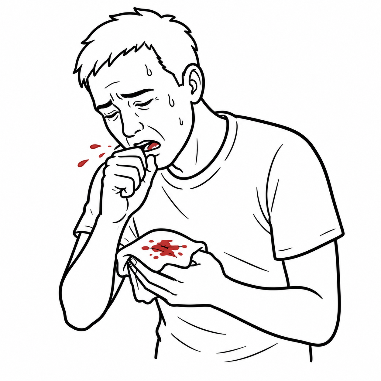
Pulmonary haemorrhage describes bleeding into the lower respiratory tract. It may arise from a focal airway lesion or occur diffusely within the alveoli. Diffuse alveolar haemorrhage (DAH) is an acute pulmonary syndrome caused by widespread bleeding from the pulmonary microcirculation into the alveolar spaces. It can rapidly impair gas exchange and progress to life-threatening hypoxaemic respiratory failure.1,2
DAH should be suspected when new bilateral pulmonary opacities occur with otherwise unexplained hypoxaemia, haemoptysis or a falling haemoglobin concentration. Haemoptysis is an important clue but may be absent, particularly when blood remains within the distal airspaces.1
DAH is not a single disease. It has immune-mediated, haematological, cardiac, infectious, toxic and drug-related causes. Identifying the underlying cause is essential because treatments differ substantially; immunosuppression may be lifesaving in pulmonary capillaritis but harmful when haemorrhage is driven by infection or coagulopathy.1,2
Definition
- Pulmonary haemorrhage
- Bleeding into the lower respiratory tract, arising from the airways or lung parenchyma.
- Diffuse alveolar haemorrhage
- Widespread bleeding from the pulmonary microcirculation into the alveolar spaces.
- Haemoptysis
- Expectoration of blood originating below the vocal cords.
- Pulmonary capillaritis
- Neutrophilic inflammation and destruction of the alveolar capillary walls, causing leakage of blood into the alveoli.
- Pulmonary–renal syndrome
- Concurrent alveolar haemorrhage and glomerulonephritis, most often associated with ANCA-associated vasculitis or anti-GBM disease.
Anatomy & Physiology
The alveolar–capillary barrier consists of alveolar epithelium, a thin interstitium and pulmonary capillary endothelium. Its small diffusion distance permits efficient oxygen and carbon dioxide exchange while normally preventing blood cells from entering the airspaces.
DAH results from disruption or increased permeability of this barrier. Blood entering multiple alveoli occupies the space normally available for ventilation, producing ventilation–perfusion mismatch, impaired diffusion and hypoxaemia.
The lungs have two circulations. The low-pressure pulmonary circulation supplies the alveolar capillary network, whereas the higher-pressure bronchial circulation supplies the conducting airways. DAH usually originates from the pulmonary microcirculation, while major focal haemoptysis more commonly arises from the bronchial circulation.1
Aetiology & Risk Factors
Aetiology
Causes can be organised by the process damaging the alveolar microcirculation.1,2
| Cause group | Important examples |
|---|---|
| Immune-mediated pulmonary capillaritis | Granulomatosis with polyangiitis, microscopic polyangiitis, anti-glomerular basement membrane (anti-GBM) disease, systemic lupus erythematosus and other connective tissue diseases |
| Non-immune or bland haemorrhage | Anticoagulants, thrombocytopenia, disseminated intravascular coagulation, mitral valve disease, pulmonary venous hypertension and idiopathic pulmonary haemosiderosis |
| Infection | Severe bacterial, viral or fungal infection; risk depends on immune status and exposure |
| Drugs and inhalational injury | Anticoagulants, certain immune-modifying or cytotoxic medicines, cocaine and other inhaled toxins |
| Diffuse lung injury | Acute respiratory distress syndrome, severe inhalational injury, transplantation and haematopoietic stem-cell transplantation |
| Focal pulmonary bleeding | Malignancy, bronchiectasis, tuberculosis, pulmonary embolism with infarction, vascular malformation or traumatic airway injury |
Risk Factors
Risk is increased by:
- Known systemic vasculitis, anti-GBM disease or connective tissue disease
- Active glomerulonephritis or a previous pulmonary–renal syndrome
- Anticoagulant, antiplatelet or thrombolytic therapy
- Thrombocytopenia or an inherited or acquired coagulation disorder
- Severe infection or immunosuppression
- Recent transplantation
- Exposure to implicated medicines, cocaine, vaping products or inhaled toxins
- Significant mitral valve disease or pulmonary venous hypertension
- Previous or recurrent DAH
Pathophysiology
Three major histopathological patterns are recognised: pulmonary capillaritis, bland alveolar haemorrhage and diffuse alveolar damage.1,2
| Pattern | Pathological process | Typical associations |
|---|---|---|
| Pulmonary capillaritis | Neutrophilic inflammation damages capillary walls, allowing erythrocytes and plasma to enter the alveoli | ANCA-associated vasculitis, anti-GBM disease, systemic lupus erythematosus and other immune-mediated disorders |
| Bland alveolar haemorrhage | Alveolar bleeding occurs without prominent inflammation or destruction of the capillary wall | Coagulopathy, anticoagulants, thrombocytopenia, pulmonary venous hypertension and some immune-mediated disorders |
| Diffuse alveolar damage with haemorrhage | Widespread epithelial and endothelial injury produces oedema, hyaline membranes and variable bleeding | Acute respiratory distress syndrome, severe infection, toxic injury and transplantation |
Once erythrocytes enter the alveoli, they interfere with gas exchange and trigger an inflammatory response. Alveolar macrophages ingest erythrocytes and convert haemoglobin iron into haemosiderin. Recurrent episodes may cause iron-deficiency anaemia, pulmonary haemosiderosis and, in some patients, fibrosis.1,2
DAH causes hypoxaemia through both alveolar filling and injury to the alveolar–capillary membrane. This explains why the physiological impairment may be severe even when little or no blood is coughed up.
Clinical Manifestations
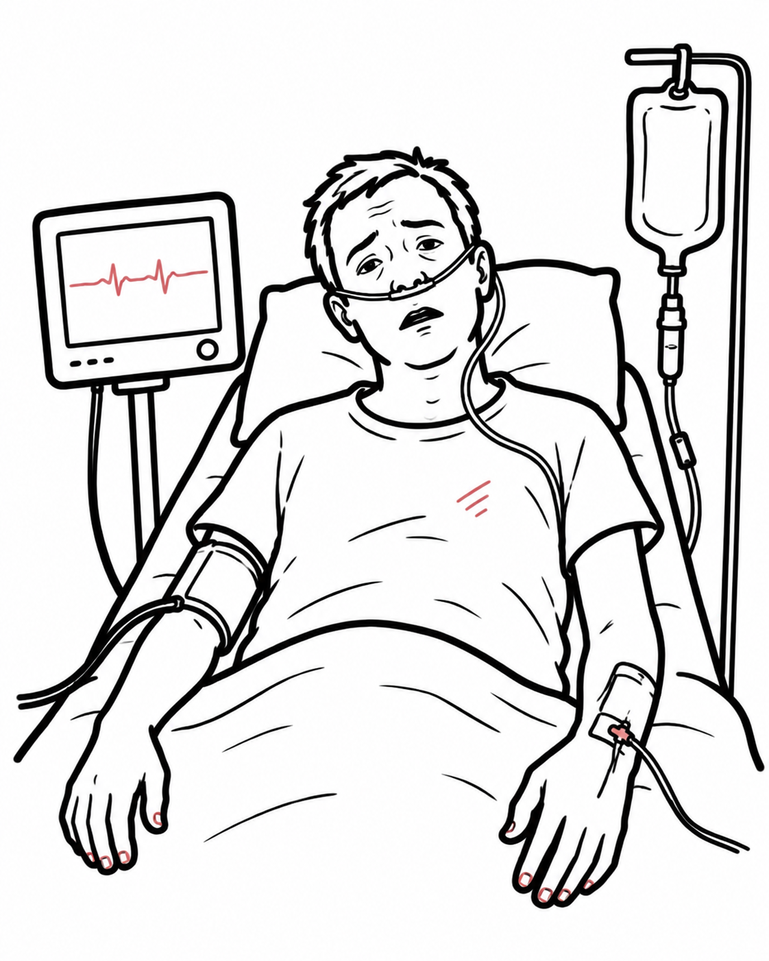
The presentation ranges from mild or recurrent bleeding to fulminant respiratory failure. Common features include:
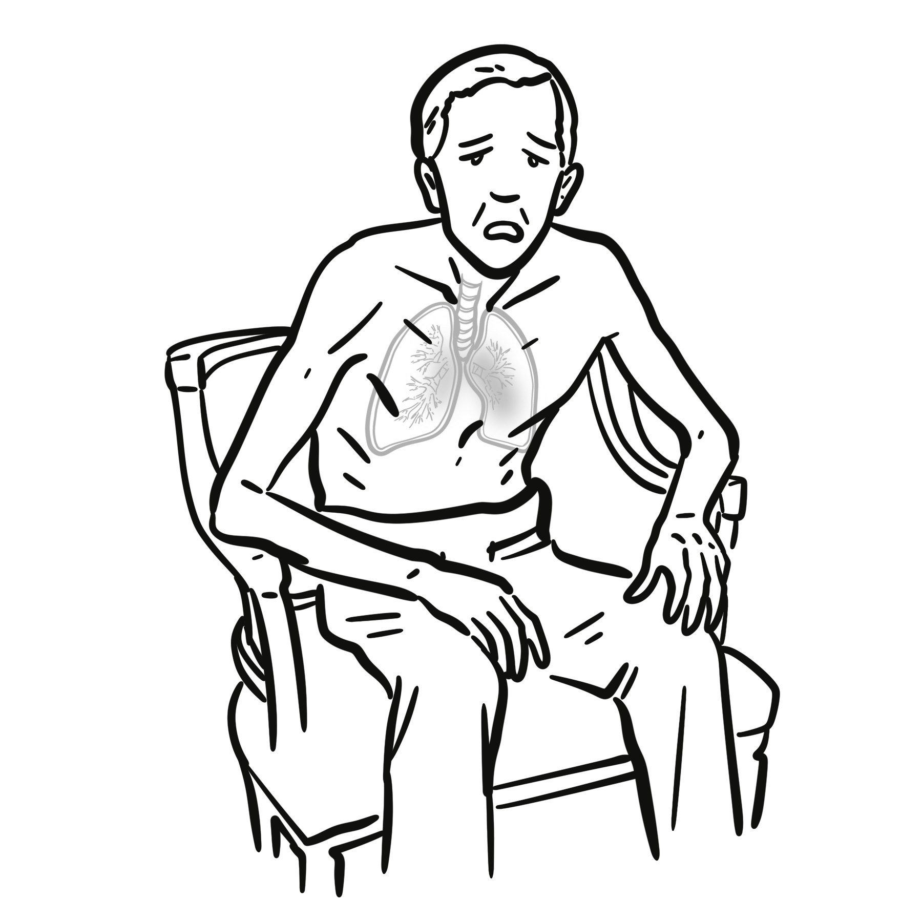
- Acute or subacute dyspnoea
- Cough
- Haemoptysis
- Hypoxaemia
- Falling haemoglobin or otherwise unexplained anaemia
- Fever or systemic inflammatory symptoms
- Tachypnoea and increased work of breathing
- Diffuse inspiratory crackles
- New bilateral airspace opacities on chest imaging
Features suggesting an underlying systemic cause include:
- Haematuria, proteinuria or acute kidney injury
- Chronic sinusitis, nasal crusting or epistaxis
- Purpura, livedo or other vasculitic skin lesions
- Inflammatory arthritis
- Oral ulcers, photosensitive rash or other connective tissue disease features
- Peripheral neuropathy
- Cardiac murmur or signs of pulmonary venous congestion
- Recent infection, medicine change or toxic exposure
The absence of haemoptysis does not exclude diffuse alveolar haemorrhage. Suspect it when otherwise unexplained hypoxaemia, bilateral pulmonary opacities and a falling haemoglobin occur together.
Diagnosis
There is no single universally accepted diagnostic criterion for DAH. Diagnosis is based on a compatible clinical and radiological syndrome, demonstration of alveolar bleeding—usually by bronchoscopy—and investigation of the underlying cause. Alternative explanations such as pulmonary oedema, infection and focal airway bleeding must be assessed simultaneously.1–3
Immediate Assessment
Assessment and stabilisation occur in parallel:
- Airway patency, work of breathing and haemodynamic status
- Continuous oxygen saturation monitoring and arterial blood gas when severe
- Full blood count, blood film and serial haemoglobin
- Coagulation studies, fibrinogen and platelet count
- Group and screen or crossmatch when bleeding is significant
- Renal function, electrolytes and liver biochemistry
- Medication review, particularly anticoagulants and antiplatelet agents
Imaging
Chest radiography commonly shows new bilateral alveolar or ground-glass opacities but is not specific. High-resolution computed tomography may demonstrate bilateral ground-glass change, consolidation or a “crazy-paving” appearance. Imaging defines the distribution and severity of disease and may identify an alternative focal source, but it cannot reliably distinguish DAH from infection, pulmonary oedema or acute respiratory distress syndrome.1,2
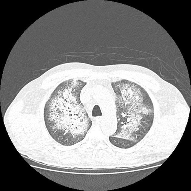
High-resolution chest CT showing bilateral perihilar ground-glass opacity with interlobular septal thickening, producing a “crazy-paving” pattern. Compatible with diffuse alveolar haemorrhage but not specific. Image: Ramos-Andrade et al., 2016, CC BY 4.0.
What to notice
Detailed image description
Source: Ramos-Andrade D, Silva F, Canelas A, Curvo-Semedo L, Caseiro-Alves F. J Belg Soc Radiol. 2016;100(1):38. doi:10.5334/jbr-btr.959. Licence: CC BY 4.0. Retrieved 5 August 2026.
Bronchoscopy
Early bronchoscopy with bronchoalveolar lavage (BAL) is the principal test for demonstrating alveolar bleeding and excluding endobronchial pathology or infection. Persistent or progressively bloodier return from sequential BAL aliquots supports DAH. BAL should also be sent for appropriate bacterial, mycobacterial, fungal and viral testing.1,2
Haemosiderin-laden macrophages support recent or recurrent alveolar bleeding but may not be present immediately after an acute event. Their absence early in the presentation therefore does not reliably exclude DAH.2
The diffusing capacity for carbon monoxide may rise during active alveolar bleeding because intra-alveolar haemoglobin binds carbon monoxide. The test is rarely practical in an acutely breathless patient and is neither sufficiently sensitive nor specific to establish the diagnosis.
Determining the Cause
Investigations are guided by the clinical context and commonly include:
- Urinalysis and urine protein quantification
- Urine microscopy for dysmorphic erythrocytes or red-cell casts
- ANCA with proteinase 3 and myeloperoxidase specificity
- Anti-GBM antibodies
- Antinuclear antibodies, anti-double-stranded DNA antibodies and complement levels
- Antiphospholipid antibodies when clinically indicated
- Blood cultures and targeted microbiological testing
- Echocardiography when mitral valve disease, left ventricular dysfunction or pulmonary venous hypertension is possible
The combination of DAH with haematuria, proteinuria or acute kidney injury should prompt urgent assessment for pulmonary–renal syndrome. Positive serology can strongly support the diagnosis, but treatment of life-threatening disease should not be delayed solely while awaiting results. Kidney biopsy is often safer and more informative than lung biopsy when glomerulonephritis is present.5–7
Bilateral pulmonary opacities plus acute kidney injury should not automatically be attributed to fluid overload. A falling haemoglobin, active urine sediment or systemic vasculitic features should trigger urgent investigation for pulmonary–renal syndrome.
Important Differentials
| Differential | Features favouring the alternative diagnosis |
|---|---|
| Cardiogenic pulmonary oedema | Elevated jugular venous pressure, peripheral oedema, cardiac dysfunction, pleural effusions or rapid improvement with treatment of congestion |
| Pneumonia or acute respiratory distress syndrome | Infectious prodrome, microbiological evidence, focal consolidation or a clear precipitating critical illness |
| Focal endobronchial bleeding | Localised radiological abnormality or a bleeding lesion identified during bronchoscopy |
| Pulmonary embolism with infarction | Pleuritic pain, venous thromboembolism risk factors and a compatible CT pulmonary angiogram |
| Upper-airway bleeding | Epistaxis or blood originating above the vocal cords |
| Gastrointestinal bleeding | Haematemesis, nausea, melaena or an identified gastrointestinal source |
Classification
DAH is most usefully classified by:
- Distribution
- Diffuse alveolar haemorrhage
- Focal pulmonary or endobronchial haemorrhage
- Underlying mechanism
- Immune-mediated capillaritis
- Non-inflammatory or bland haemorrhage
- Diffuse alveolar damage with secondary haemorrhage
- Clinical context
- Pulmonary-limited disease
- Pulmonary–renal syndrome
- Haematological or coagulopathic disease
- Cardiac disease
- Infection, toxic exposure or transplantation
This classification guides the investigation and treatment strategy more effectively than the volume of expectorated blood alone.1,2
Treatment
DAH is a medical emergency. Management requires simultaneous respiratory support, control of bleeding and treatment of the cause.2
Immediate Supportive Management
- Provide supplemental oxygen and escalate respiratory support when required.
- Involve respiratory and intensive-care teams early when hypoxaemia is severe or worsening.
- Establish intravenous access and monitor haemoglobin, coagulation and haemodynamics.
- Stop suspected causative medicines where clinically appropriate.
- Reverse anticoagulation and correct severe thrombocytopenia, hypofibrinogenaemia or other coagulopathy using cause-specific protocols.
- Obtain microbiological samples and treat suspected serious infection promptly.
- Avoid delays in cause-specific treatment when immune-mediated DAH is strongly suspected and life-threatening.
Cause-Specific Treatment
| Cause | Treatment principles |
|---|---|
| ANCA-associated vasculitis | Glucocorticoids with rituximab or cyclophosphamide for remission induction. The 2021 ACR/Vasculitis Foundation guideline conditionally recommends against routinely adding plasma exchange solely for alveolar haemorrhage.5 |
| Hypoxaemic ANCA-associated DAH | KDIGO 2024 states that plasma exchange may be considered when diffuse alveolar bleeding causes hypoxaemia, acknowledging high early mortality and continuing uncertainty about benefit.6 |
| Anti-GBM disease | Urgent glucocorticoids, cyclophosphamide and plasma exchange are standard initial therapy unless treatment is considered futile because of irreversible disease and there is no pulmonary haemorrhage. Plasma exchange is generally continued until circulating anti-GBM antibodies are no longer detectable.7 |
| ANCA and anti-GBM overlap | Manage with an anti-GBM strategy that includes plasma exchange.6,7 |
| Systemic lupus erythematosus or another connective tissue disease | High-dose glucocorticoids and additional specialist-directed immunosuppression; exclude or concurrently treat infection when clinically suspected. Evidence is predominantly observational.2 |
| Coagulopathy or medicine-related bleeding | Remove the precipitant, reverse anticoagulation when indicated and correct the haemostatic abnormality. Immunosuppression is not routinely indicated unless there is a separate inflammatory cause. |
| Infection-associated haemorrhage | Targeted antimicrobial therapy and supportive care. Uncritical immunosuppression may worsen the underlying infection. |
| Cardiac or pulmonary venous hypertension | Treat the cardiac lesion and pulmonary venous congestion. |
| Idiopathic pulmonary haemosiderosis | Specialist-directed immunosuppression after exclusion of immune, infectious, cardiac and haemostatic causes. |
The role of plasma exchange in ANCA-associated DAH remains uncertain. In the DAH subgroup of PEXIVAS, plasma exchange did not have a statistically clear effect on mortality, although the confidence intervals could not exclude benefit or harm.3 Consequently, current guidelines differ: ACR advises against its routine use for alveolar haemorrhage, whereas KDIGO permits consideration in hypoxaemic DAH.5,6
Plasma exchange is cause-specific. It is standard initial therapy for anti-GBM disease, but it is not routinely recommended for every patient with ANCA-associated alveolar haemorrhage; hypoxaemia, kidney involvement, anti-GBM overlap and guideline differences must be considered.
Complications & Prognosis
Complications
Potential complications include:
- Acute hypoxaemic respiratory failure
- Acute respiratory distress syndrome
- Requirement for mechanical ventilation or extracorporeal support
- Severe anaemia and transfusion requirement
- Recurrent alveolar haemorrhage
- Pulmonary haemosiderosis and fibrosis after repeated episodes
- Acute kidney injury or kidney failure in pulmonary–renal syndromes
- Secondary infection related to critical illness or immunosuppression
- Treatment-related cytopenia, haemorrhage, thrombosis or infection
- Death
Prognosis
Prognosis depends on the cause, severity of hypoxaemia, extent of extrapulmonary organ involvement, speed of diagnosis and response to treatment. Recurrent or untreated bleeding increases the risks of chronic anaemia and fibrotic lung damage.1,2
DAH associated with ANCA-associated vasculitis carries substantial early risk. In the PEXIVAS cohort, 12.0% of participants with DAH died within one year, compared with 6.6% of participants without DAH; the study population had severe ANCA-associated vasculitis, so these figures should not be generalised to every cause of pulmonary haemorrhage.3
References
- Park MS. Diffuse alveolar hemorrhage. Tuberc Respir Dis (Seoul). 2013;74(4):151–162. doi:10.4046/trd.2013.74.4.151
- Park JA. Treatment of diffuse alveolar hemorrhage: controlling inflammation and obtaining rapid and effective hemostasis. Int J Mol Sci. 2021;22(2):793. doi:10.3390/ijms22020793
- Fussner LA, Flores-Suárez LF, Cartin-Ceba R, Specks U, Cox PG, Jayne DRW, et al. Alveolar hemorrhage in antineutrophil cytoplasmic antibody–associated vasculitis: results of an international randomized controlled trial (PEXIVAS). Am J Respir Crit Care Med. 2024;209(9):1141–1151. doi:10.1164/rccm.202308-1426OC
- Ramos-Andrade D, Silva F, Canelas A, Curvo-Semedo L, Caseiro-Alves F. Diffuse alveolar hemorrhage due to malignant arterial hypertension—an unusual manifestation of a common disease. J Belg Soc Radiol. 2016;100(1):38. doi:10.5334/jbr-btr.959
- Chung SA, Langford CA, Maz M, Abril A, Gorelik M, Guyatt G, et al. 2021 American College of Rheumatology/Vasculitis Foundation guideline for the management of antineutrophil cytoplasmic antibody–associated vasculitis. Arthritis Care Res (Hoboken). 2021;73(8):1088–1105. doi:10.1002/acr.24634
- Kidney Disease: Improving Global Outcomes (KDIGO) ANCA-Associated Vasculitis Work Group. KDIGO 2024 clinical practice guideline for the management of antineutrophil cytoplasmic antibody–associated vasculitis. Kidney Int. 2024;105(3 Suppl)–S116. Available from: https://kdigo.org/wp-content/uploads/2024/02/KDIGO-2024-ANCA-Vasculitis-Guideline.pdf
- Kidney Disease: Improving Global Outcomes (KDIGO) Glomerular Diseases Work Group. KDIGO 2021 clinical practice guideline for the management of glomerular diseases. Kidney Int. 2021;100(4 Suppl)–S276. doi:10.1016/j.kint.2021.05.021









Members only discussions coming soon…