

Knee Pain
Overview
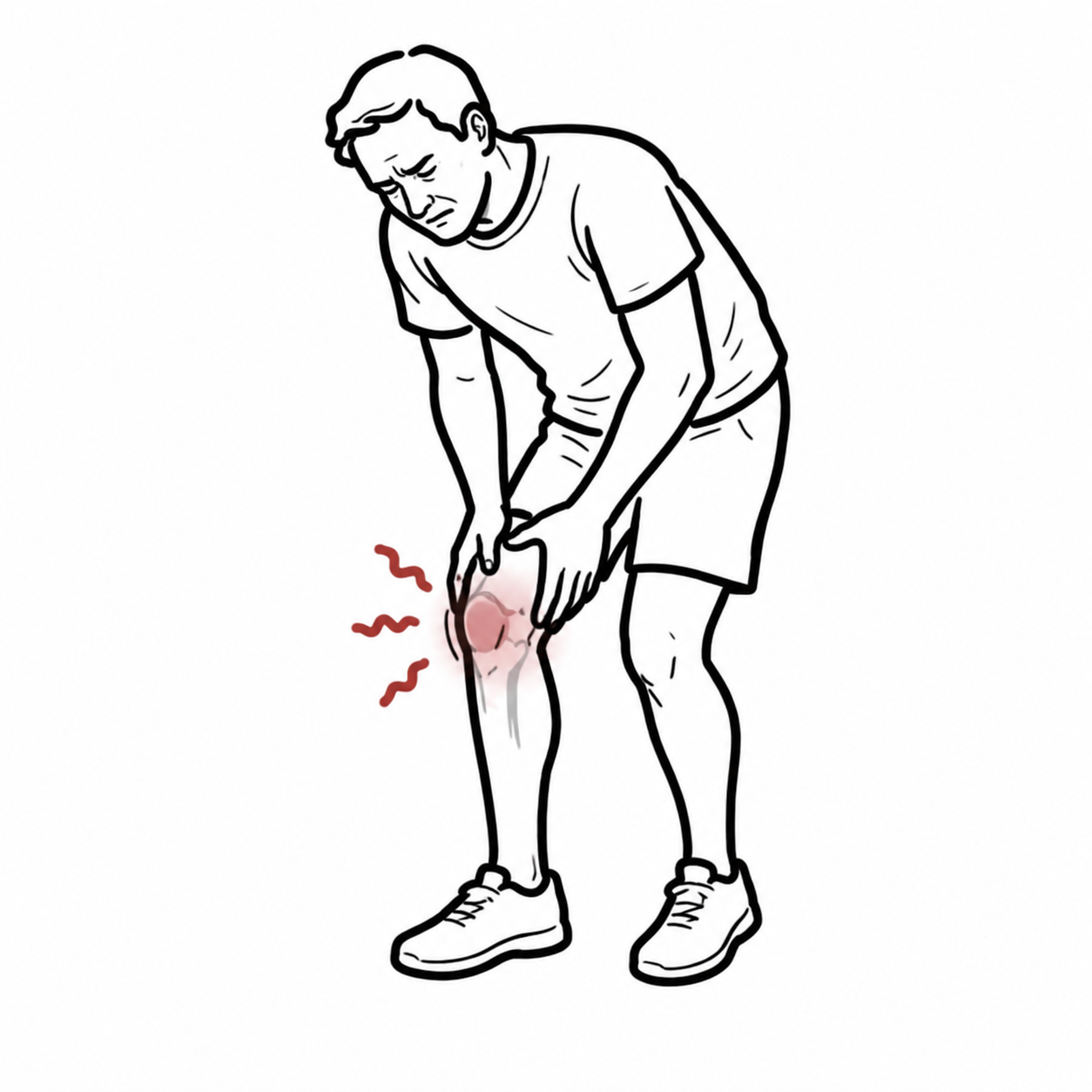
Acute or chronic pain originating from the knee joint or surrounding periarticular soft tissue structures (ligaments, menisci, tendons, bursae).
Differentiate intra-articular vs. extra-articular disease, assess for mechanical instability, and immediately rule out acute surgical or infectious emergencies (Septic Arthritis, Acute Hemarthrosis, Neurovascular Compromise).
In any acute monoarthritis or hot, swollen joint, Septic Arthritis is the primary can’t-miss emergency. Perform joint aspiration (arthrocentesis) for synovial fluid analysis before starting antibiotics, unless the patient is in frank septic shock.
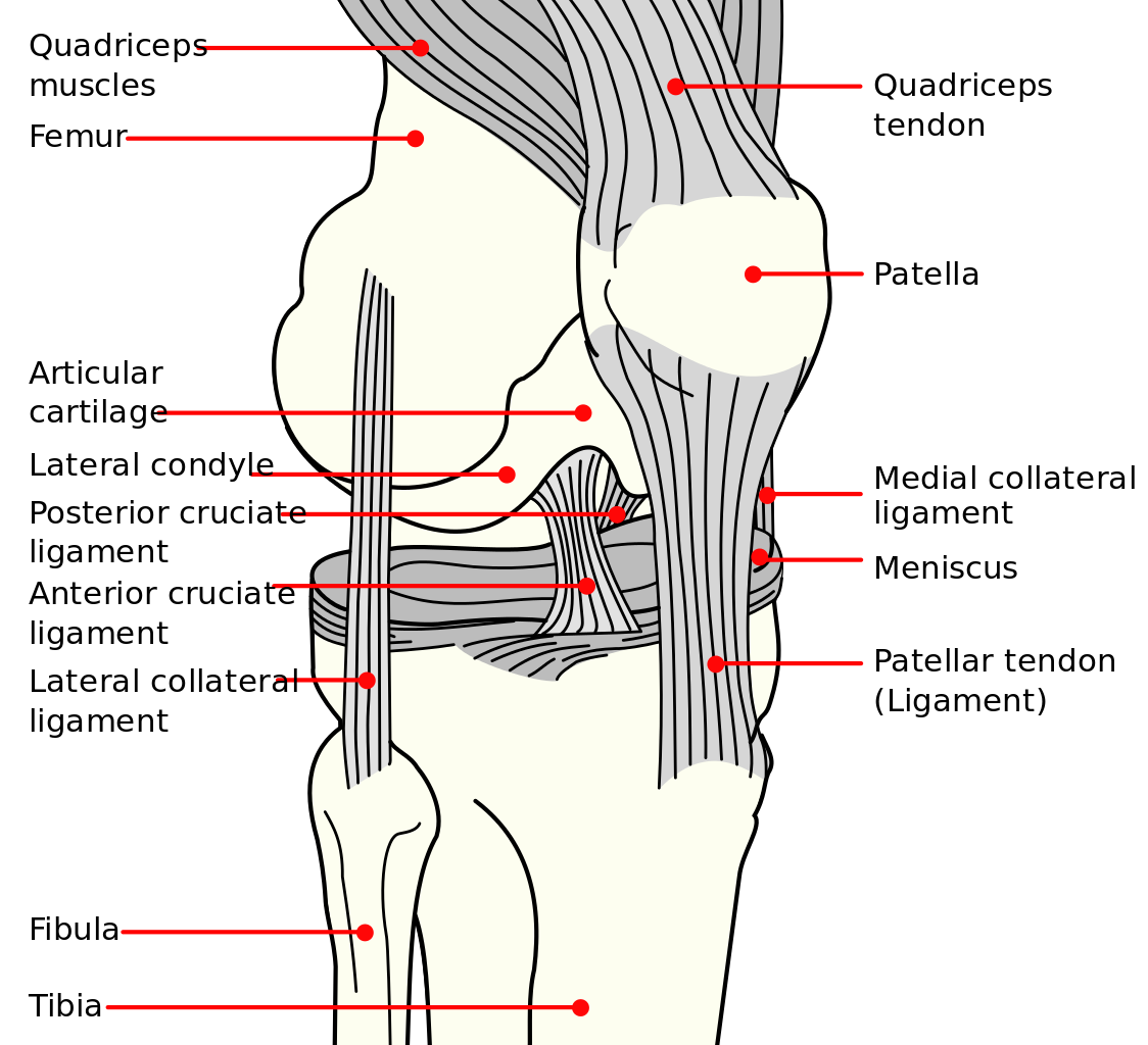
Use the labelled structures to localise pain and relate instability to ligament injury. The menisci lie between femur and tibia; the patella and patellar ligament form part of the anterior extensor mechanism.
What to notice
Detailed image description
Use the labelled structures to localise pain and relate instability to ligament injury. The menisci lie between femur and tibia; the patella and patellar ligament form part of the anterior extensor mechanism.
Triage & Approach
Step 1: Emergency Triage & Red Flag Screening
- Hot, Swollen, Monoarticular Joint + Fever/Systemic Unwellness: -> Suspect Septic Arthritis or Crystal Arthropathy (Gout/CPPD). Urgent arthrocentesis required.
- Acute Trauma with Inability to Bear Weight / Inability to Flex to 90 degrees / Bony Tenderness: -> Apply Ottawa Knee Rules to determine need for X-ray.
- Distal Neurovascular Deficit / Pale, Cold Foot post-trauma: -> Suspect Knee Dislocation with Popliteal Artery Disruption. Emergency reduction and ABI / CT Angiography required.
- Inability to Actively Extend Knee / High-Riding Patella: -> Suspect Quadriceps or Patellar Tendon Rupture.
Step 2: Clinical Decision Branching (Trauma vs. Non-Trauma)
- Acute Trauma + Rapid Onset Effusion (< 2h): -> Hemarthrosis (ACL tear > 70%, Patellar dislocation, Tibial plateau fracture, Osteochondral fracture).
- Acute Trauma + Delayed Onset Effusion (12–24h): -> Meniscal tear or Collateral ligament sprain.
- Non-Traumatic Pain: -> Categorize by anatomical pain localization and presence/absence of inflammatory features (warmth, swelling, morning stiffness > 30 min).
The Ottawa Knee Rules state that a knee X-ray is required ONLY if trauma is present PLUS any of: Age >= 55, Isolated patella tenderness, Tenderness at head of fibula, Inability to flex to 90 degrees, or Inability to bear weight immediately AND in the ED for 4 steps.
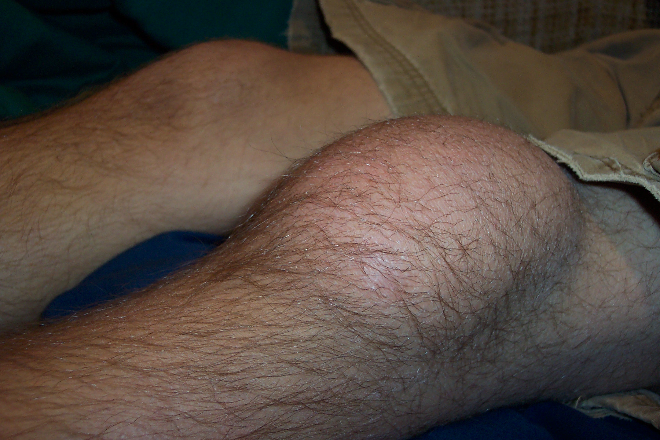
Localised swelling directly over the patella illustrates prepatellar bursitis, an extra-articular cause of anterior knee swelling. Compare its superficial distribution with a joint effusion. Appearance alone cannot determine whether a bursa is infected.
What to notice
Detailed image description
Localised swelling directly over the patella illustrates prepatellar bursitis, an extra-articular cause of anterior knee swelling. Compare its superficial distribution with a joint effusion. Appearance alone cannot determine whether a bursa is infected.
Differential Diagnosis
Acute Traumatic Injuries:
- Anterior Cruciate Ligament (ACL) Tear:
- Distinguishing features: Non-contact pivoting/deceleration injury; classic “pop” heard/felt; rapid hemarthrosis (< 2h); positive Lachman test (most sensitive) and Anterior Drawer test.
- Meniscal Tear (Medial or Lateral):
- Distinguishing features: Twisting injury on loaded flexed knee; mechanical locking, catching, or joint line tenderness; positive McMurray test and Thessaly test.
- Collateral Ligament (MCL / LCL) Sprain:
- Distinguishing features: Direct valgus (MCL) or varus (LCL) blow; localized collateral line tenderness; instability or pain on valgus/varus stress at 30 degrees flexion.
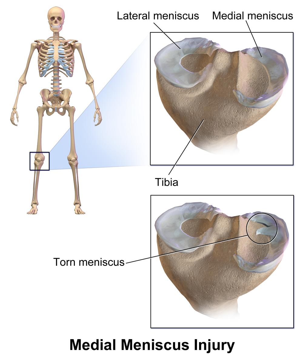
The medial and lateral menisci sit on the tibial plateau. The lower panel illustrates a medial meniscal tear, helping connect the anatomical lesion with joint-line pain and mechanical symptoms described above.
What to notice
Detailed image description
The medial and lateral menisci sit on the tibial plateau. The lower panel illustrates a medial meniscal tear, helping connect the anatomical lesion with joint-line pain and mechanical symptoms described above.
Non-Traumatic / Inflammatory & Infectious (Acute/Subacute):
- Septic Arthritis:
- Distinguishing features: Severe acute pain, inability to bear weight or tolerate passive ROM, fever, joint effusion/warmth. Synovial fluid WBC > 50,000 x 10^6/L with > 90% PMNs.
- Crystal Arthropathy (Gout / Pseudogout [CPPD]):
- Distinguishing features: Sudden onset severe pain, intense erythema/warmth; synovial fluid shows negatively birefringent needle crystals (Gout) or positively birefringent rhomboid crystals (CPPD).
Chronic & Overuse Etiologies:
- Osteoarthritis (OA):
- Distinguishing features: Age > 50; insidious onset, activity-related pain, brief morning stiffness (< 30 min), crepitus, bony enlargement, loss of joint space on weight-bearing X-ray.
- Patellofemoral Pain Syndrome (PFPS):
- Distinguishing features: Young females/runners; anterior knee pain worse with prolonged sitting (“theater sign”), squatting, or descending stairs; patellar apprehension/compression test positive.
- Patellar Tendinopathy (“Jumper’s Knee”):
- Distinguishing features: Focal tenderness at inferior pole of patella; pain aggravated by jumping or explosive knee extension.
Lachman test is significantly more sensitive (approx. 85-95%) for acute ACL disruption than the Anterior Drawer test, because muscle guarding around 90 degrees of flexion can cause false negatives in the Anterior Drawer test.
Investigations
First-Line / Bedside & Emergency Diagnostics:
- Synovial Fluid Aspiration (Arthrocentesis): Mandatory in any acute unexplained effusion. Evaluate WBC, Gram stain, culture, and crystal microscopy.
- Plain X-Rays (Weight-Bearing AP, Lateral, Skyline/Patellar views): Assess for fractures, joint space narrowing, subchondral sclerosis/cysts, osteophytes (OA), or Segond fracture (pathognomonic for ACL tear).
- Inflammatory Markers & Bloods: FBC, CRP, ESR, Serum Urate (Note: Normal serum urate does NOT rule out acute gout attack).
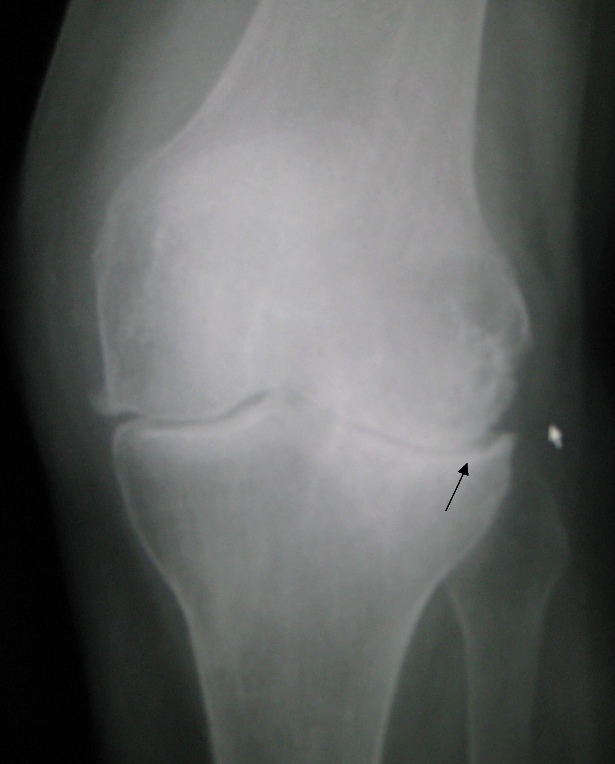
Look for asymmetric medial joint-space narrowing, increased subchondral bone density and marginal osteophytes. The arrow highlights the narrowed, sclerotic medial compartment. Interpret radiographic changes alongside the clinical presentation.
What to notice
Detailed image description
Look for asymmetric medial joint-space narrowing, increased subchondral bone density and marginal osteophytes. The arrow highlights the narrowed, sclerotic medial compartment. Interpret radiographic changes alongside the clinical presentation.
Targeted / Diagnostic Imaging Strategy:
- MRI Knee (Gold Standard for Soft Tissues): Indicated for evaluation of meniscal tears, cruciate/collateral ligament tears, osteochondral lesions, bone stress injuries, or occult fractures.
- Ultrasound Knee: Rapid identification of joint effusions, Baker’s cyst rupture, patellar/quadriceps tendon pathology, or guiding arthrocentesis.
A Segond Fracture (a tiny avulsion fracture off the lateral tibial plateau) on plain X-ray is highly pathognomonic for an underlying Anterior Cruciate Ligament (ACL) tear.
Critical Management
Septic Arthritis (Emergency Intervention):
- Joint Decompression: Urgent orthopedic consult for joint aspiration, formal arthroscopic/open washout, and debridement.
- Empiric Antibiotics: Administer IV empiric antibiotics (e.g., Flucloxacillin or Cefazolin; add Vancomycin if MRSA suspected) after synovial fluid aspiration.
Acute Hemarthrosis / Ligamentous Injury:
- PRICE / POLICE Protocol: Protection, Optimal Loading, Ice, Compression, Elevation.
- Analgesia: Paracetamol, short-term NSAIDs (avoid in hyperacute bleeding phase if fracture/severe tissue tear suspected).
- Rehabilitation: Early physical therapy for range of motion, quadriceps strengthening, and joint stability.
Do NOT administer intra-articular corticosteroid injections if there is any suspicion of infection or in the presence of an undiagnosed joint effusion. Steroids in a septic joint can cause catastrophic tissue damage.
References
- National Institute for Health and Care Excellence. Osteoarthritis in over 16s: diagnosis and management (NG226) [Internet]. 2022 [cited 2026 Aug 20]. Available from: https://www.nice.org.uk/guidance/ng226
- American College of Radiology. ACR Appropriateness Criteria® Chronic Knee Pain [Internet]. [cited 2026 Aug 20]. Available from: https://acsearch.acr.org/docs/69432/Narrative/
Image sources and licences
- Knee anatomy: ligaments, menisci and extensor mechanism. U.S. Federal Government illustration; PNG rendering by Mikael Häggström, via Wikimedia Commons. Public domain.
- Prepatellar bursitis: localised anterior knee swelling. Atropos235, via Wikimedia Commons; reproduced without alteration. CC BY-SA 3.0.
- Medial meniscal tear: anatomical illustration. BruceBlaus / Blausen Medical Communications, via Wikimedia Commons; reproduced without alteration. CC BY-SA 4.0.
- Left knee osteoarthritis: AP radiograph. James Heilman, MD, via Wikimedia Commons; reproduced without alteration. CC BY-SA 3.0.













Members only discussions coming soon…