

Hip Pain
OVERVIEW
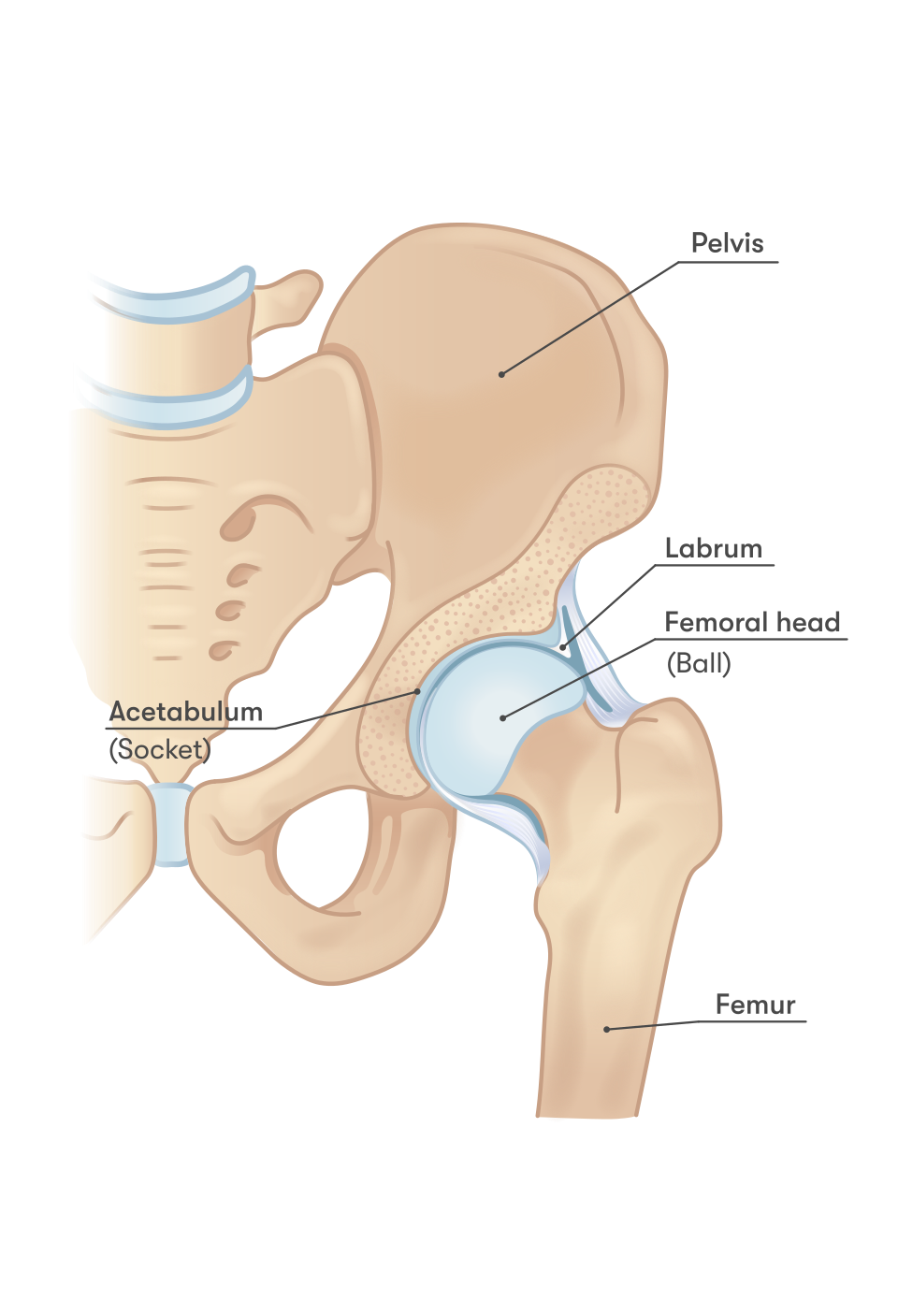
Hip pain is discomfort arising from the acetabulofemoral articulation or surrounding periarticular soft tissues (tendons, bursae, musculature, neurovascular structures).
Anatomical Mapping & Referral Patterns:
- Anterior / Groin Pain: Most specific for true intra-articular pathology (Osteoarthritis, Labral Tear, Femoroacetabular Impingement [FAI], Avascular Necrosis, Septic Arthritis).
- Lateral Hip Pain: Periarticular soft tissue pathology, predominantly Greater Trochanteric Pain Syndrome / gluteal tendinopathy.
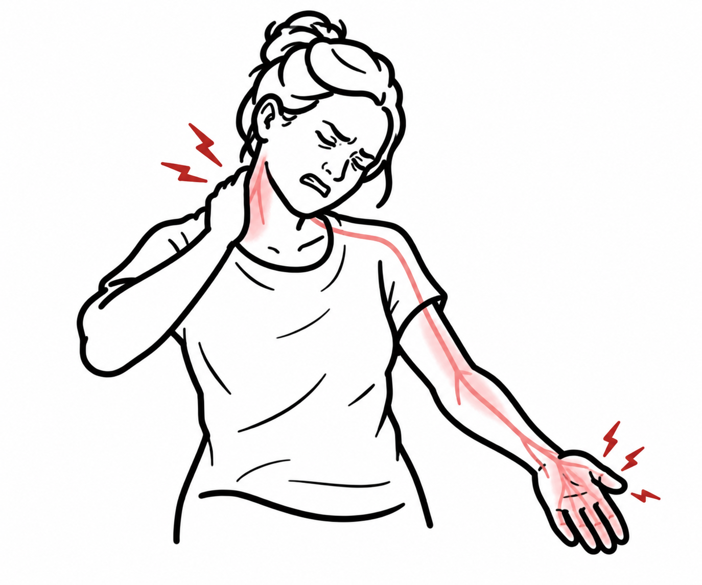
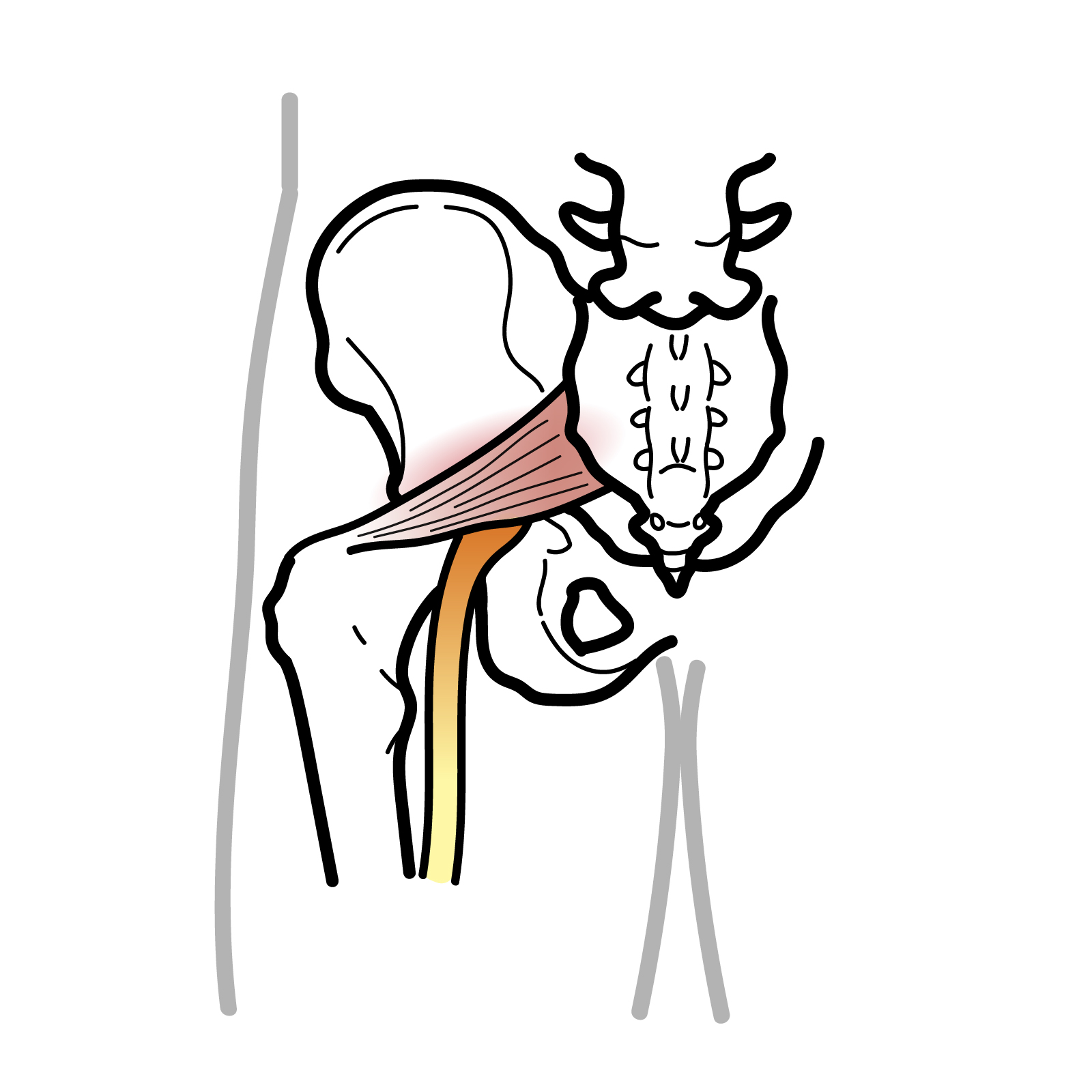
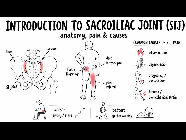
- Posterior Hip / Buttock Pain: Referred pain from the lumbar spine (L4–S1 radiculopathy), sacroiliac joint, or deep gluteal syndrome (piriformis syndrome).
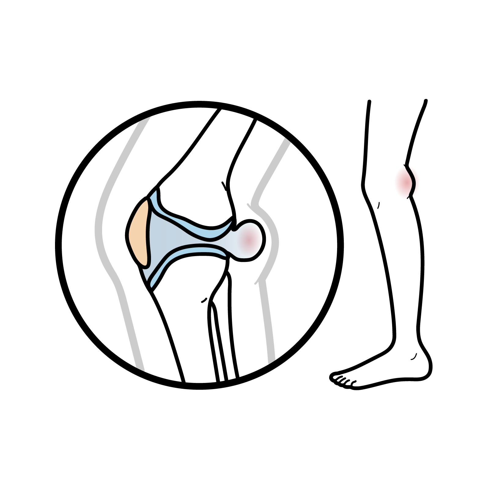
Knee pain is a classic referred presentation of hip pathology (via the obturator and femoral nerves); every patient presenting with isolated knee pain—especially children and adolescents—requires a mandatory hip examination.
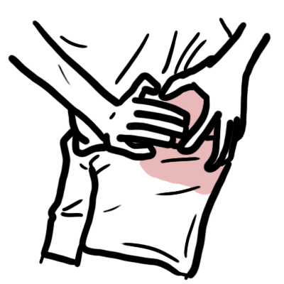
True intra-articular hip pathology typically presents as anterior groin pain (often demonstrating the “C-sign,” where the patient cups their hand above the greater trochanter with thumb and index finger). Pain localized strictly to the posterior buttock is lumbar spine or sacroiliac pathology until proven otherwise.
TRIAGE & ALGORITHM
Step 1: Emergency Triage & Red Flag Screening
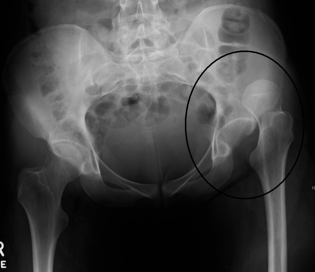
- Trauma + Inability to Bear Weight + Shortened & Externally Rotated Leg
- Trauma + Flexed, Adducted, Internally Rotated Leg
- -> Suspect Posterior Hip Dislocation (High-energy MVA; emergency closed reduction required within 6 hours to avoid avascular necrosis).
- Fever + Severe Inability to Bear Weight + Refusal to Tolerate Passive ROM
- -> Suspect Septic Arthritis (Apply Kocher criteria in children; urgent joint aspiration required).
- Adolescent / Child with Limp or Isolated Knee/Thigh Pain:
- -> Suspect Slipped Capital Femoral Epiphysis (orthopedic emergency: strict non-weight-bearing) or Perthes Disease.
Step 2: Anatomical Pain Localization
- Anterior Groin Pain
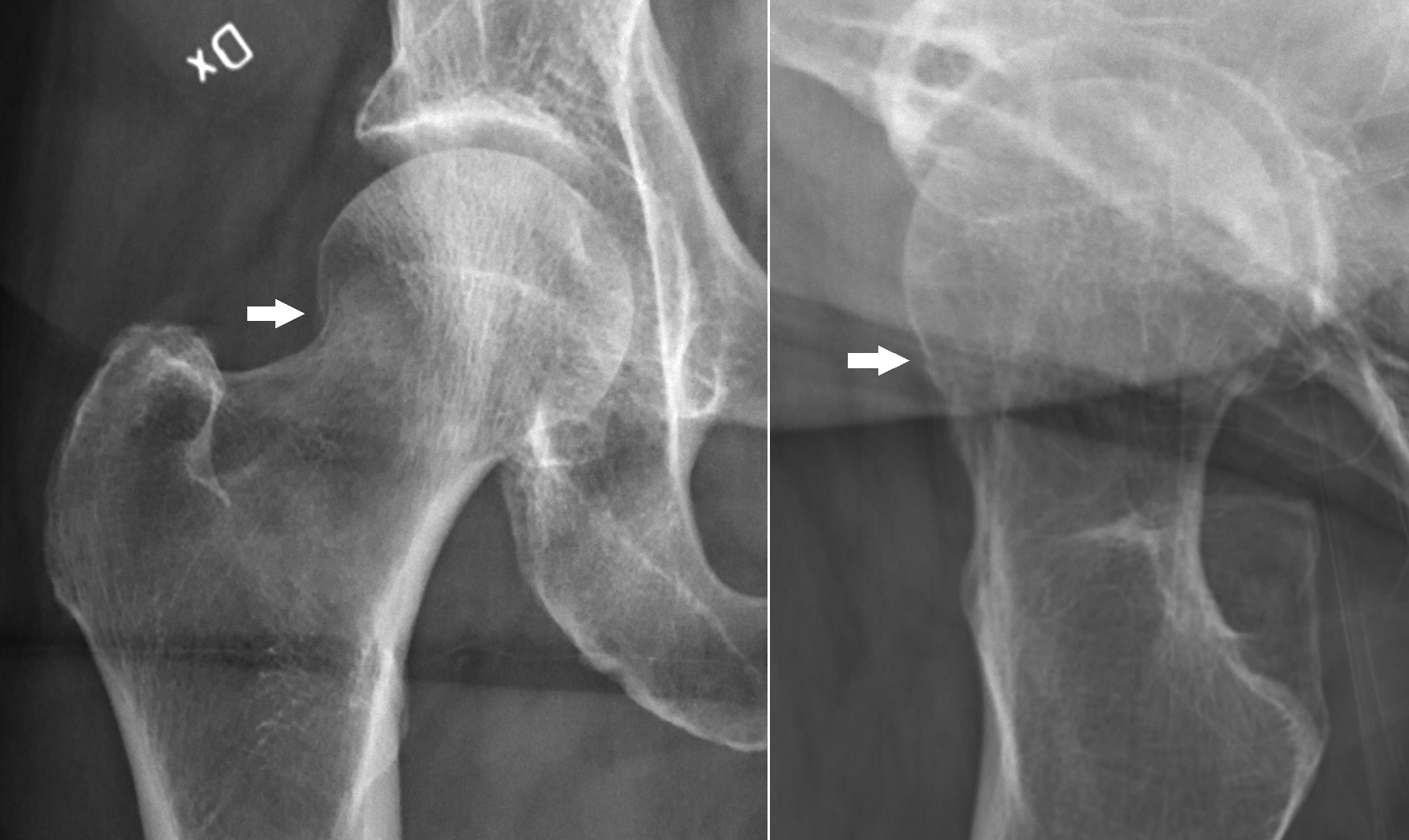
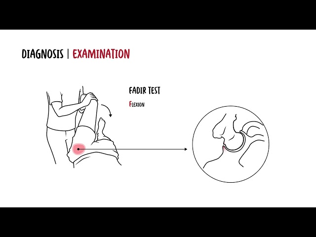
- Worse with internal rotation/flexion (FADIR test positive): Labral Tear / FAI.
- Chronic, activity-related, morning stiffness < 30 min: Hip Osteoarthritis.
- High-dose corticosteroid use or alcohol excess: Avascular Necrosis (AVN).
- Lateral Hip Pain
- Focal tenderness over greater trochanter, worse lying on affected side: Greater Trochanteric Pain Syndrome (GTPS) / Gluteal Tendinopathy.
- Posterior / Buttock Pain
- Dermatomal radiation down the leg, positive straight leg raise: Lumbar Radiculopathy / Sciatica.
Step 3: Diagnostic Decision Branching
- Acute Traumatic Suspicion + Normal Plain Radiographs: -> Obtain an urgent MRI Hip (or CT Pelvis/Hip) to rule out an occult femoral neck fracture or stress fracture.
- Non-Traumatic Acute Monoarthritis: -> Urgent Ultrasound-guided hip aspiration to differentiate Septic Arthritis from Crystal Arthropathy.
In elderly patients who present with hip pain and an inability to walk following a fall, a normal initial plain radiograph does NOT exclude a fracture. Approximately 5–10% of hip fractures are radiographically occult on standard X-rays; an MRI Hip (or thin-slice CT) is mandatory.
DIFFERENTIAL DIAGNOSIS
Acute & Traumatic Emergencies
- Neck of Femur Fracture
- Distinguishing features: Elderly, osteoporotic, minor fall; shortened and externally rotated lower limb; groin/hip pain on active straight leg raise and gentle axial loading.
- Septic Arthritis of the Hip
- Distinguishing features: Rapid onset of severe groin pain, high fever, systemic toxicity, refusal to bear weight; joint held in flexion, abduction, and external rotation (position of maximal intracapsular volume); synovial WBC > 50,000 x 10^6/L with > 90% polymorphs.
Pediatric & Adolescent Hip Conditions
- Slipped Capital Femoral Epiphysis (SCFE / SUFE)
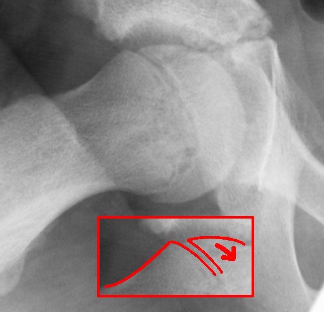
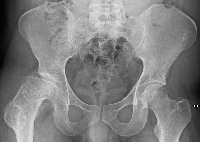
- Distinguishing features: Overweight/obese adolescent (age 10–16); presents with an antalgic limp and dull ache referred to the thigh or knee; obligate external rotation during passive hip flexion; frog-leg lateral X-ray shows failure of Klein’s line to intersect the lateral epiphysis.
- Legg-Calvé-Perthes Disease (Avascular Necrosis of Femoral Epiphysis)
- Distinguishing features: Boys aged 4–8 years; insidious painless or mildly painful limp; limited hip abduction and internal rotation; X-ray shows femoral head flattening, fragmentation, and sclerosis.
- Transient Synovitis (Irritable Hip)
- Distinguishing features: Young child (age 3–8); mild limp following a viral URTI; child is afebrile or low-grade; inflammatory markers normal/mildly elevated; self-limiting within 7–10 days.
Chronic & Degenerative Intra-Articular Conditions
- Hip Osteoarthritis
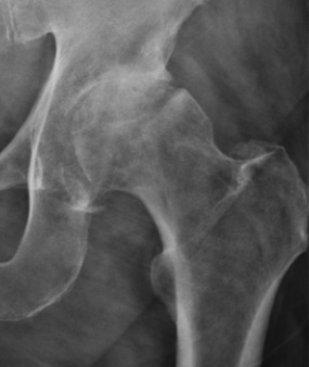
- Distinguishing features: Age > 50; insidious groin/anterior thigh pain; morning stiffness < 30 minutes; loss of internal rotation is the earliest clinical sign; weight-bearing X-ray shows joint space narrowing, subchondral sclerosis, osteophytes, and subchondral cysts.
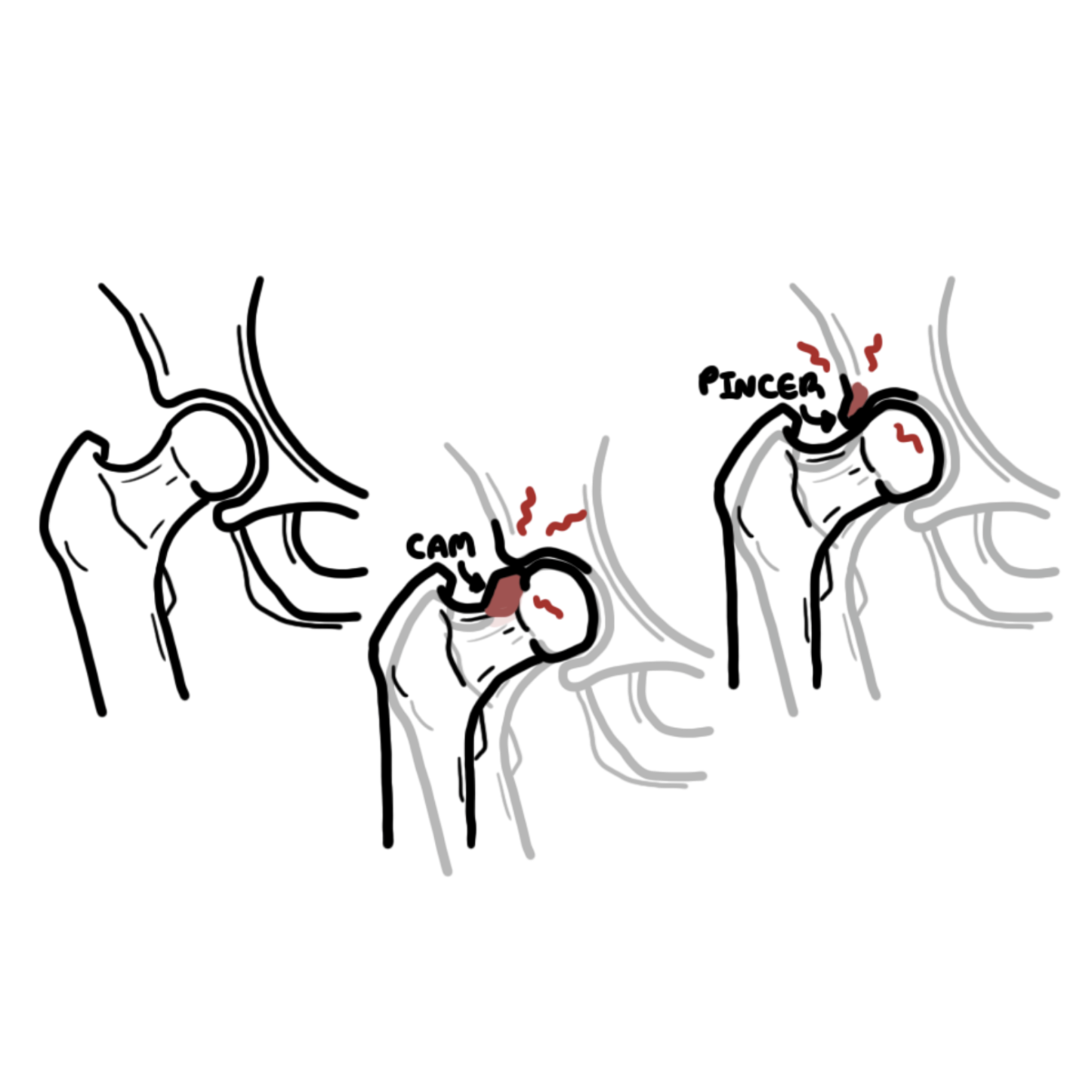
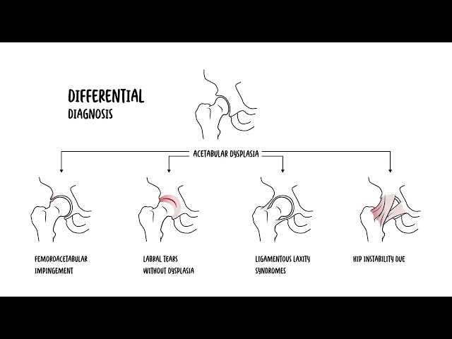
- Femoroacetabular Impingement & Acetabular Labral Tears
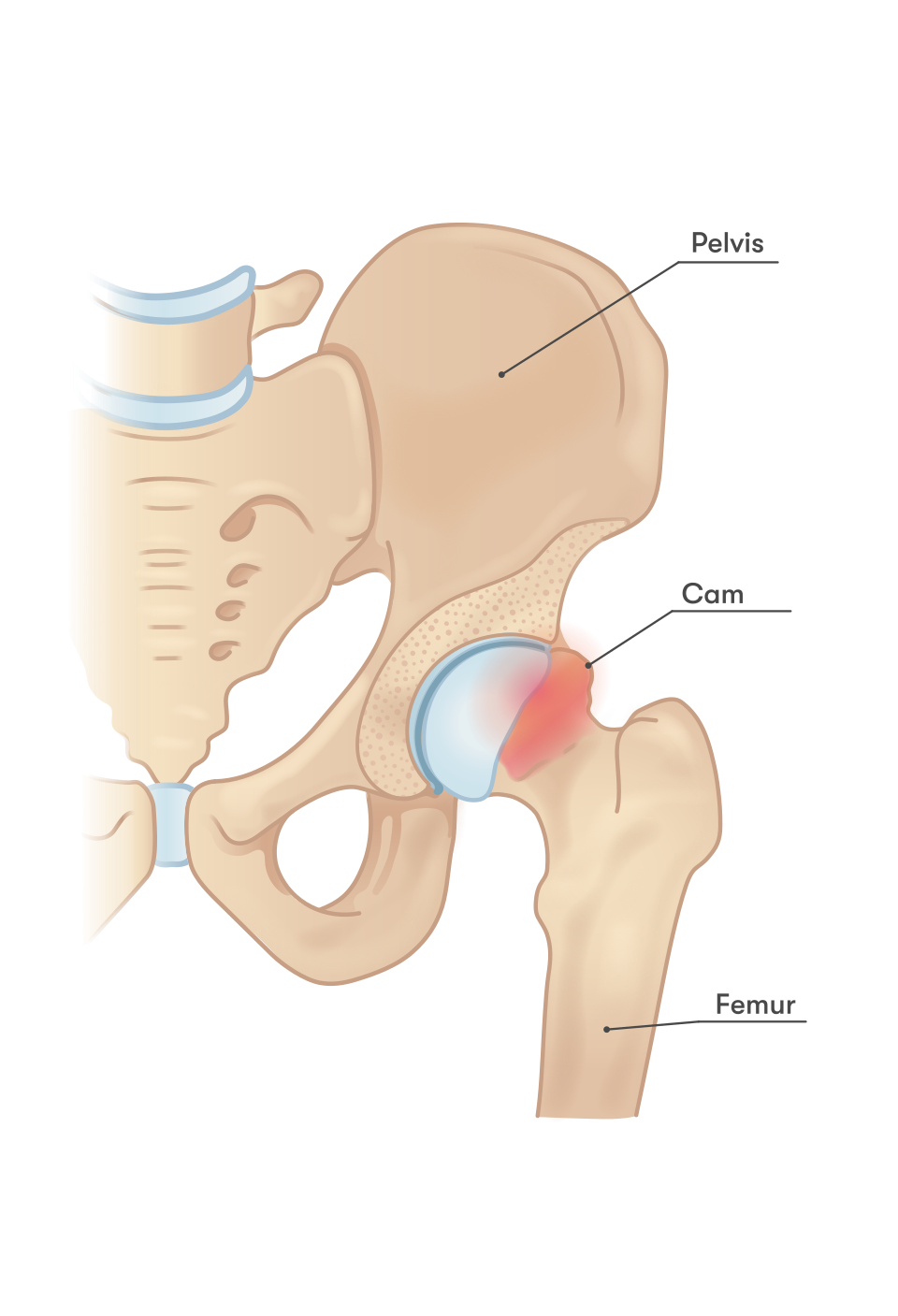
- Distinguishing features: Young active athletes; deep anterior groin pain provoked by prolonged sitting, pivoting, or deep squatting; positive FADIR test (Flexion, Adduction, Internal Rotation reproduces sharp pain).
- Avascular Necrosis (AVN / Osteonecrosis) of the Femoral Head
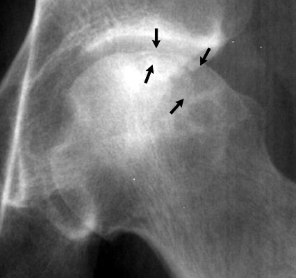
- Distinguishing features: History of systemic steroid therapy, chronic alcohol excess, sickle cell disease, or previous trauma; progressive groin pain, normal early X-ray followed by the pathognomonic “crescent sign” (subchondral collapse) on plain film; MRI is gold standard.
- Extra-Articular / Soft Tissue Disorders:
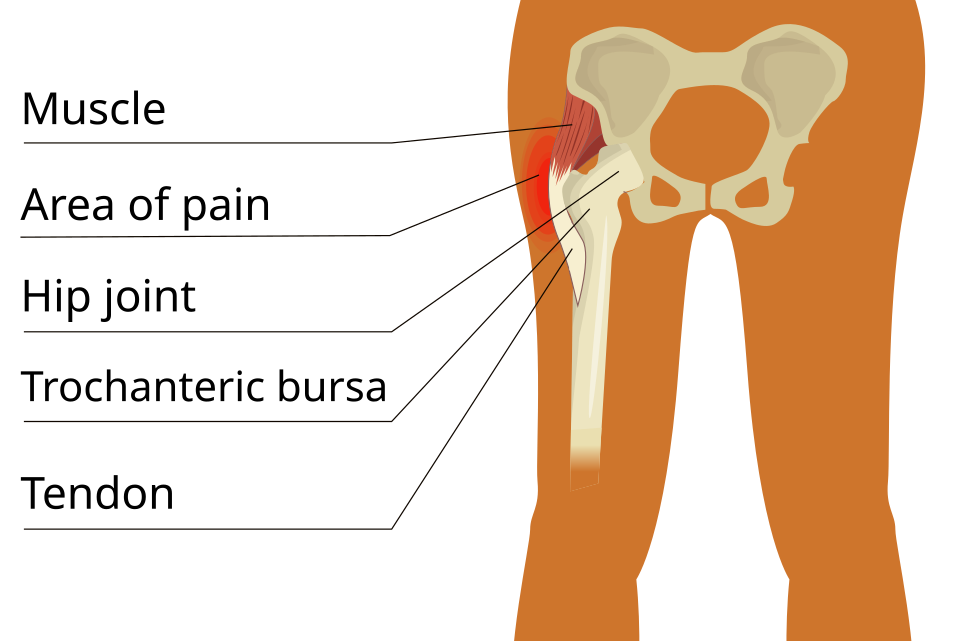
- Greater Trochanteric Pain Syndrome / Gluteal Tendinopathy:
- Distinguishing features: Middle-aged females; lateral hip/thigh pain, point tenderness over the greater trochanter; pain aggravated by lying on the affected side, climbing stairs, or single-leg standing for 30 seconds.
- Greater Trochanteric Pain Syndrome / Gluteal Tendinopathy:
The Kocher Criteria differentiate pediatric Septic Arthritis from Transient Synovitis: (1) Inability to bear weight, (2) Temperature > 38.5°C, (3) ESR > 40 mm/h, (4) WBC > 12.0 x 10^9/L. If 4/4 criteria are met, the probability of Septic Arthritis exceeds 90%, requiring emergency joint aspiration.
INVESTIGATIONS
First-Line / Emergency Diagnostics
- Plain Radiographs (AP Pelvis + Dedicated Lateral of Affected Hip):
- Adults: Weight-bearing AP Pelvis, cross-table lateral of affected hip (assesses fractures, osteoarthritis, AVN).
- Pediatrics: AP Pelvis + Bilateral Frog-Leg Lateral (Dunlap) views (mandatory to detect subtle posterior displacement in SCFE).
- Inflammatory Markers & Bloods: FBC, ESR, CRP, Blood cultures (mandatory if septic arthritis or osteomyelitis is suspected).
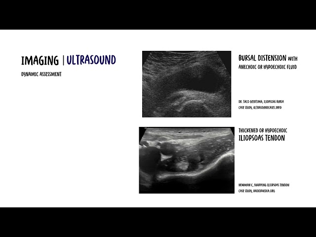
- Diagnostic Ultrasound of the Hip: Evaluates hip joint effusion; guides urgent percutaneous arthrocentesis for synovial fluid Gram stain, culture, and crystal analysis.
Targeted / Specialized Imaging
- MRI Hip (Non-Contrast):
- Indications: Gold standard for diagnosing occult femoral neck fractures, early Avascular Necrosis (before X-ray changes appear), stress fractures, osteitis pubis, and transient osteoporosis of the hip.
- MR Arthrogram (MRA Hip with Intra-Articular Gadolinium):
- Indications: Diagnostic modality of choice for acetabular labral tears and articular cartilage defects.
- CT Pelvis / Hip: Best for defining complex acetabular fractures, preoperative planning for arthroplasty, or assessing bony morphology in FAI (cam/pincer lesions).
In suspected Slipped Capital Femoral Epiphysis (SCFE), never order only an AP view. A mild posterior slip is frequently invisible on a standard AP view and can ONLY be seen on the frog-leg lateral radiograph.
CRITICAL MANAGEMENT
Fractured Neck of Femur (#NOF):
- Emergency Resuscitation: Fast-track analgesia (Fascia Iliaca Compartment Block [FICB]), IV fluids, pressure area care.
- Early Surgical Fixation: Operative management within 24–48 hours (Cannulated screws for undisplaced intracapsular; Hemiarthroplasty / Total Hip Arthroplasty for displaced intracapsular; Cephalomedullary nail or Dynamic Hip Screw for extracapsular/intertrochanteric fractures).
Slipped Capital Femoral Epiphysis Emergency Protocol:
- Immediate Non-Weight-Bearing: Patient must NOT walk or bear any weight (admit directly to hospital in a wheelchair/bed).
- Urgent Surgical Pinning: Urgent in situ single cannulated screw fixation to prevent further slip progression and catastrophic avascular necrosis of the femoral head.
Septic Arthritis of the Hip
- Emergency Decompression: Immediate arthroscopic or open arthrotomy washout by Orthopedics.
- Empiric IV Antibiotics: Administer IV Flucloxacillin or Cefazolin (+ Vancomycin if MRSA suspected) only after synovial fluid aspiration has been performed.
Greater Trochanteric Pain Syndrome
- Conservative Therapy: Avoid aggressive stretching of the ITB; activity modification (avoid crossing legs, sleep with pillow between knees); targeted physical therapy focusing on isometric gluteal strengthening.
- Second-Line: Ultrasound-guided corticosteroid injection around the trochanteric bursa (short-term relief only; not a long-term cure).
A Fascia Iliaca Compartment Block (FICB) should be administered early in the Emergency Department for patients with acute hip fractures. It provides profound opioid-sparing analgesia, minimizes delirium in the elderly, and facilitates pre-operative nursing and radiological positioning.







Members only discussions coming soon…