

Lower Limb Ulcer
OVERVIEW
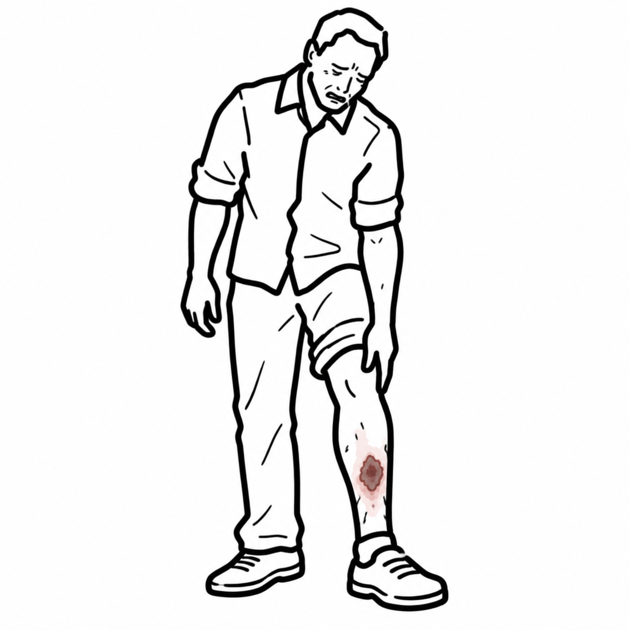
A lower limb ulcer is a breach in the epidermis and dermis of the leg or foot that fails to heal spontaneously through organized tissue repair, persisting for > 4 to 6 weeks.
Etiology
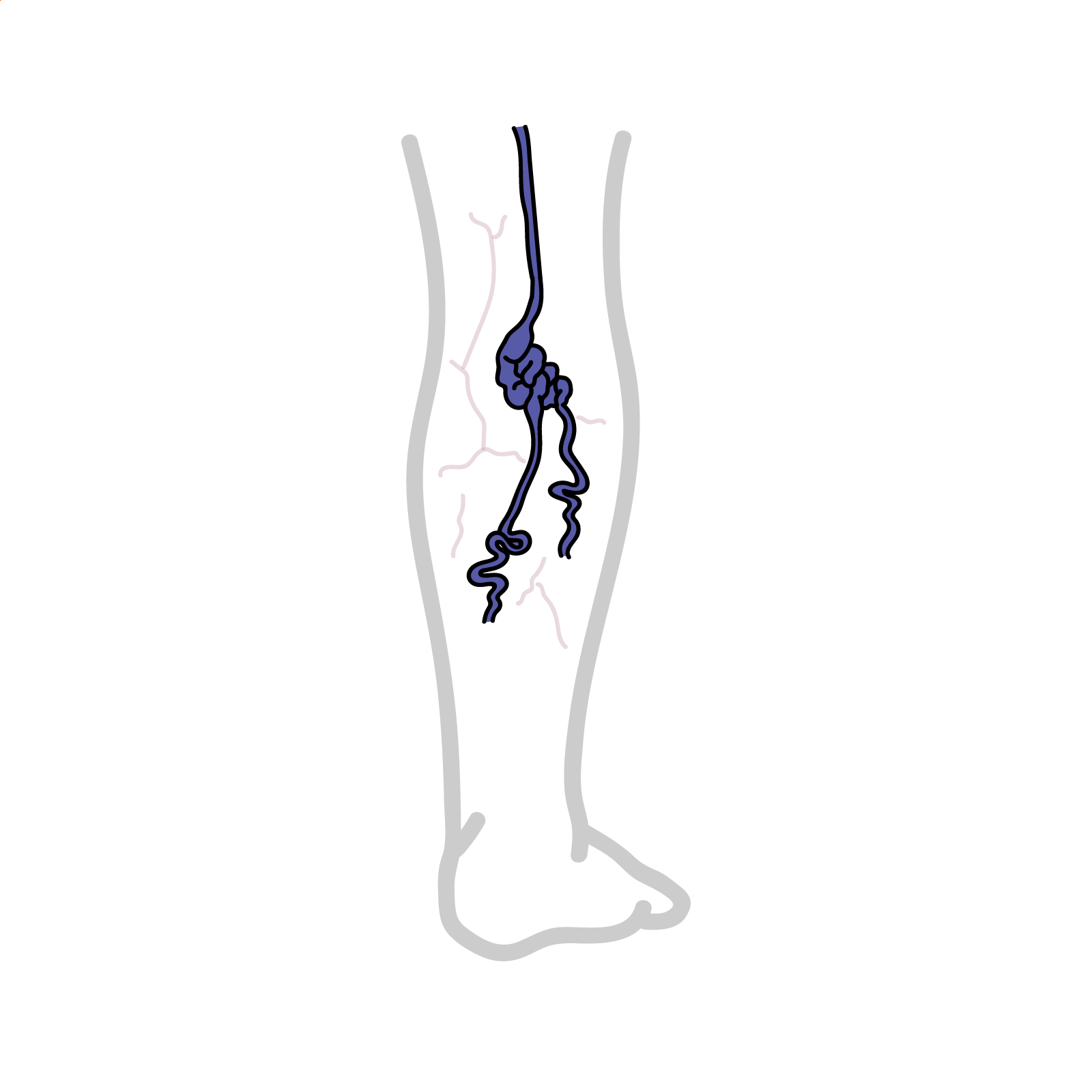
- Venous Disease (~70%): Ambulatory venous hypertension due to valvular incompetence or deep vein thrombosis.
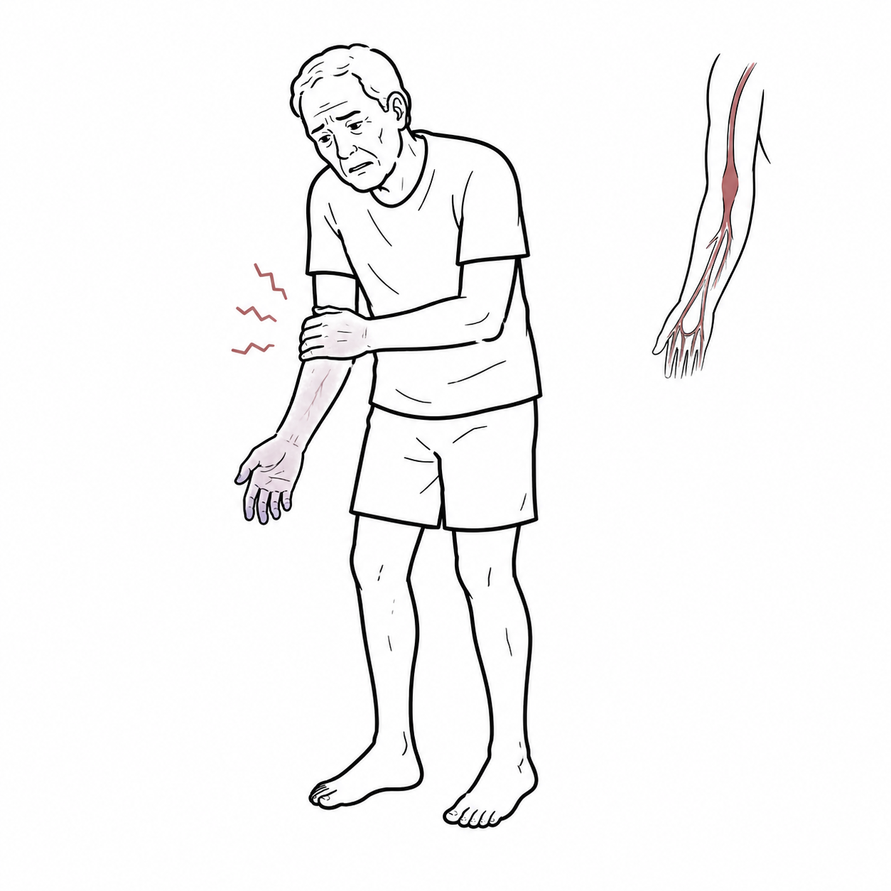
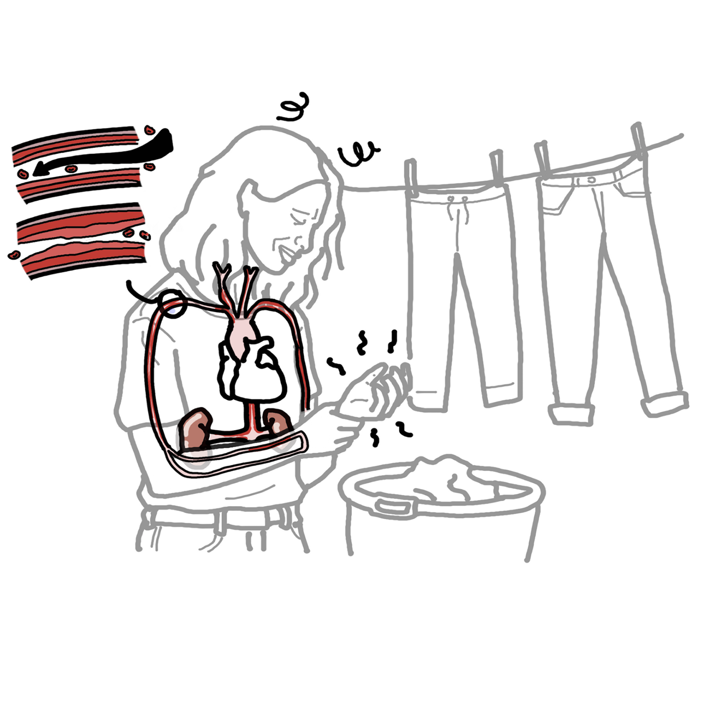
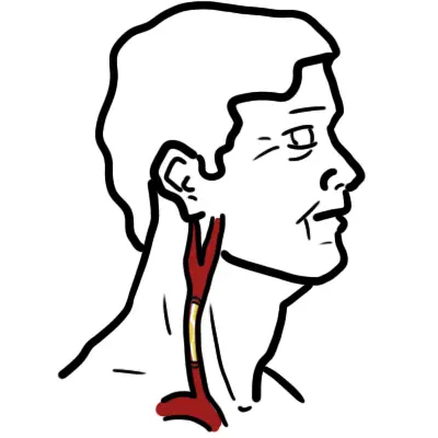
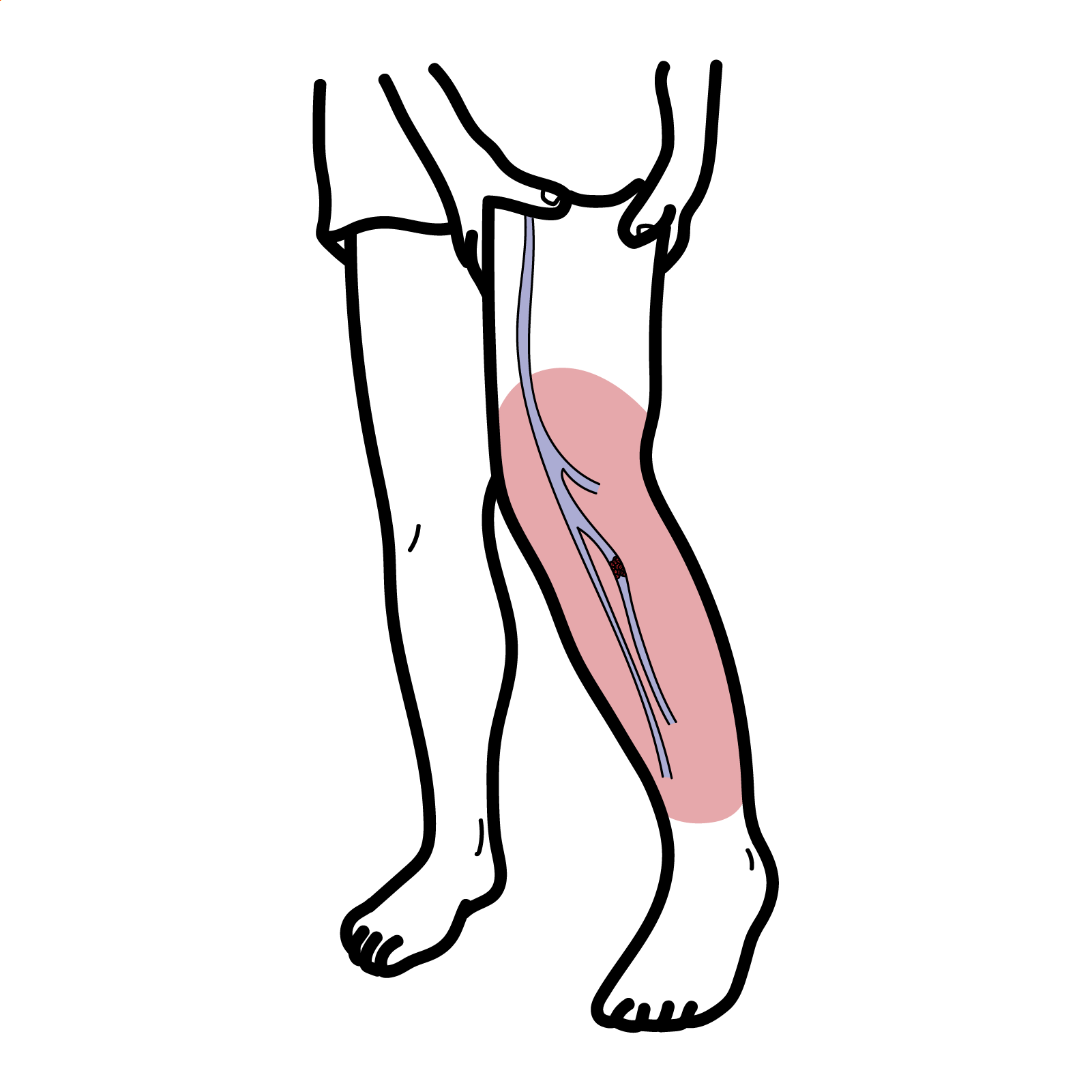
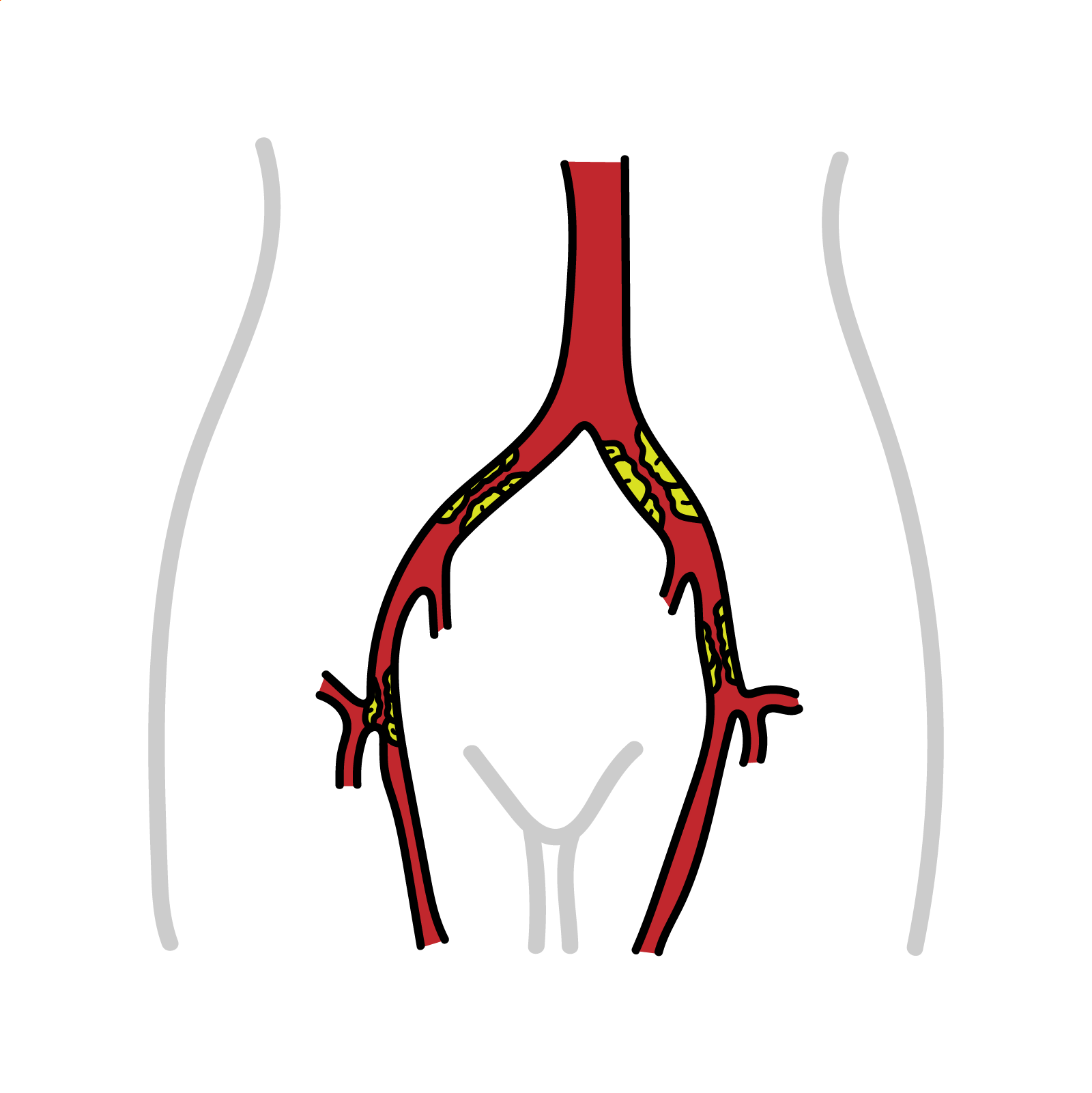
- Arterial Disease (~10–15%): Peripheral artery disease causing tissue hypoperfusion and critical limb-threatening ischemia.
- Neuropathic / Diabetic Foot (~10–15%): Loss of protective sensation, autonomic dysfunction, repetitive microtrauma, and microvascular disease.
- Other (~5%): Vasculitis (e.g., rheumatoid arthritis, systemic lupus erythematosus), hematological disorders, malignancy (Marjolin ulcer), and Pyoderma Gangrenosum.
Never apply high-compression bandaging to a lower limb ulcer without first measuring the Ankle-Brachial Index (ABI). Applying compression therapy to a severely ischemic limb (ABI < 0.5 to 0.8) precipitates acute arterial occlusion, extensive tissue necrosis, and limb loss.
TRIAGE & APPROACH
Step 1: Emergency Triage & Red Flag Screening
- Critical Limb-Threatening Ischemia:
- Rest pain (worse supine, relieved by hanging the foot over the edge of the bed), ischemic ulceration, or gangrene persisting for > 2 weeks -> Immediate Vascular Surgery consultation for emergency revascularization.
- Acute Spreading Infection / Sepsis:
- Ascending cellulitis > 2 cm around ulcer margin, lymphangitis, systemic fever, hypotension, or gas in soft tissue on radiography -> Urgent surgical debridement + parenteral antibiotics.
- Necrotizing Soft Tissue Infection:
- Severe out-of-proportion pain, cutaneous bullae, dishwater fluid drainage, crepitus -> Emergency surgical exploration.
Step 2: Bedside Vascular & Neurological Screening
- Palpate femoral, popliteal, dorsalis pedis, and posterior tibial pulses.
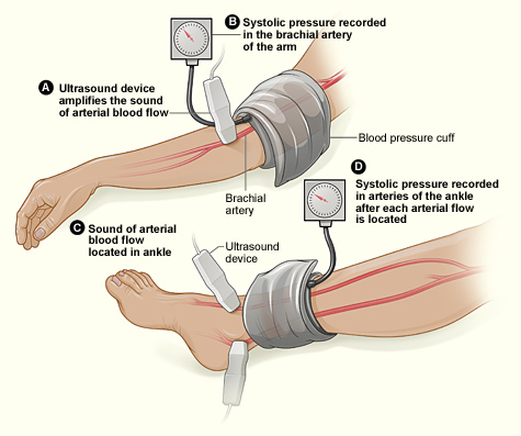
- Perform Handheld Doppler Ankle-Brachial Index (ABI):
- ABI > 1.3: Incompressible calcified vessels (common in diabetes and end-stage renal disease; proceed to Toe-Brachial Index [TBI]).
- ABI 0.9–1.2: Normal arterial perfusion.
- ABI 0.5–0.8: Moderate arterial disease (caution: modified/reduced compression only under vascular specialist guidance).
- ABI < 0.5: Severe arterial disease / critical ischemia (compression therapy strictly contraindicated).
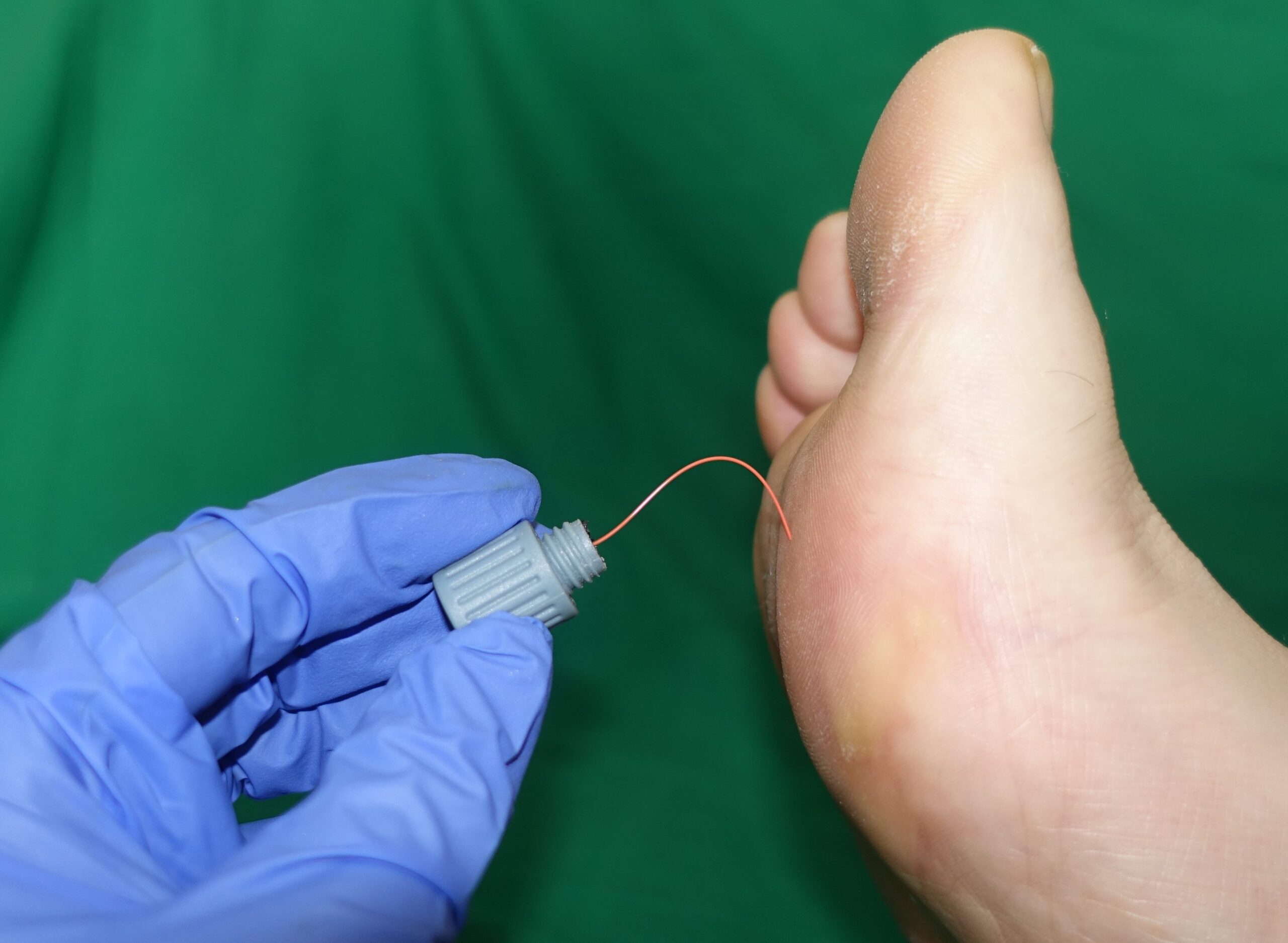
- Monofilament testing: Inability to detect a 10 g Semmes-Weinstein monofilament indicates loss of protective sensation (neuropathic ulcer risk).
Step 3: Branching Decision Tree by Ulcer Morphology & Location
- Medial Malleolus / Gaiter Area + Irregular Margins + Heavy Exudate:
- -> Venous Leg Ulcer (Initiate multilayer compression bandaging if ABI >= 0.8).
- Toes / Lateral Malleolus / Heel + “Punched-Out” Borders + Pale, Dry, Necrotic Base:
- -> Arterial Ulcer (Strictly avoid compression; refer for arterial duplex ultrasound and revascularization).
- Plantar Foot / Metatarsal Heads + Surrounding Hyperkeratosis / Callus:
- -> Neuropathic / Diabetic Foot Ulcer (Offload pressure, debride surrounding callus, optimize glycemic control).
- Rapidly Expanding Violaceous Undermined Border + Pathergy:
- -> Pyoderma Gangrenosum (Avoid surgical debridement; initiate systemic immunosuppression).
- Chronic Non-Healing Ulcer with Rolled / Everted Borders:
- -> Marjolin Ulcer (Squamous Cell Carcinoma) (Perform punch biopsy).
Pyoderma Gangrenosum is characterized by intense pathergy—minor surgical trauma or debridement causes rapid expansion of the ulceration. If a non-healing ulcer with an undermined purple-violaceous border fails to respond to antibiotics and worsens after surgical debridement, suspect Pyoderma Gangrenosum and stop all invasive procedures.
DIFFERENTIAL DIAGNOSIS
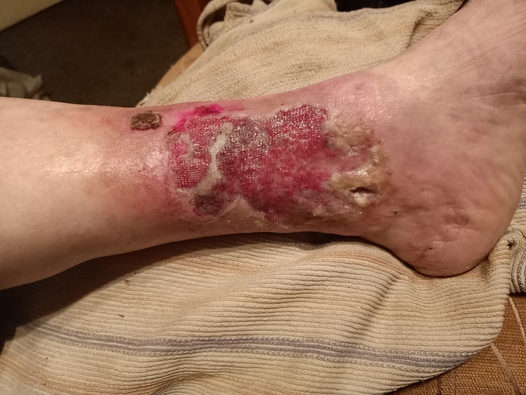
Venous Leg Ulcers (Most Common)
- Location: Gaiter area of the lower leg, particularly proximal to the medial malleolus.
- Morphology: Shallow, irregular margins, moist/ruddy granulation tissue bed, heavy serous exudate.
- Associated skin changes:Stasis dermatitis, hemosiderin hyperpigmentation, lipodermatosclerosis (fibrosing panniculitis causing an “inverted champagne bottle” leg), atrophie blanche, pitting edema.
- Symptoms: Dull aching heaviness exacerbated by prolonged standing, relieved by elevation.
Arterial (Ischemic) Ulcers
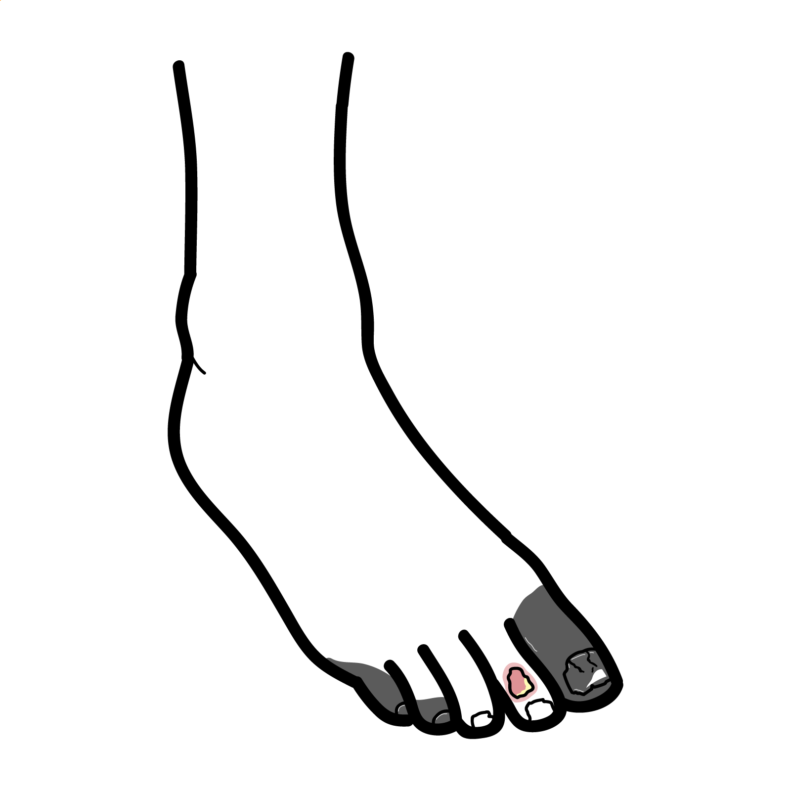
- Location: Distal extremities, tips of toes, interdigital web spaces, heel, or lateral malleolus.
- Morphology:“Punched-out” well-demarcated margins, pale/dry necrotic or black slough base, minimal exudate or bleeding.
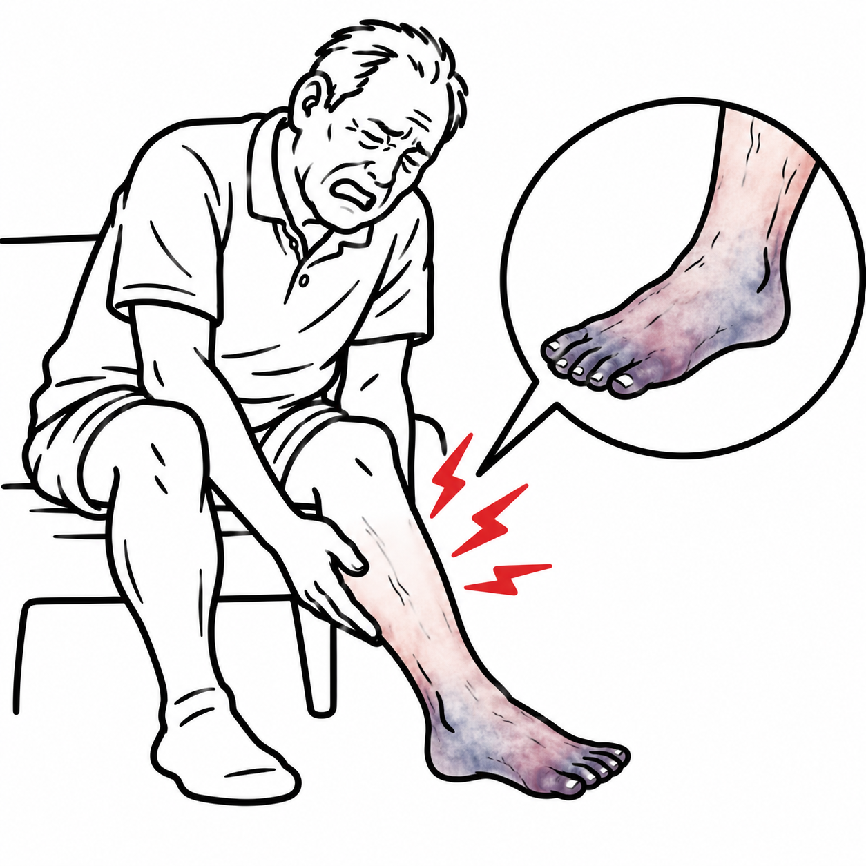
- Associated skin changes: Cold, pale, shiny skin, loss of hair, thickened brittle nails, absent or diminished peripheral pulses, dependent rubor with pallor on elevation.
- Symptoms: Severe pain, particularly nocturnal and worsened by leg elevation.
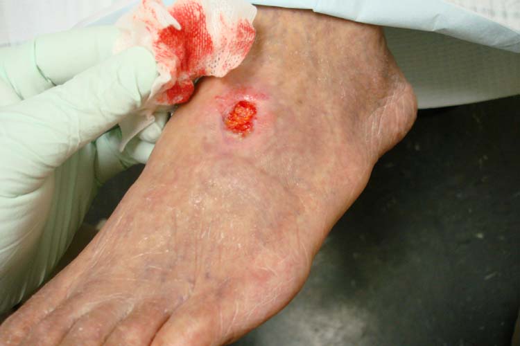
Neuropathic (Diabetic) Ulcers
- Location: Plantar pressure points (metatarsal heads, heel, plantar surface of interphalangeal joints).
- Morphology: Deep, punched-out ulcer bed surrounded by a thick rim of hyperkeratotic callus; often painless due to sensory neuropathy.
- Associated features: “Rocker-bottom” foot deformity (Charcot neuroarthropathy), clawing of toes, dry cracked skin due to sympathetic autonomic failure.
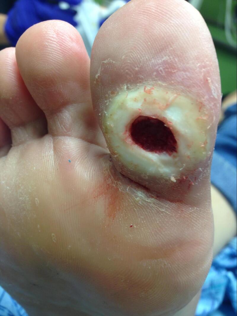
Malignant & Inflammatory Ulcers
- Marjolin Ulcer (Squamous Cell Carcinoma in Chronic Wounds):
- Chronic ulcer persisting for months to years that develops everted/raised borders, excessive friable bleeding, foul odor, or failure to respond to appropriate wound management.
- Pyoderma Gangrenosum:
- Associated with Inflammatory Bowel Disease (Crohn disease, Ulcerative Colitis), rheumatoid arthritis, or hematological malignancies. Starts as a sterile pustule or nodule that rapidly breaks down into an agonizing ulcer with undermined gunmetal-violaceous edges.
Any chronic ulcer that fails to show measurable healing after 8 to 12 weeks of optimized standard therapy—or exhibits progressive fungation, heaped-up margins, or excessive bleeding—requires an edge-wedge or punch biopsy to definitively rule out a Marjolin Ulcer (Squamous Cell Carcinoma).
INVESTIGATIONS
First-Line / Bedside Diagnostics:
- Handheld Doppler ABI / TBI: Mandatory baseline test. If ABI > 1.3 due to arterial medial sclerosis, a Toe-Brachial Index (TBI < 0.7 confirms arterial disease) must be obtained.
- 10 g Semmes-Weinstein Monofilament & 128 Hz Tuning Fork: Evaluates protective touch sensation and vibratory sense.
- Ulcer Depth & Probe-to-Bone Test: Palpate ulcer base with a sterile metallic probe. A positive “probe-to-bone” test in high-risk diabetic ulcers has a > 85% positive predictive value for underlying osteomyelitis.
- Wound Swabs: Indicated ONLY if clinical signs of active local or spreading infection are present (erythema, warmth, edema, purulence, foul odor). Swab the wound base after cleansing and debriding; never culture superficial dry slough or exudate.
- Baseline Bloods: Full Blood Count (leukocytosis), ESR, C-reactive protein, HbA1c (diabetes control), Serum Albumin (nutritional healing reserve), Renal function.
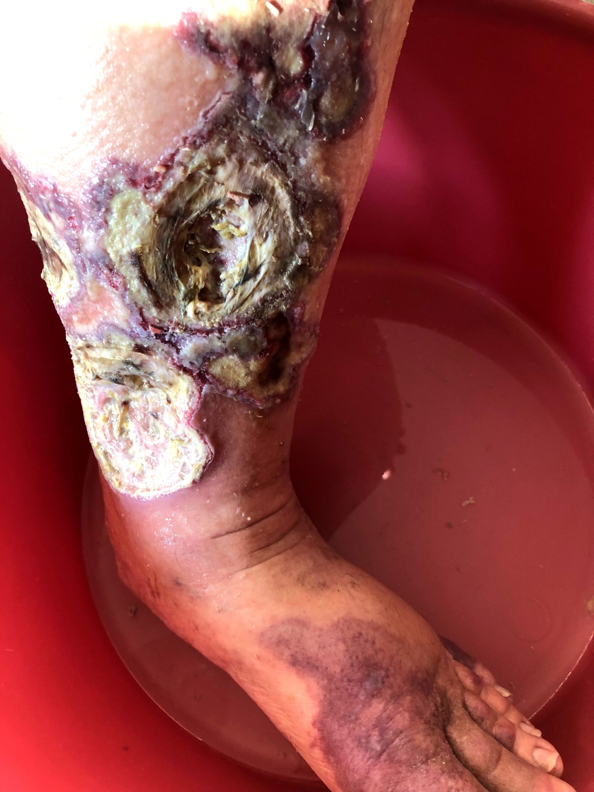
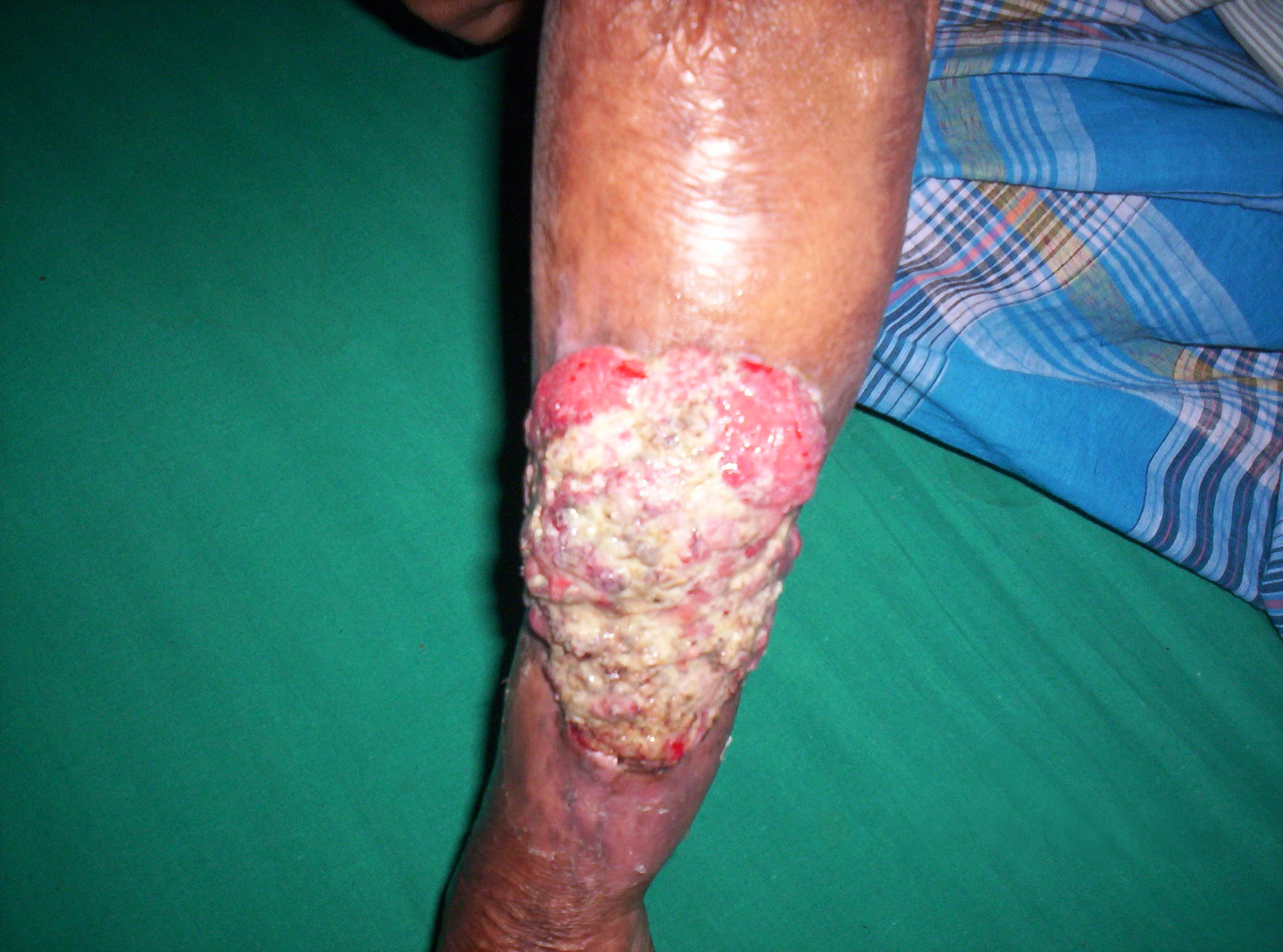
Targeted / Specialized Imaging & Procedures:
- Duplex Ultrasound of Lower Extremity (Venous & Arterial):
- Venous Duplex: Assesses superficial and deep venous reflux (> 0.5 seconds in superficial veins, > 1.0 second in deep veins) and excludes deep vein thrombosis.
- Arterial Duplex: Identifies arterial occlusions, stenosis, and flow velocities from the iliac arteries to the digital vessels.
- Plain Radiography (Foot / Ankle): Evaluates for cortical bone erosion, periosteal reaction (osteomyelitis), Charcot neuroarthropathy, or soft tissue radio-opaque foreign bodies.
- Magnetic Resonance Imaging (MRI): Gold standard imaging modality to confirm osteomyelitis, deep fascial space abscesses, or sinus tracts in the diabetic foot.
- Punch Biopsy (Wound Margin): Mandatory for atypical, deteriorating, or non-healing ulcers to exclude vasculitis or malignancy.
Do NOT swab clinically uninfected chronic ulcers. All chronic wounds are colonized with polymicrobial flora. Culturing a non-infected ulcer leads to inappropriate systemic antibiotic prescribing, accelerating antimicrobial resistance without aiding wound healing.
CRITICAL MANAGEMENT
Venous Leg Ulcer Protocol (Cornerstone Therapy)
- Multilayer Compression Bandaging: High-compression (30–40 mmHg at the ankle, graduated up to the calf) via multi-layer bandage systems or short-stretch bandages.
- Prerequisite: ABI must be >= 0.8 (or >= 0.6–0.8 for reduced/modified compression under specialist guidance).
- Endovenous Ablation: Early surgical endovenous ablation of incompetent superficial veins (e.g., radiofrequency ablation or endovenous laser therapy) accelerates ulcer healing and drastically reduces ulcer recurrence rates (EVRA trial protocol).
- Adjunctive Pharmacotherapy:Pentoxifylline 400 mg PO TDS (improves microcirculatory blood flow; enhances venous ulcer healing when added to compression).
- Leg Elevation: Elevate legs above the level of the heart for at least 30 minutes 3–4 times daily to reduce ambulatory venous pressure.
Arterial Ulcer Protocol
- Strict Avoidance of Compression Bandaging.
- Urgent Revascularization: Endovascular angioplasty/stenting or surgical bypass to restore pulsatile arterial flow.
- Cardiovascular Risk Optimization: High-dose statin, antiplatelet therapy (Aspirin or Clopidogrel), smoking cessation, and tight blood pressure control.
- Local Wound Care: Keep ischemic wounds dry and clean to prevent conversion of dry gangrene into wet gangrene prior to revascularization.
Neuropathic / Diabetic Foot Ulcer Protocol
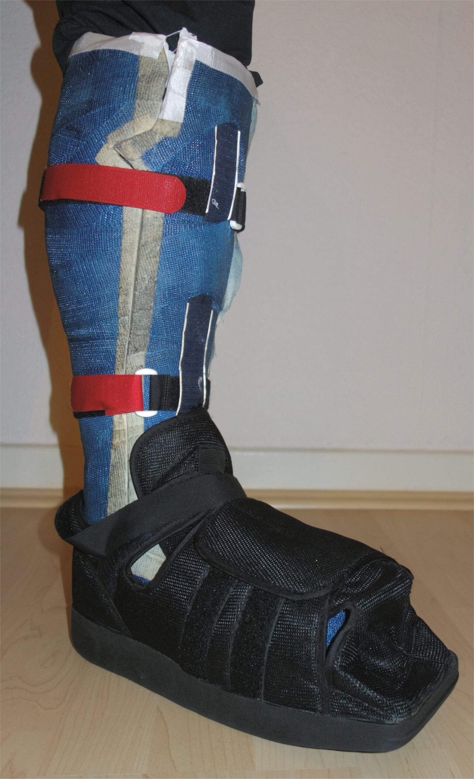
- Offloading (CRITICAL): Non-removable Total Contact Cast (TCC) or fixed ankle offloading boot is the gold standard for plantar neuropathic ulcers.
- Sharp Debridement: Removal of hyperkeratotic callus, necrotic slough, and non-viable tissue to promote granulating margins.
- Moist Wound Dressings: Hydrogels, alginates, or foams selected based on exudate level.
Infection & Osteomyelitis Management
- Systemic broad-spectrum oral or IV antibiotics (e.g., Amoxicillin-Clavulanate, or Piperacillin-Tazobactam +/- Vancomycin for limb-threatening infections) directed by deep tissue/bone cultures, combined with surgical debridement of infected bone.
The Total Contact Cast (TCC) is the gold-standard treatment for plantar diabetic neuropathic ulcers because it redistributes weight away from the ulcer site and enforces patient compliance. Standard orthopedic shoes and standard dressings without effective offloading have failure rates exceeding 70%.
REFERENCES
- European Society for Vascular Surgery. Clinical Practice Guidelines on the Management of Chronic Venous Disease of the Lower Limbs (2022).
- NICE. Peripheral arterial disease: diagnosis and management (CG147).
- NICE. Leg ulcer infection: antimicrobial prescribing (NG152).
- International Working Group on the Diabetic Foot. Practical guidelines on the prevention and management of diabetes-related foot disease: offloading (2023).



.jpg)


Members only discussions coming soon…