

Acute Mesenteric Ischaemia
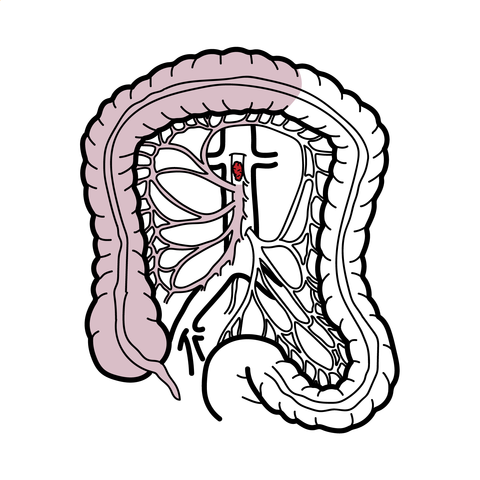
AF with abdominal pain think mesenteric ischaemia.
Definition
Acute mesenteric ischaemia: sudden reduction in blood supply to the intestine, with risk of bowel infarction.
Mesenteric arterial embolism: an embolus, usually cardiac in origin, acutely obstructing a mesenteric artery.
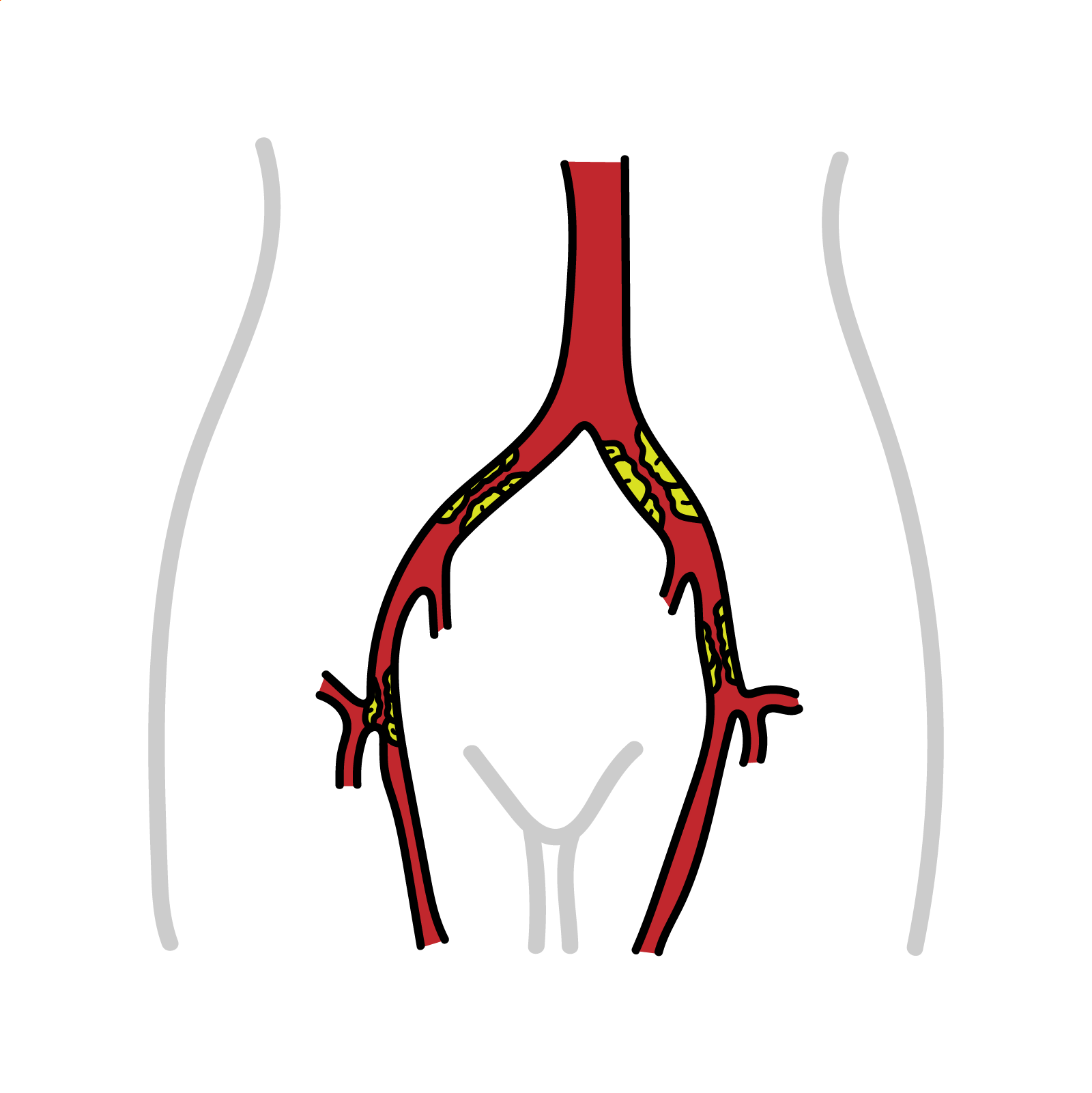
Mesenteric arterial thrombosis: acute thrombosis developing within a diseased mesenteric artery, usually over an atherosclerotic stenosis.
Non-occlusive mesenteric ischaemia—NOMI: intestinal ischaemia caused by severe hypoperfusion and mesenteric vasoconstriction without a major vascular obstruction.
Mesenteric venous thrombosis—MVT: thrombosis of mesenteric venous drainage causing venous congestion, bowel-wall oedema and impaired arterial inflow.
Intestinal infarction: irreversible, usually transmural bowel necrosis caused by prolonged ischaemia.
Aetiology & Risk Factors
Aetiology
Acute mesenteric ischaemia (AMI) is a life-threatening vascular emergency caused by a sudden reduction in intestinal blood flow. Inadequate perfusion rapidly produces mucosal injury, bowel-wall necrosis, bacterial translocation, sepsis and multiorgan failure. AMI is uncommon—accounting for approximately 0.09–0.2% of acute surgical admissions—but mortality remains around 50%, largely because early symptoms are non-specific and diagnosis is frequently delayed. The principal types are arterial embolism, arterial thrombosis, non-occlusive mesenteric ischaemia and mesenteric venous thrombosis.
- Mesenteric arterial embolism
- Accounts for approximately 50% of AMI cases
- Common embolic sources:
- Atrial fibrillation
- Recent myocardial infarction
- Left-ventricular aneurysm or severe ventricular dysfunction
- Infective endocarditis
- Emboli commonly lodge several centimetres distal to the SMA origin and may occur with renal or splenic emboli.
- Mesenteric arterial thrombosis
- Accounts for approximately 15–25% of cases and usually develops over chronic atherosclerotic disease.
- Risk factors:
- Generalised atherosclerosis
- Smoking
- Hypertension
- Mesenteric artery dissection
- Vasculitis
- Thrombosis commonly occurs near the SMA origin and may affect a larger bowel territory than a distal embolus.
- Non-occlusive mesenteric ischaemia
- Accounts for approximately 20% of cases and is caused by critically reduced mesenteric perfusion and vasoconstriction
- Causes: shock, cardiac arrest
- Mesenteric venous thrombosis
- Accounts for approximately 5–15% of AMI.
Pathophysiology
Arterial occlusion
- An embolus or thrombus obstructs mesenteric arterial inflow.
- Reduced oxygen delivery initially damages the metabolically active intestinal mucosa.
- Mucosal barrier failure causes oedema, ulceration and gastrointestinal bleeding.
- Continued ischaemia progresses from the mucosa through the submucosa and muscular wall.
- Full-thickness injury causes bowel necrosis, perforation and peritonitis.
- Bacterial translocation and release of inflammatory mediators cause sepsis and multiorgan failure.
Mesenteric venous thrombosis
- Venous outflow is obstructed.
- Venous pressure rises, causing bowel-wall congestion and oedema.
- Increasing tissue pressure reduces arterial perfusion.
- Haemorrhagic infarction and bowel necrosis may develop if thrombosis progresses.
Non-occlusive mesenteric ischaemia
- Low cardiac output or systemic hypotension reduces splanchnic perfusion.
- Compensatory mesenteric vasoconstriction further limits blood flow.
- Patchy or diffuse bowel ischaemia develops despite patent major vessels.
- Vasopressors may aggravate vasoconstriction, particularly when hypovolaemia remains uncorrected.
Ischaemia–reperfusion injury
- Restoration of blood flow may release reactive oxygen species and inflammatory mediators.
- Capillary leak, systemic inflammation and haemodynamic instability may worsen after revascularisation.
- Reperfusion injury can contribute to acute kidney injury, respiratory failure and multiorgan dysfunction.
Clinical Manifestation
The hallmark of the diagnosis of acute mesenteric ischaemia is abrupt onset of intense cramping epigastric and periumbilical pain out of proportion to the findings on abdominal examination.
- Diarrhea
- Vomiting
- Bloating
- Melena
Classical Triad: Acute severe abdominal pain, no abdominal signs, rapid hypovolemic.
Examination
- Acutely ill, but the presentation may be subtle
- Shock is present in about 25% of cases
Diagnosis
- FBC – may show decreased haemoglobin due to plasma loss and increase in WCC
- Amylase -elevated
- ABG – metabolic acidosis
- Abdominal X-ray – early may show gasless bowels.
- CT/MRI
Severe abdominal pain with metabolic acidosis is bowel ischemia until proven otherwise.
References
UptoDate
Best Practice
Oxford Handbook Clinical Surgery
Oxford Handbook Clinical Medicine


Members only discussions coming soon…