

Amyloidosis (Overview)

Overview
Amyloidosis encompasses a diverse group of protein-folding disorders characterized by the extracellular deposition of insoluble, toxic protein aggregates known as amyloid fibrils. Under physiological conditions, precursor proteins remain soluble and functional. In amyloidosis, these precursor proteins undergo conformational changes that cause them to misfold, self-assemble, and deposit within the extracellular matrix of various tissues and organs.
As these rigid, cross-beta-sheet fibrillar structures accumulate, they disrupt tissue architecture, induce direct local cellular toxicity, and progressively compromise organ function. The clinical presentation varies dramatically depending on whether the disease is localized to a single organ or systemic in nature, as well as the specific precursor protein involved. Left untreated, systemic amyloidosis leads to progressive organ failure, with cardiac involvement serving as the primary driver of mortality.
Definition
Amyloid Fibril: An insoluble, non-branching fibrillar protein aggregate (7.5–10 nm in diameter) formed from misfolded monomeric precursors.
Cross-beta-Sheet Structure: The characteristic super-secondary folding arrangement shared by all amyloid fibrils, which confers extreme chemical stability and resistance to normal proteolytic degradation.
Congo Red Stain: The definitive histological dye used to identify amyloid; exhibits pathognomonic apple-green birefringence when viewed under cross-polarized light.
Serum Amyloid P Component (SAP): A normal plasma glycoprotein that binds universally to all amyloid deposits, protecting them from degradation and serving as a target for diagnostic nuclear imaging (123I-SAP scintigraphy).
Birefringence: The optical property of a material having a refractive index that depends on the polarization and propagation direction of light.
What to notice
Detailed image description
Classification
Amyloidosis is classified using the prefix “A” followed by an abbreviation for the specific precursor protein (e.g., AL, AA, ATTR).
1. By Extent of Organ Involvement
- Systemic Amyloidosis: Widespread deposition affecting multiple organs and tissues (e.g., heart, kidneys, liver, peripheral nerves). Includes AL, AA, and ATTR types.
- Localized Amyloidosis: Deposition restricted to a single organ or tissue site without systemic involvement (e.g., Abeta in Alzheimer’s disease, IAPP in Type 2 Diabetes, or localized AL in the urinary bladder or larynx).
2. Major Systemic Types
| Type | Precursor Protein | Primary Underlying Pathology |
| AL (Primary) | Immunoglobulin Light Chains (lambda > kappa) | Clonal plasma cell dyscrasias (e.g., MGUS, Multiple Myeloma). |
| AA (Secondary) | Serum Amyloid A (SAA) | Sustained high SAA levels during chronic inflammatory states. |
| ATTRwt (Wild-Type) | Unmutated Transthyretin | Age-related destabilization of normal hepatic transthyretin (formerly “Senile Systemic”). |
| ATTRm (Hereditary) | Mutated Transthyretin | Autosomal dominant point mutations in the TTR gene. |
| Abeta2M | beta-2-Microglobulin | Long-term hemodialysis with reduced renal clearance of beta-2M. |
The precursor protein determines the disease subtype, but the organ distribution determines the clinical presentation and prognosis.
Aetiology & Risk Factors
1. AL Amyloidosis (Primary Systemic)
- Aetiology: Proliferation of a low-grade clonal plasma cell population secreting abnormal free light chains that misfold.
- Risk Factors: Age > 60 years, preexisting Monoclonal Gammopathy of Undetermined Significance (MGUS), or Multiple Myeloma.
2. AA Amyloidosis (Secondary / Reactive)
- Aetiology: Overproduction of Serum Amyloid A (SAA)—an acute-phase reactant synthesized by the liver—driven by persistent elevated IL-1, IL-6, and TNF-alpha.
- Risk Factors:
- Autoimmune: Rheumatoid arthritis, Ankylosing Spondylitis, Inflammatory Bowel Disease.
- Chronic Infections: Tuberculosis, Bronchiectasis, Chronic Osteomyelitis.
- Autoinflammatory Disorders: Familial Mediterranean Fever (FMF).
3. ATTR Amyloidosis (Transthyretin)
- Aetiology: Transthyretin (a homotetrameric transport protein for thyroxine and retinol) dissociates into unstable monomers that aggregate.
- ATTRwt Risk Factors: Advanced age (predominantly males > 70 years).
- ATTRm Risk Factors: Inheritance of TTR gene mutations (e.g., Val30Met for polyneuropathy; Val122Ile for cardiomyopathy).
AA amyloidosis has become significantly less common in developed nations due to modern biologic therapies targeting rheumatoid arthritis and inflammatory bowel disease.
Pathophysiology
- Misfolding & Oligomerization: Soluble proteins lose their native tertiary structure due to mutation, structural modification, or extreme concentration, forming toxic soluble oligomers.
- Fibrillogenesis: Oligomers assemble into protofilaments and cross-beta-sheet fibrils that deposit within the extracellular matrix.
- Dual Mechanisms of Damage:
- Physical Infiltration: Massive fibril accumulation expands tissues, stiffens organs (e.g., restrictive cardiomyopathy), and disrupts architecture.
- Direct Cytotoxicity: Circulating pre-fibrillar light chain oligomers directly cause oxidative stress, mitochondrial dysfunction, and apoptosis in cardiomyocytes independent of bulk tissue infiltration.
In AL cardiac amyloidosis, functional impairment occurs before massive fibril deposition because circulating light chain oligomers cause direct cellular toxicity to cardiomyocytes.
Clinical Manifestations
Clinical signs and symptoms reflect organ infiltration and functional impairment. Systemic amyloidosis often presents as a constellation of multi-organ findings.
Cardiac Manifestations (Most Critical)
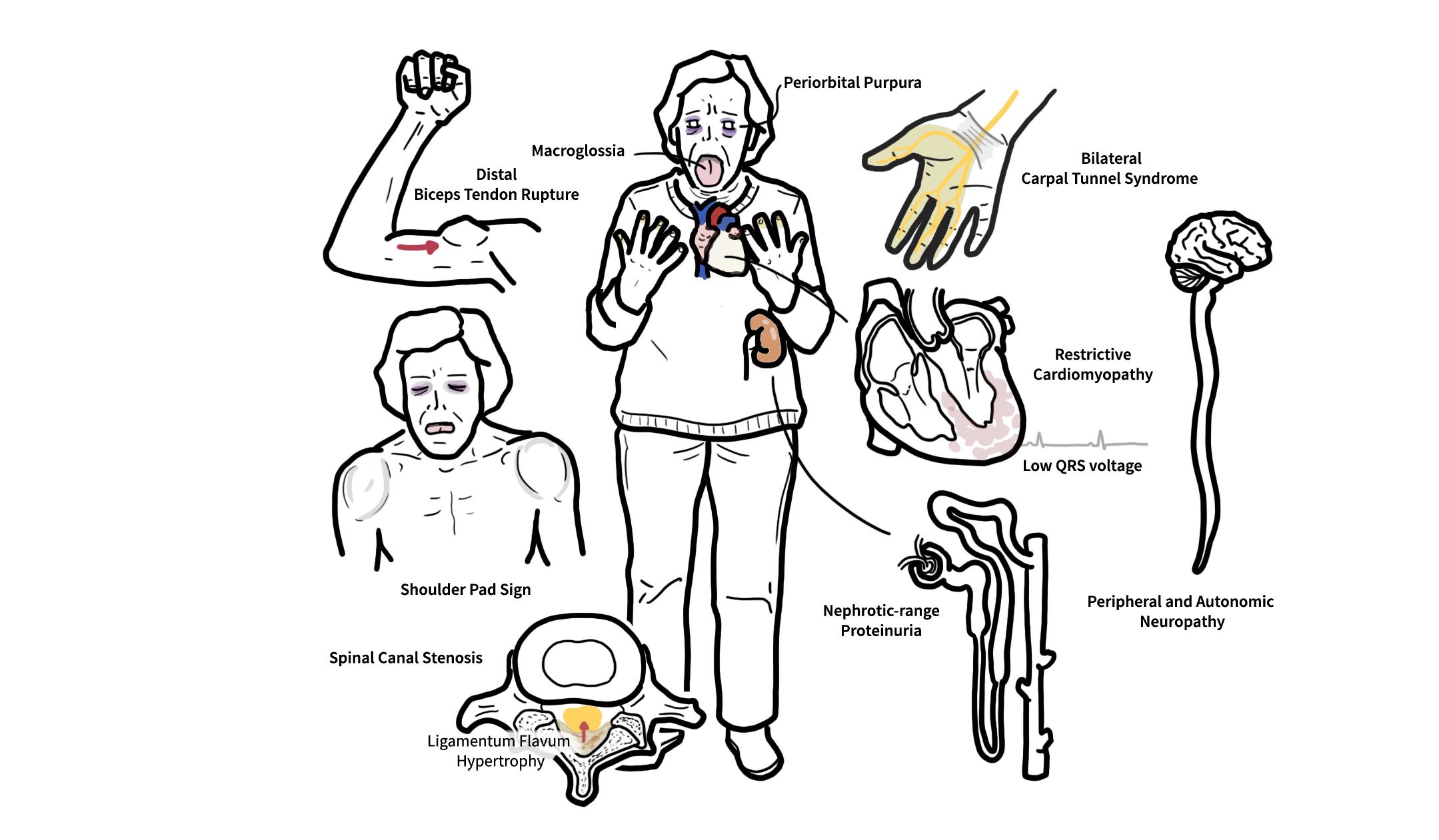
- Restrictive Cardiomyopathy: Progressive exertional dyspnoea, fatigue, peripheral oedema, elevated JVP, and ascites (right heart failure signs often predominate).
- Arrhythmias & Conduction Defects: Atrial fibrillation, AV blocks, sinus node dysfunction, and fatal ventricular arrhythmias.
- Angina-like Chest Pain: Caused by intramyocardial coronary artery amyloid infiltration (microvascular ischemia without obstructive CAD).
Renal Manifestations (Most Common Initial Presentation in AA & AL)
- Nephrotic Syndrome: Heavy proteinuria (> 3.5 g/day), hypoalbuminemia, severe peripheral oedema, and hypercholesterolemia.
- Progressive Renal Insufficiency: Azotemia and declining eGFR, leading to End-Stage Renal Disease (ESRD).
Neurological Manifestations
- Peripheral Polyneuropathy: Symmetrical, distal length-dependent axonal neuropathy starting with pain/paresthesia (small-fibre loss) followed by motor weakness.
- Autonomic Neuropathy: Severe orthostatic hypotension, neurogenic bladder, erectile dysfunction, and gastrointestinal dysmotility (alternating diarrhea and constipation).
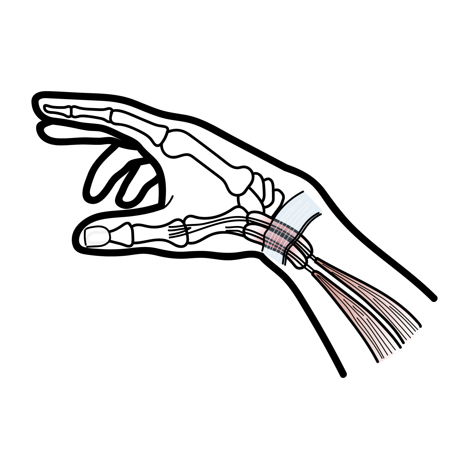
- Entrapment Neuropathies: Bilateral Carpal Tunnel Syndrome (frequently precedes cardiac manifestations of ATTRwt by 5–10 years).
Soft Tissue, Musculoskeletal & Vascular Manifestations
- Macroglossia: Enlarged, rigid, indented tongue (causes airway compromise and obstructive sleep apnea); highly specific to AL type.
- Periorbital Purpura (“Raccoon Eyes”): Spontaneous or post-coughing eyelid ecchymosis due to vascular capillary fragility and Factor X adsorption onto amyloid fibrils.
- Submandibular & Lymph Node Enlargement: “Bull neck” appearance due to soft tissue deposition.
- Spontaneous Biceps Tendon Rupture: “Popeye sign” highly characteristic of ATTR amyloidosis.
Gastrointestinal & Hepatic Manifestations
- Gastrointestinal Tract: Dysphagia, early satiety, malabsorption, pseudo-obstruction, and GI bleeding.
- Hepatomegaly: Firm, non-tender enlargement with marked elevation of Serum Alkaline Phosphatase (ALP) out of proportion to bilirubin/transaminases.
Bilateral Carpal Tunnel Syndrome in an elderly male is a major red flag for ATTRwt Amyloidosis and should prompt an evaluation for occult cardiac amyloidosis.
Diagnosis & Investigations
Histopathology & Biopsy
- Screening Biopsy: Abdominal fat pad aspirate (low-risk, sensitive for AL) or minor salivary gland biopsy.
- Organ Biopsy: Done if screening biopsies are negative but suspicion remains high (Renal or Endomyocardial biopsy).
- Staining: Congo Red stain demonstrating apple-green birefringence under cross-polarized light.
- Electron Microscopy: Reveals classic non-branching, rigid 7.5–10 nm fibrils.
Apple-green birefringence on Congo Red staining confirms the presence of amyloid, but it does NOT tell you the type. You MUST perform typing before starting treatment.
Amyloid Subtype Identification
- Mass Spectrometry (LC-MS/MS): Gold standard for definitive protein identification in tissue.
- Immunohistochemistry / Immunofluorescence: Used to identify light chain (kappa/lambda) or protein specific deposits.
Diagnostic Workup by Organ & Subtype
- AL Amyloidosis Screening:
- Serum Free Light Chain (sFLC) assay (kappa/lambda ratio).
- Serum Protein Electrophoresis with Immunofixation (SPEP/IFE).
- 24-hour Urine Protein Electrophoresis with Immunofixation (UPEP/IFE).
- Cardiac Investigations:
- ECG: Low QRS voltage in limb leads despite concentric left ventricular thickening on Echo (classic voltage-to-mass discrepancy).
- Echocardiogram: Granular “sparkling” myocardium, biatrial enlargement, and “apical sparing” pattern on strain imaging.
- Cardiac MRI: Diffuse, transmural, or subendocardial Late Gadolinium Enhancement (LGE).
- Bone Scintigraphy (99mTc-PYP / DPD / HMDP): Strongly positive (Grade 2 or 3 uptake) in ATTR cardiac amyloidosis.
Non-invasive diagnosis of ATTR cardiac amyloidosis can be made without a heart biopsy if bone scintigraphy shows Grade 2/3 myocardial uptake AND serum/urine monoclonal protein screening is completely negative.
Treatment
Subtype-Directed Therapies
AL Amyloidosis
- Goal: Eradicate the abnormal plasma cell clone to stop light chain production.
- First-line Regimen: Daratumumab (anti-CD38 monoclonal antibody) + Bortezomib + Cyclophosphamide + Dexamethasone (CyBorD-Dara).
- Consolidation: High-dose Melphalan with Autologous Stem Cell Transplantation (ASCT) for eligible candidates.
AA Amyloidosis
- Goal: Suppress the underlying chronic inflammatory state to reduce SAA levels below 10 mg/L.
- Targeted Agents: Anti-IL-1 agents (Anakinra, Canakinumab for FMF), Anti-TNF-alpha agents, or disease-modifying anti-rheumatic drugs (DMARDs).
ATTR Amyloidosis
- Transthyretin Stabilizers: Tafamidis binds the TTR tetramer, preventing its breakdown into misfolded monomers (slows disease progression in ATTRwt and ATTRm cardiomyopathy).
- TTR Silencers (RNAi / Antisense): Patisiran, Vutrisiran, and Inotersen suppress hepatic synthesis of transthyretin.
- Transplantation: Liver transplantation for select young patients with ATTRm to eliminate mutant TTR production.
Supportive Management
- Heart Failure: Gentle loop diuretics combined with aldosterone antagonists for volume control.
- Autonomic Neuropathy: Midodrine, Fludrocortisone, or Compression stockings for symptomatic orthostatic hypotension.
Traditional heart failure drugs—specifically Digoxin, Calcium Channel Blockers (e.g., Diltiazem, Verapamil), and Beta-Blockers—are generally CONTRAINDICATED in cardiac amyloidosis. Digoxin binds to amyloid fibrils causing severe toxicity, while CCBs and Beta-blockers cause profound hypotension and heart block due to fixed stroke volume.
Complications & Prognosis
- Cardiac: Restrictive cardiomyopathy, heart failure with preserved ejection fraction (HFpEF), severe conduction blocks, ventricular arrhythmias, and sudden cardiac death.
- Renal: Nephrotic syndrome (heavy proteinuria, hypoalbuminemia, hyperlipidemia, edema), progressive renal insufficiency, and End-Stage Renal Disease (ESRD).
- Neurological: Sensorimotor peripheral polyneuropathy, autonomic dysfunction (severe orthostasis, neurogenic bladder, gastroparesis), and bilateral Carpal Tunnel Syndrome.
- Soft Tissue & Vascular: Macroglossia (airway risk, highly characteristic of AL), periorbital purpura (“raccoon eyes” from capillary fragility and Factor X adsorption), hepatosplenomegaly, and bleeding diathesis.
Periorbital purpura (“raccoon eyes”) and macroglossia are highly specific pathognomonic signs for AL Amyloidosis and are almost never seen in ATTR or AA types.
Prognosis
- Primary Determinant: Presence and severity of cardiac involvement (staged via cardiac troponin and NT-proBNP levels).
- AL Amyloidosis: Untreated median survival is < 6 months in patients presenting with advanced heart failure. Modern Daratumumab-based combinations yield overall response rates > 90% and significantly improve long-term survival.
- AA Amyloidosis: Course depends on control of the underlying inflammatory disease; renal failure is the primary cause of mortality.
- ATTR Amyloidosis: Generally slower disease progression than AL, but cardiac involvement remains the major driver of morbidity and mortality.
References
- Merlini G, Bellotti V. Molecular mechanisms of amyloidosis. N Engl J Med. 2003;349(6):583-596. doi:10.1056/NEJMra023144
- Gillmore JD, Hawkins PN. Pathophysiology and treatment of systemic amyloidosis. Nat Rev Nephrol. 2013;9(10):574-586. doi:10.1038/nrneph.2013.171
- Merlini G, Dispenzieri A, Sanchorawala V, Schönland SO, Palladini G, Hawkins PN, Gertz MA. Systemic immunoglobulin light chain amyloidosis. Nat Rev Dis Primers. 2018;4(1):38. doi:10.1038/s41572-018-0034-3











Members only discussions coming soon…