

Caplan Syndrome
Overview
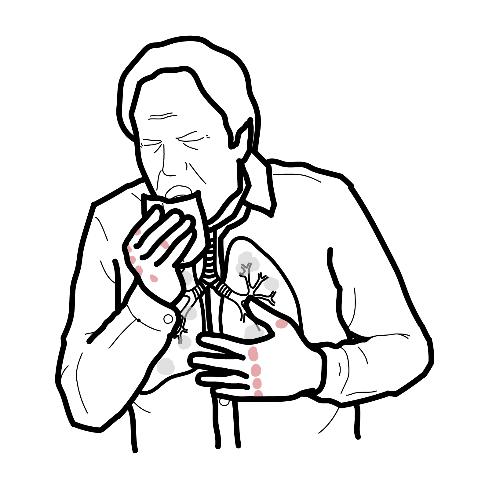
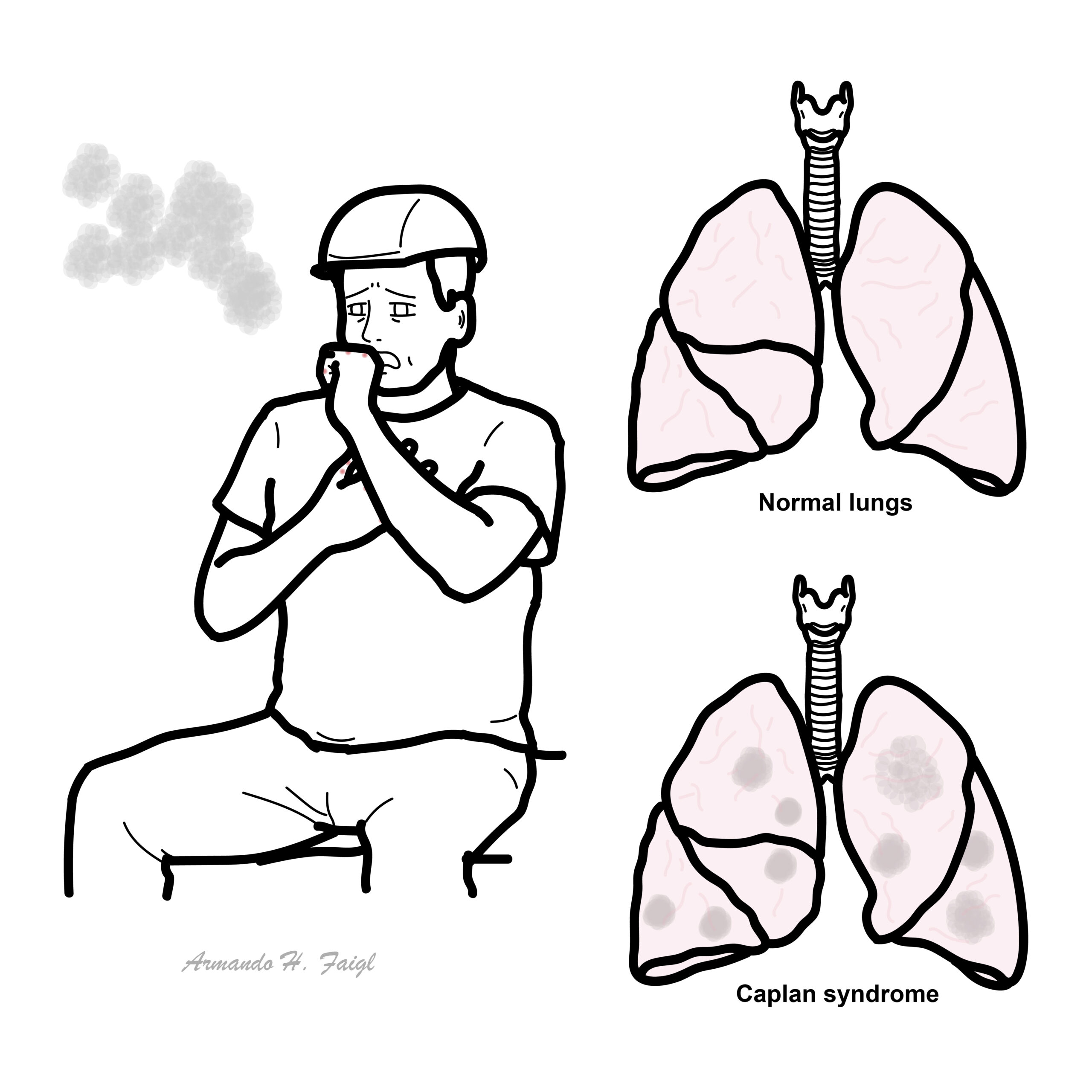
Caplan syndrome, also known as rheumatoid pneumoconiosis, is a rare condition seen in patients with rheumatoid arthritis (RA) who have had occupational exposure to inhaled dust, particularly coal dust or silica. It is characterised by multiple, rounded pulmonary nodules and underlying pneumoconiosis. It primarily affects middle-aged male coal workers and is now rare in developed countries due to improved occupational safety. Nodules are typically asymptomatic but may be associated with cough, dyspnoea, or constitutional symptoms in active RA.
Definition
Rheumatoid arthritis (RA): Chronic autoimmune inflammatory disorder targeting synovial joints with systemic manifestations.
Pneumoconiosis: Lung disease caused by inhalation of dust particles (e.g., coal, silica), leading to fibrosis.
Caplan nodules: Rounded, necrobiotic lung nodules in patients with RA and dust exposure.
Silicosis: Progressive lung disease caused by inhalation of silica dust, often seen in mining or construction workers.
What to notice
Detailed image description
Anatomy & Physiology
- Site of Caplan nodules, primarily upper lobes; composed of alveoli and interstitium
- RA-related autoimmunity can lead to granuloma-like nodule formation in the lungs
- Inhaled particles normally cleared by mucociliary apparatus or alveolar macrophages; failure leads to fibrosis and nodules
Aetiology & Risk Factors
Aetiology
- Interaction between RA autoimmunity and inhaled dust exposure (coal, silica)
- Immunological reaction to retained dust in lungs leads to granulomatous nodule formation
- Seropositive RA (especially RF or anti-CCP positive)
- Occupational exposure to silica, coal, asbestos, kaolin
- Male sex
- >10 years working in mining or construction
- Smoking (may exacerbate lung disease)
Caplan syndrome occurs only in patients with RA + dust exposure — both are required1.
Pathophysiology
- Inhalation of dust (silica, coal) → phagocytosed by alveolar macrophages
- Macrophages activate and release cytokines → chronic inflammation and fibrosis
- Patients with RA develop immune-mediated granulomas around dust particles
- Necrobiotic nodules form, often with central necrosis and peripheral inflammation
- Nodules may enlarge or coalesce and mimic malignancy or infection
Caplan syndrome differs from simple pneumoconiosis by the presence of RA and immunologically driven nodules.
Clinical Manifestations
- Often asymptomatic in early stages
- Pulmonary: dry cough, dyspnoea, wheeze
- Constitutional: fever, weight loss, fatigue (often from active RA)
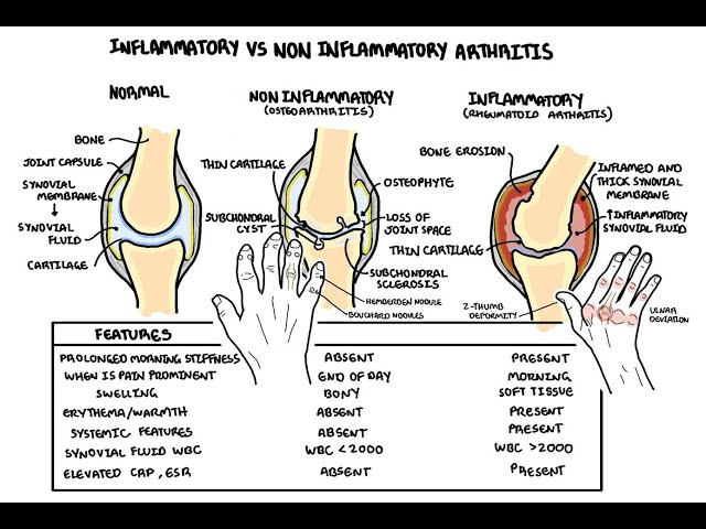
- Signs of RA: including joint swelling, morning stiffness, rheumatoid nodules
- Crackles or wheeze may be heard on chest auscultation.
Diagnosis
No formal diagnostic criteria
• Known RA
• History of relevant dust exposure
• Radiographic findings: multiple round nodules (0.5–5 cm), typically in upper lobes
Investigations
• Chest X-ray or CT: rounded peripheral nodules, usually bilateral and upper lobe predominant
• Pulmonary function tests: may show restrictive defect or normal
• Serology: positive RF or anti-CCP
• BAL (bronchoalveolar lavage) or biopsy if nodules are atypical or rapidly enlarging (to exclude malignancy or infection)
• ESR/CRP: may be elevated with RA activity
Differential diagnoses
| Condition | Key Features | Differentiating Points |
| Silicosis | Nodules + exposure | No RA, no serology positivity |
| Sarcoidosis | Bilateral hilar lymphadenopathy | Non-caseating granulomas, systemic involvement |
| Lung cancer | Solitary nodule or mass | Irregular margins, smoking history |
| Rheumatoid lung disease | Interstitial pattern | Diffuse fibrosis, not rounded nodules |
Triad: Rheumatoid arthritis, Occupational dust exposure and Pulmonary nodules on imaging.
Treatment
RA management:
- Methotrexate
- Leflunomide, sulfasalazine, biologics
Avoid further dust exposure
Symptomatic pulmonary care:
- Inhaled bronchodilators (if airway involvement)
- Corticosteroids may help with nodule inflammation
Surgical intervention: rarely needed; biopsy may be performed if diagnosis unclearMonitor for superimposed infection, nodule growth, malignancy.
Withdrawal from exposure is crucial — lung nodules often stabilise or regress once exposure ceases2.
Complications & Prognosis
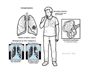
• Nodule cavitation, rupture
• Secondary infection
• Misdiagnosis as TB or malignancy
• Interstitial lung disease (in advanced cases)
• Prognosis generally good if exposure is discontinued and RA controlled
References
- Shaw M, Collins BF, Ho LA, Raghu G. Rheumatoid arthritis–associated lung disease. Eur Respir Rev. 2015;24(135):1–16.
- Blanc PD, Annesi-Maesano I, Balmes JR, et al. The occupational burden of nonmalignant respiratory diseases. An official American Thoracic Society and European Respiratory Society statement. Am J Respir Crit Care Med. 2019;199(11):1312–34.
- Wells AU, et al. Rheumatoid lung disease: Clinical, radiological, and histological patterns. Semin Respir Crit Care Med. 2019;40(2):194–203.









Members only discussions coming soon…