

Paget's Disease of the Breast

Overview
Paget’s disease of the breast is a rare cutaneous manifestation of an underlying breast malignancy, accounting for approximately 1% to 4% of all breast cancers. It is characterized by the intraepithelial infiltration of adenocarcinoma cells (known as Paget cells) into the epidermis of the nipple-areolar complex.
Over 85% to 95% of patients with Paget’s disease of the breast have an underlying ipsilateral breast carcinoma. This is most commonly Ductal Carcinoma In Situ (DCIS) or Invasive Ductal Carcinoma (IDC). Because the clinical presentation closely mimics benign dermatological conditions like eczema or contact dermatitis, diagnostic delays are common. Recognizing the characteristic unilateral distribution and failure to respond to topical therapy is critical for early biopsy and diagnosis.
Definition
Paget Cells: Large, atypical epithelial cells with pale or clear cytoplasm, large hyperchromatic nuclei, and prominent nucleoli residing within the epidermal layer of the skin.
Epidermotropism Theory: The predominant pathophysiological theory stating that malignant cells migrate from an underlying ductal carcinoma up through the lactiferous ducts and into the epidermis of the nipple.
Nipple-Areolar Complex (NAC): The central superficial anatomical structure of the breast comprising the nipple and the surrounding pigmented skin (areola).
Aetiology & Pathophysiology
- Epidermotropic Migration: Adenocarcinoma cells originating in the epithelium of underlying lactiferous ducts migrate upward along the ductal system into the basal layer of the nipple epidermis.
- Immunohistochemical Markers: Paget cells characteristically overexpress the HER2/neu oncoprotein in up to 80% to 90% of cases. They test positive for Cytokeratin 7 (CK7), Carcinoembryonic Antigen (CEA), and mucin (Mucicarmine stain).
- Epidermal Barrier Disruption: Infiltration of Paget cells disrupts normal keratinocyte cell-to-cell adhesion (desmosomes), causing extracellular fluid transudation, scaling, hyperkeratosis, and chronic superficial erosion.
Paget cells are HER2/neu positive in the vast majority of cases (80–90%), reflecting their origin from high-grade underlying ductal carcinoma.
Clinical Manifestations
Primary Cutaneous Features
- Eczematous Nipple Lesion
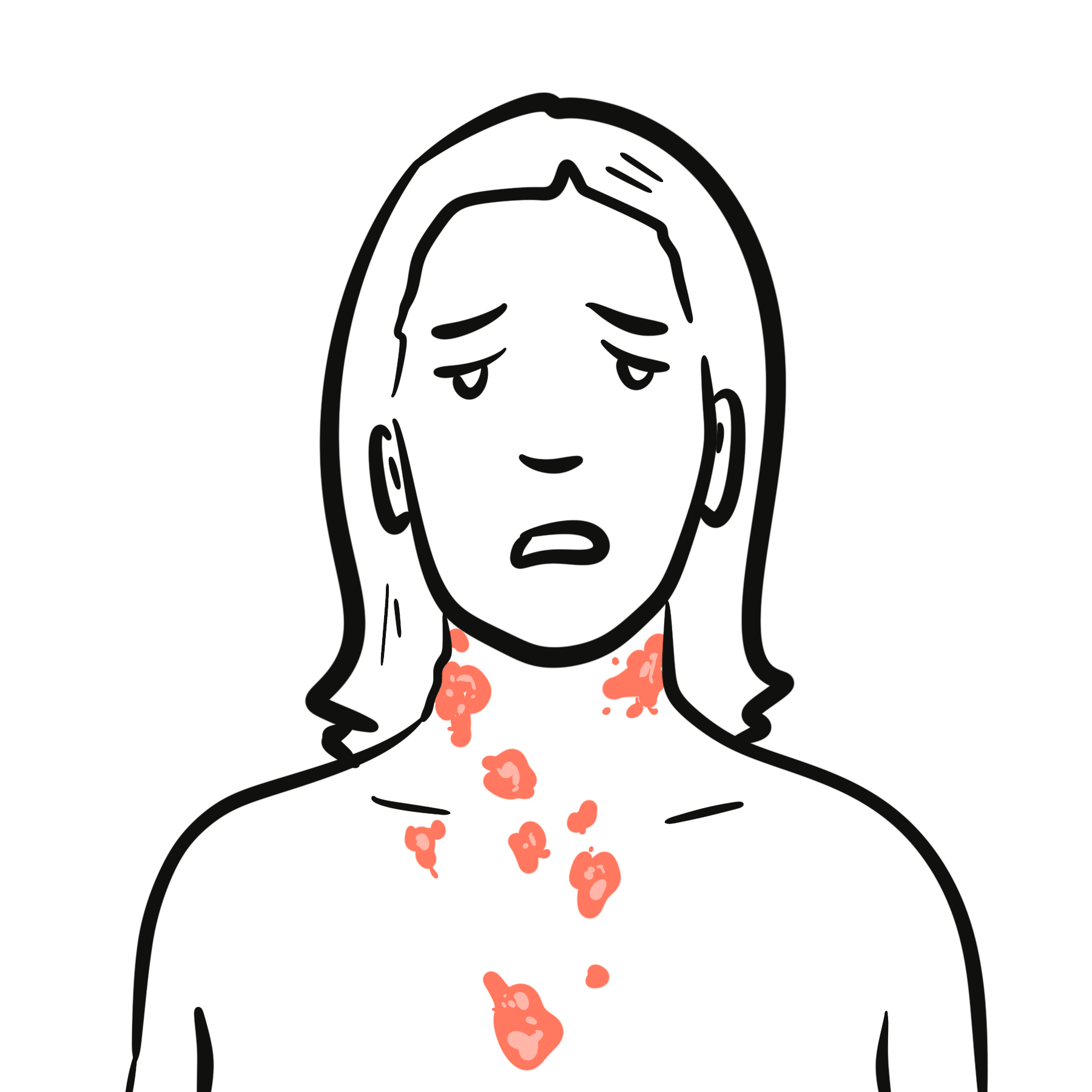
- Unilateral, slow-growing, erythematous, scaly, or crusty plaque
- Characteristically starts on the nipple and extends centrifugally onto the surrounding areola.
- Itching (pruritus), burning, tingling, or localized pain at the nipple.
- Structural Nipple Changes
- Flattening
- Retraction
- Inversion, or
- Frank erosion/ulceration of the nipple.
- Nipple Discharge: Serosanguinous or bloody discharge from the affected nipple.
Palpable Findings
- Palpable Subareolar Mass: Present in approximately 50% of patients upon clinical breast examination.
Clinical Differentiation: Paget’s Disease vs. Benign Eczema
| Clinical Feature | Paget’s Disease of the Breast | Benign Eczema / Dermatitis |
| Laterality | Almost strictly Unilateral | Frequently Bilateral |
| Primary Site of Origin | Nipple first, then spreads to areola | Areola first, often spares the nipple |
| Nipple Structure | Often destroyed, retracted, or eroded | Nipple structure remains intact |
| Palpable Mass | May be present (~50%) | Absent |
| Topical Steroid Response | No response | Rapid improvement |
Any unilateral “eczema” of the nipple that fails to resolve after a short trial (2–3 weeks) of topical corticosteroids is Paget’s disease until proven otherwise and requires a punch biopsy.
Diagnosis
Diagnostic Biopsy (Gold Standard)
- Full-Thickness Nipple Punch Biopsy / Wedge Biopsy: Gold standard diagnostic procedure. Histology reveals intraepidermal infiltration of single or clustered Paget cells with abundant pale cytoplasm.
- Immunohistochemistry (IHC) Panel:
- Positive: CK7, HER2/neu, CEA, Mucicarmine.
- Negative: S100, HMB-45, and Melan-A (essential to rule out cutaneous malignant melanoma).
Imaging Studies
- Bilateral Diagnostic Mammography: Evaluates for underlying microcalcifications (indicative of DCIS), soft tissue masses, or architectural distortion.
- Targeted Breast Ultrasound: Used to evaluate subareolar masses or suspicious findings identified on mammography.
- Breast MRI: Highly sensitive test indicated when mammography and ultrasound are negative to detect occult underlying DCIS or multifocal invasive disease prior to surgical planning.
A negative mammogram does NOT rule out Paget’s disease or an underlying malignancy; a full-thickness nipple biopsy is still mandatory.
Treatment
Surgical Management
- Breast-Conserving Surgery (BCS): Central wide local excision incorporating complete resection of the entire nipple-areolar complex (NAC) with clear surgical margins, followed by mandatory whole-breast radiation therapy.
- Total Mastectomy: Indicated for patients with extensive underlying DCIS, multifocal/multicentric invasive cancer, large tumor-to-breast ratio, or patient preference.
- Axillary Staging: Sentinel Lymph Node Biopsy (SLNB) is indicated if an invasive carcinoma component is identified, or if a total mastectomy is performed (to avoid missing invasive disease that would prevent future SLNB).
Systemic Adjuvant Therapy
Guided by the stage and receptor status of the underlying breast carcinoma:
- Endocrine Therapy: Tamoxifen or Aromatase Inhibitors if Estrogen Receptor (ER) positive.
- HER2-Targeted Therapy: Trastuzumab (Herceptin) if HER2 positive with invasive disease.
- Systemic Chemotherapy: Administered based on tumor size, grade, and axillary lymph node status.
Complications & Prognosis
The overall prognosis of Paget’s disease is not determined by the nipple lesion itself, but rather by the presence, stage, and biological characteristics of the underlying breast carcinoma.
Survival Rates
- Without Palpable Mass / Pure DCIS: Excellent prognosis; 5-year overall survival exceeds 90% to 95%.
- With Palpable Mass / Invasive Carcinoma: Overall 5-year survival drops to ~60% to 70% (and further declines if axillary lymph node metastases are present).
References
- Kanitakis J. Mammary and extramammary Paget’s disease. J Eur Acad Dermatol Venereol. 2007;21(5):581-590. doi:10.1111/j.1468-3083.2007.02154.x
- Dixon AR, Galea MH, Ellis IO, Elston CW, Blamey RW. Paget’s disease of the nipple. Br J Surg. 1991;78(6):722-723. doi:10.1002/bjs.1800780628
- Marshall JK, Griffith KA, Haffty BG, et al. Conservative management of Paget disease of the breast with radiotherapy: 10-year results. Cancer. 2003;97(9):2142-2149. doi:10.1002/cncr.11337















Members only discussions coming soon…