Pneumonia
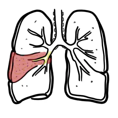

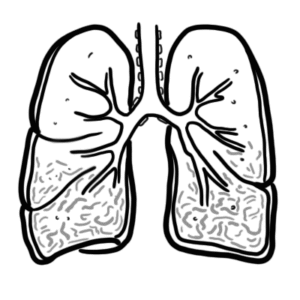
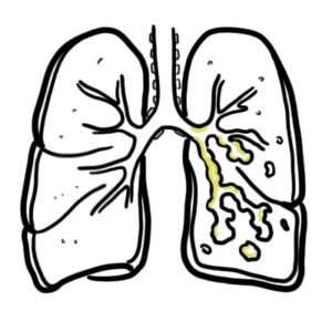
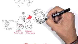
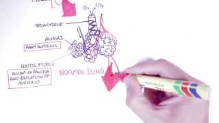
Pneumonia is infection in the lower respiratory tract and is characterised by fevers, purulent sputum, leucocytosis, low oxygen levels and new lung infiltrates (consolidation) on radiographic imaging. The infection is identified by the site of consolidation. Consolidation is where the lung tissue is filled with fluid (ie. water, blood, pus, microbes). Types of pneumonia include lobar pneumonia (infection affects a whole lobe of a lung) and bronchopneumonia (affects the bronchioles through out a lung lobe).
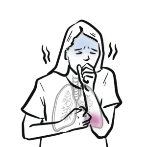
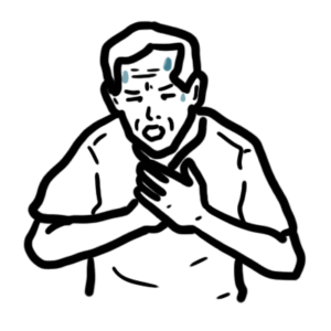
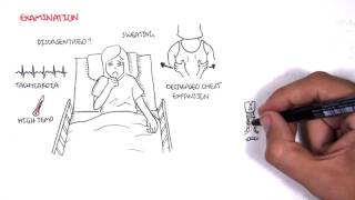
Cardinal symptoms: Cough (productive), Dyspnoea, Pleuritic chest pain, Fever.
Examination findings include signs of consolidation on affect side and lobe
Causative agent
| S. pneumoniae | H. influenzae | S. aureus | M. pneumoniae | M. catarrhalis | Gram – Enterobacteriae | Klebsiella spp. | Legionalla spp. | |
| Community-acquired Pneumonia | ||||||||
| Hospital-acquired Pneumonia | ||||||||
| Health-care Pneumonia |
Classification by location acquired
Hospital acquired pneumonia is lower respiratory infection that was not incubating at the time of hospital admission and that presents clinically 2 or more days after hospitalization.
Classification by microorganism
Classification by lung involvement
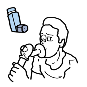
Management depends on where the pneumonia was acquired and what the causative agent is or might be. The standard is antibiotic therapy.
Assessment of severity
There are scoring criteria’s that exists to help identify whether a patient should be admitted to hospital. These include PSI and CURB-65 (CORB-65)
Some patients with mild illness, good social circumstances and no significant co-morbidities may be safely discharges with oral antibiotics, simple analgesia for pleuritic chest pain and follow up with doctors.
For patients with a high severity score (CURB-65 or SMART-COP or PSI index) admission to hospital is advised with commencement of IV antibiotics.
Summary of Pneumonia
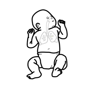
Pneumonia involves symptoms and sign of lower respiratory tract infections usually associated with chest x-ray abnormalities (consolidation). The majority of causes are bacterial (80%). Finding on examination may show signs of consolation. Risk factors include smoking, young and old age and having an underlying respiratory disease. Treatment is antibiotic therapy.


Discussion