

Pulmonary Embolism
Overview
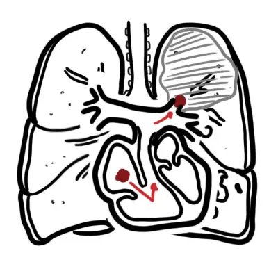
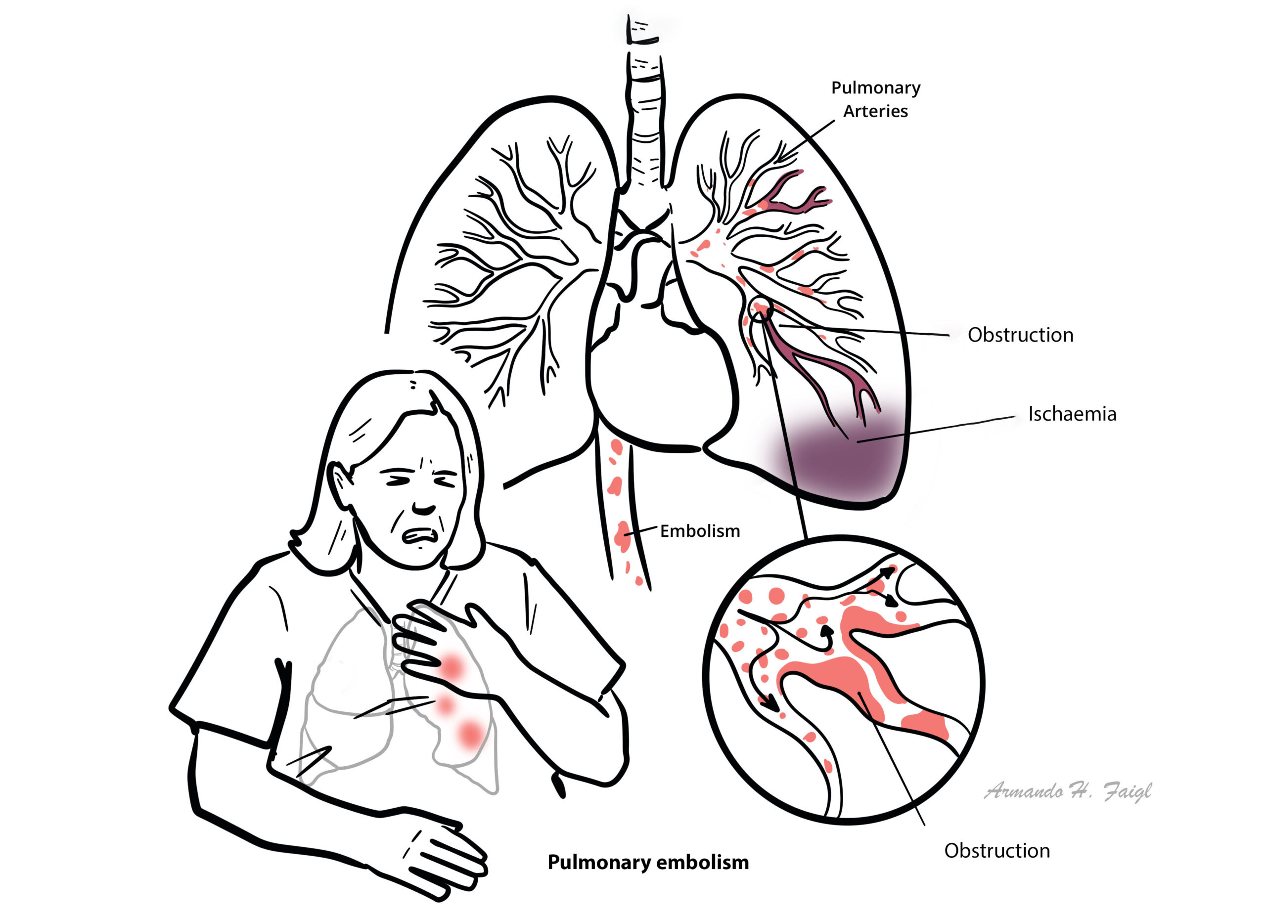
Pulmonary embolism occurs most often from a dislodged thrombus from the lower limb (DVT). The embolus travels towards the lung and becomes lodged in the pulmonary artery resulting in infarction of the lung tissue.
What to notice
Detailed image description
Definition
Pulmonary Embolism:
Deep Vein Thrombosis (DVT): Blood clot in the deep venous system that usually affects the lower extremities or pelvic veins.
Summary of Pulmonary Embolism
The mortality of diagnosed and treated PE is 7%. Many more people die from undiagnosed PE. Most patients present with dyspnea +/- chest pain. The main risk factors is the development of DVT from immobility; surgery, pregnancy, chronic disease, increasing age. There are two forms of imaging for PE: CT pulmonary angiography and V/Q scanning. Other investigations include bloods taking a d-dimer test. General treatment includes IV heparin followed by oral warfarin therapy. For massive (serious) PE thrombolytic therapy is initiated.
Aetiology & Risk Factors
Pulmonary Embolism usually arise from Deep Vein Thrombosis (DVT). DVT is the most common in patient over 40 years of age who undergo major surgery.
Genetic predisposition to hypercoagulability accounts for approximately 20% of PEs. The most common inherited conditions are the factor V Leiden mutation and the prothrombin gene mutations.
Clinical Manifestations
- Sudden or worsening dyspnoea
- Pleuritic chest pain
- Cough ± haemoptysis
- Palpitations
- Dizziness, presyncope or syncope
- Unexplained anxiety or reduced exercise tolerance
- May be asymptomatic and found incidentally
Always suspect pulmonary embolism (PE) in sudden collapse 1-2weeks after surgery.
Examination findings
- Tachypnoea
- Tachycardia
- Hypoxaemia
- Low-grade fever
- Signs of DVT: unilateral leg swelling, tenderness or erythema
- Pleural rub or reduced breath sounds
- Severe/high-risk PE:
- Hypotension
- Raised JVP
- Cyanosis
- Acute right-heart failure
- Obstructive shock or cardiac arrest.
Diagnosis
- ECG: commonly sinus tachycardia; may show right-heart strain or S1Q3T3, but cannot confirm or exclude PE
- Chest X-ray: often normal or non-specific; mainly excludes alternative diagnoses
- Blood tests:
Diagnostic pathway
- Assess clinical probability using the Wells score
- Very low clinical probability: consider PERC
- PE unlikely: D-dimer
- Negative → PE generally excluded
- Positive → imaging
- Consider age-adjusted D-dimer in patients aged over 50
- PE likely: proceed directly to imaging
- CT pulmonary angiography: first-line diagnostic imaging
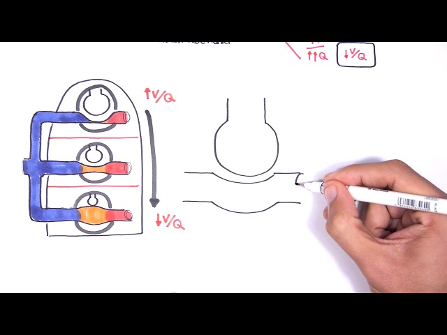
- V/Q scan: alternative with contrast allergy, significant renal impairment or where radiation considerations favour it
- Echocardiography: assesses RV dysfunction, particularly in unstable PE; a normal echo does not exclude PE in stable patients
PE is incorrectly diagnosed in almost 75% if patients. Acute onset of dyspnea or hypoxemia with a normal chest x-ray should be considered a pulmonary embolism until proven otherwise.
Treatment
Immediate management
- ABC assessment and cardiac monitoring
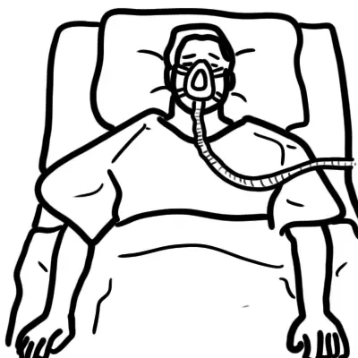
- Oxygen if hypoxaemic
- Analgesia
- Treat hypotension/shock with cautious IV fluids and vasopressors where required
- Begin therapeutic anticoagulation while awaiting imaging when clinical suspicion is significant and diagnostic testing is delayed, provided bleeding risk is acceptable.
Haemodynamically stable PE
- Anticoagulation is the main treatment
- DOACs generally preferred:
- Apixaban
- Rivaroxaban
- Dabigatran or edoxaban after initial parenteral anticoagulation
- Alternatives:
- LMWH
- Warfarin after LMWH/UFH bridging
- LMWH is preferred over UFH when initial parenteral treatment is needed in most stable patients.
Haemodynamically unstable/high-risk PE
- IV unfractionated heparin
- Urgent reperfusion:
- Systemic thrombolysis
- Catheter-directed thrombolysis or thrombectomy
- Surgical embolectomy
- Consider extracorporeal life support in selected refractory cases at specialist centres.
Duration
- Anticoagulation for at least 3 months
- Provoked PE with resolved transient risk factor: consider stopping after 3 months
- Unprovoked PE or persistent risk factor: consider extended or indefinite anticoagulation, balancing recurrence and bleeding risk
- Low-risk patients may be suitable for early discharge or outpatient treatment using a validated risk tool.
Special considerations
- Pregnancy: LMWH generally preferred
- Triple-positive antiphospholipid syndrome: warfarin preferred over a DOAC
- IVC filter only when anticoagulation is contraindicated or PE recurs despite adequately managed anticoagulation.
Complications & Prognosis
- Acute right-ventricular failure
- Obstructive shock
- Cardiac arrest and sudden death
- Pulmonary infarction, pleurisy or haemoptysis
- Recurrent DVT or PE
- Persistent dyspnoea and reduced exercise capacity—post-PE syndrome
- Chronic thromboembolic pulmonary disease
- Chronic thromboembolic pulmonary hypertension
- Treatment complications:
- Major bleeding
- Intracranial haemorrhage
- Heparin-induced thrombocytopenia.
References
- Creager MA, Barnes GD, Giri J, et al. 2026 AHA/ACC/ACCP/ACEP/CHEST/SCAI/SHM/SIR/SVM/SVN guideline for the evaluation and management of acute pulmonary embolism in adults. Circulation. 2026. doi:10.1161/CIR.0000000000001415.
- National Institute for Health and Care Excellence. Venous thromboembolic diseases: diagnosis, management and thrombophilia testing. NICE guideline NG158. London: NICE; 2020. Updated 2023.
- Konstantinides SV, Meyer G, Becattini C, et al. 2019 ESC guidelines for the diagnosis and management of acute pulmonary embolism. Eur Heart J. 2020;41(4):543–603.
- Ortel TL, Neumann I, Ageno W, et al. American Society of Hematology 2020 guidelines for management of venous thromboembolism: treatment of DVT and PE. Blood Adv. 2020;4(19):4693–4738.






Members only discussions coming soon…