

COPD
Overview
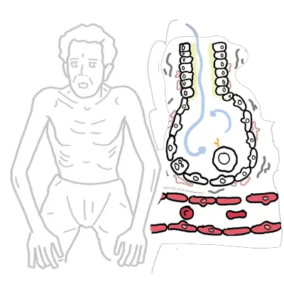
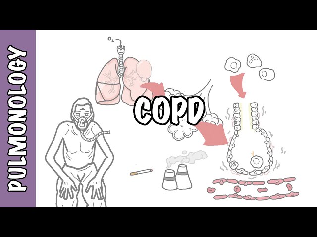
The diagnosis of COPD is usually limited to individuals who have chronic airflow obstruction associated with tobacco smoke or some other noxious inhalant, and it is usually not difficult to distinguish it from other causes of chronic airflow obstruction. The most commonly associated clinical disorders associated with COPD are emphysema and chronic bronchitis.
Definition
Chronic Obstructive Lung Disease: A serious, progressive and disabling condition that limits airflow in the lungs. It includes emphysema and chronic bronchitis. Strongly associated with smoking
Interstitial Lung Disease: Includes a diverse group of respiratory conditions characterised by inflammation and fibrosis of the interstitium. Worsening hypoxia and respiratory failure may develop with disease progression. The disease is considered a restrictive lung disease
Restrictive Lung Disease: Unable to fully fill the lungs with air. Their lungs are restricted from fully expanding.
Risk Factors
- Smoking
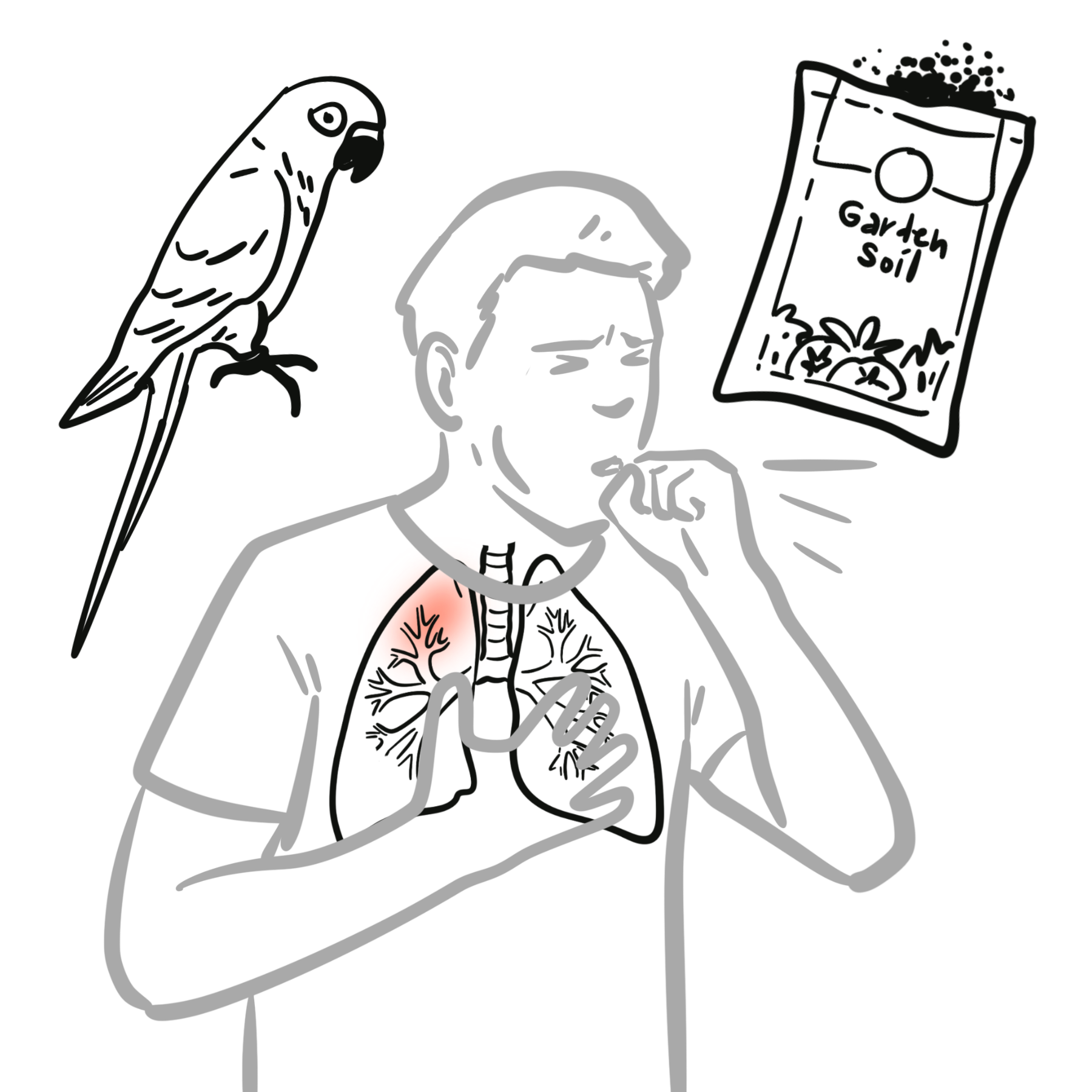
- Air-pollution
- α1-antitrypsin deficiency
Clinical Manifestations
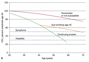
COPD results from an increase in the rate of decline in lung function over time.
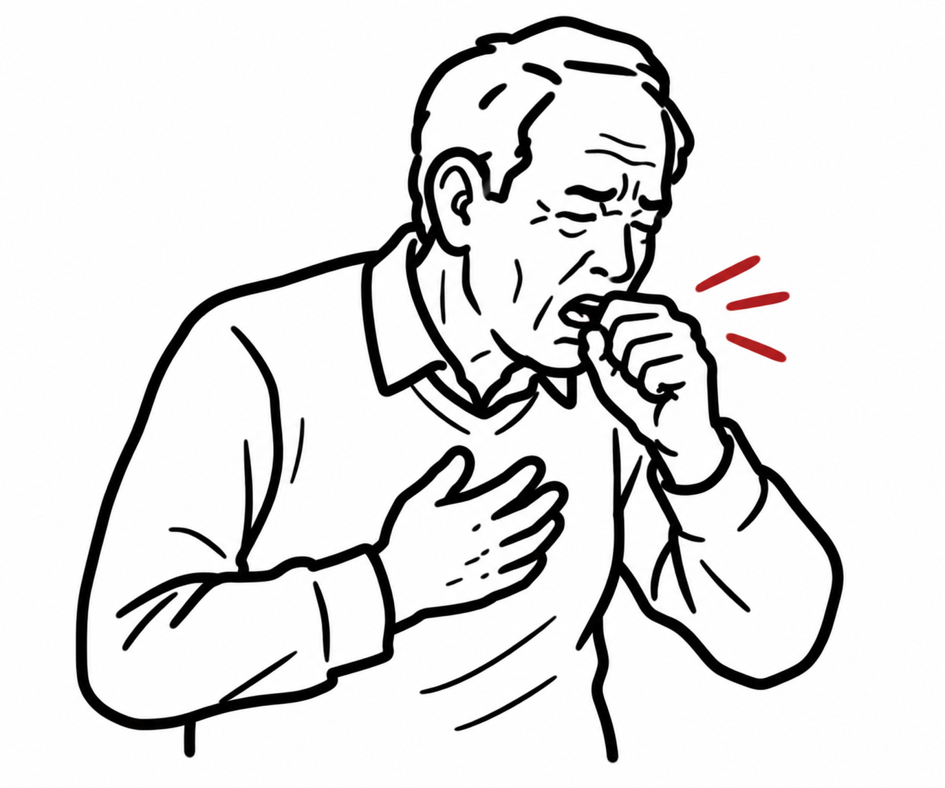
Cardinal Triad of COPD: (exertional) Dyspnoea, Chronic cough and sputum production.
Clinical manifestations
- Progressive exertional dyspnoea — most characteristic symptom
- Chronic cough, which may initially be intermittent
- Sputum production
- Wheeze or chest tightness
- Reduced exercise tolerance and activity limitation
- Fatigue
- Recurrent lower respiratory tract infections
- Acute worsening of dyspnoea, cough and/or sputum during exacerbations.
Clinical Examination
Examination may be normal in early disease. In established COPD:
- Tachypnoea
- Prolonged expiration
- Expiratory wheeze
- Reduced breath sounds
- Hyperresonant percussion
- Use of accessory respiratory muscles
- Pursed-lip breathing
- Barrel-shaped/hyperinflated chest
- Reduced chest expansion
- Tripod positioning
- Low body weight and muscle wasting in advanced emphysema
- Cyanosis in severe hypoxaemia
- Signs of cor pulmonale:
- Raised JVP
- Peripheral oedema
- Parasternal heave
- Loud pulmonary component of S2
“Pink puffers” and “blue bloaters”
These are historical terms and are no longer recommended in modern clinical practice because COPD is heterogeneous and most patients have overlapping airway disease and emphysema. They are not used for current diagnosis or treatment classification.
- “Pink puffer” traditionally described emphysema-predominant COPD:
- Severe dyspnoea
- Thin/cachectic appearance
- Pursed-lip breathing
- Hyperinflation
- Relatively preserved oxygenation until later disease
- “Blue bloater” traditionally described chronic-bronchitis-predominant COPD:
- Chronic productive cough
- Cyanosis and hypoxaemia
- Peripheral oedema/cor pulmonale
- Hypercapnia
- Often described as having a higher body weight
Use “emphysema-predominant COPD” or “chronic bronchitis phenotype” instead
Investigations
- Spirometry—essential diagnostic test
- Perform pre- and post-bronchodilator spirometry.
- Persistent post-bronchodilator FEV₁/FVC <0.70 confirms airflow obstruction consistent with COPD.
- Repeat spirometry if results are borderline or inconsistent with the clinical presentation.
- Assess severity and impact
- Chest X-ray
- Excludes alternatives such as pneumonia, heart failure, lung cancer or pneumothorax.
- May show hyperinflation but does not diagnose COPD.
- Blood tests
- FBC: anaemia, polycythaemia, infection and eosinophil count.
- UEC/creatinine and LFTs before some treatments.
- Consider BNP/troponin if cardiac disease suspected.
- Alpha-1 antitrypsin testing should be considered in all patients with chronic airflow obstruction, particularly younger patients, non-smokers or those with a family history.
- Arterial blood gas
- Severe COPD, low oxygen saturation, suspected hypercapnia, acute respiratory failure or assessment for long-term oxygen.
- Full pulmonary function tests
- Lung volumes and gas transfer/DLCO where symptoms are disproportionate, diagnosis is uncertain or surgery is being considered.
- CT chest—not routine: consider for emphysema distribution, bullae, bronchiectasis, lung cancer, unexplained symptoms or lung-volume-reduction assessment.
- Other investigations as indicated
- ECG ± echocardiogram: cardiac disease, pulmonary hypertension or cor pulmonale.
- Six-minute walk test: exercise limitation and desaturation.
- Sputum microscopy/culture: treatment failure, recurrent infection or suspected resistant organisms; not routinely required in stable COPD.
| Emphysema | Chronic Bronchitis |
| Defined Pathologically Dilation and destruction of air spaces distal to the terminal bronchiole without obvious fibrosis. Decreased elastic recoil of alveoli causing decreased expiratory driving pressure, airway collapse and air trapping. | Defined Clinically Productive cough on most days for at least 3 consecutive months in 2 successive years. Obstruction is due to narrowing of the airway lumen by mucosal thickening and excess mucus |
Differential Diagnosis
| COMMON CAUSES OF CHRONIC COUGH |
| COPD |
| Asthma |
| GORD |
| Post nasal drip |
| COMMON CAUSES OF CHRONIC DYSPNOEA | |
| Respiratory Disease | Cardiovascular Disease |
| COPD | Myocardial Dysfunction (Heart Failure) |
| Asthma | Obesity/de-conditioning |
| Interstitial lung disease | |
Classification
| COPD Severity is based on FEV1 (COPD is diagnosed by FEV1/FVC) |
| Mild FEV1 <.7 |
| Moderate FEV1 0.5-0.3 |
| Severe FEV1 <0.3 |
The hallmark of restrictive lung disease is decreased lung capacities, particularly the TLC but also the vital capacity.
In both obstructive and restrictive lung disease, the FEV1 is decreased, the FEV1 /FVC is decreased in obstructive processes and normal in restrictive processes.
| MRC Dyspnea score | |
| 1 | Not troubled by breathlessness except on streous exercise |
| 2 | Dyspnea when hurrying or walking up a slight hill |
| 3 | Walks slower than contemprorores on level ground due to dyspnea, or has to stop for breath when talking at own pace |
| 4 | Stops for breath after walking 100m or less on ground level |
| 5 | Too breathlesss to leave house, or when getting dressed |
Treatment
Non-pharmacological treatment
- Smoking cessation
- Vaccination:
- Influenza
- Pneumococcal
- COVID-19
- RSV where eligible.
- Encourage regular physical activity
- Pulmonary rehabilitation for exertional breathlessness, reduced exercise tolerance or following hospitalisation.
- Optimise nutrition and manage anxiety, depression and comorbidities.
- Check inhaler technique and adherence at every review.
- Provide a written COPD action plan.
Smoking cessation and supplemental oxygen to treat chronic hypoxemia are the only medical therapies shown to decrease mortality among persons with chronic obstructive pulmonary disease.
Inhaled treatment
- Reliever: short-acting bronchodilator as required:
- SABA, e.g. salbutamol.
- ± SAMA, e.g. ipratropium.
- Persistent symptoms: commence a long-acting bronchodilator:
- LAMA or LABA.
- Ongoing breathlessness despite monotherapy: use LABA/LAMA dual therapy.
- Recurrent exacerbations despite long-acting bronchodilators:
- Consider adding an inhaled corticosteroid, particularly with raised blood eosinophils or coexisting asthma.
- Usually given as LABA/LAMA/ICS triple therapy.
- ICS should generally not be used alone in COPD.
- ICS adverse effects include oral candidiasis, dysphonia and increased pneumonia risk.
Selected advanced treatments
- Mucolytic therapy in selected patients with chronic productive cough or frequent exacerbations.
- Long-term macrolide therapy for selected frequent exacerbators under specialist supervision.
- Roflumilast may be considered for severe COPD with chronic bronchitis and recurrent exacerbations.
- Long-term oxygen therapy for stable severe resting hypoxaemia:
- PaO₂ <55 mmHg, or SpO₂ <88%.
- Also consider PaO₂ 55–59 mmHg with pulmonary hypertension, right-heart failure or secondary polycythaemia.
- Selected severe emphysema: bullectomy, bronchoscopic or surgical lung-volume reduction, or lung transplantation.
- Avoid routine long-term oral corticosteroids.
Acute COPD exacerbation
- Increase SABA ± SAMA by inhaler and spacer or nebuliser.
- Controlled oxygen targeting SpO₂ 88–92%.
- Prednisolone 30–50 mg daily for 5 days for moderate–severe exacerbations.
- Antibiotics, usually for approximately 5 days, when there is:
- Purulent sputum, particularly with increased volume or dyspnoea.
- Pneumonia.
- Requirement for mechanical ventilation.
- Obtain blood gas in severe exacerbations.
- Non-invasive ventilation for persistent hypercapnic respiratory acidosis despite optimal medical treatment.
- Treat the precipitating cause and exclude pneumonia, pulmonary embolism, pneumothorax, arrhythmia and heart failure
Complications & Prognosis
Complications
- Acute exacerbations
- Pulmonary hypertension due to chronic hypoxia.
- Cor pulmonale: right-sided heart failure secondary to pulmonary hypertension.
- Pneumothorax, particularly with bullous emphysema.
- Secondary polycythaemia from chronic hypoxaemia.
- Weight loss, muscle wasting, frailty and reduced exercise capacity.
- Anxiety, depression, social isolation and impaired quality of life.
- Increased risk of cardiovascular disease, osteoporosis, lung cancer and venous thromboembolism.
Prognosis
- COPD is a chronic, progressive disease; damaged lung tissue is generally irreversible.
- The rate of progression varies considerably, and lung-function decline may be slowed by smoking cessation and appropriate management.
- Frequent exacerbations and previous hospital admissions predict further exacerbations and poorer survival.
- Hospitalisation with hypercapnic respiratory failure, pneumonia or significant cardiac disease indicates a poor prognosis.
- Prognosis is better assessed using several factors—such as the BODE index: BMI, airflow obstruction, dyspnoea and exercise capacity—rather than FEV₁ alone.
References
- Global Initiative for Chronic Obstructive Lung Disease. Global strategy for prevention, diagnosis and management of COPD: 2026 report. GOLD; 2026. (goldcopd.org)
- Yang IA, George J, McDonald CF, Disler R, Ordman R, Goodwin A, et al. The COPD-X Plan: Australian and New Zealand guidelines for the management of chronic obstructive pulmonary disease 2025. Version 2.78. Lung Foundation Australia; 2025. (COPD-X Guidelines)
- Nici L, Mammen MJ, Charbek E, Alexander PE, Au DH, Boyd CM, et al. Pharmacologic management of chronic obstructive pulmonary disease: an official American Thoracic Society clinical practice guideline. Am J Respir Crit Care Med. 2020;201(9):e56–e69. doi:10.1164/rccm.202003-0625ST. (PMC)
- Rochester CL, Alison JA, Carlin B, Jenkins AR, Cox NS, Bauldoff G, et al. Pulmonary rehabilitation for adults with chronic respiratory disease: an official American Thoracic Society clinical practice guideline. Am J Respir Crit Care Med. 2023;208(4):e7–e26. doi:10.1164/rccm.202306-1066ST. (PubMed)
- Wedzicha JA, Miravitlles M, Hurst JR, Calverley PMA, Albert RK, Anzueto A, et al. Management of COPD exacerbations: a European Respiratory Society/American Thoracic Society guideline. Eur Respir J. 2017;49(3):1600791. doi:10.1183/13993003.00791-2016. (PubMed)
Members only discussions coming soon…