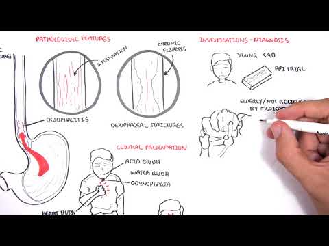
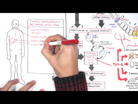
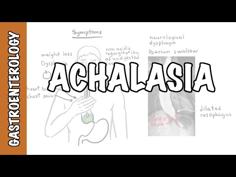
0:00 Hello, in this video we're going to talk about dysphagia. 0:09 Dysphagia is the sensation of difficulty or abnormality of swallowing. 0:14 It is due to a structural or a motility abnormality in the passage of solids or 0:19 liquids from the 0:19 mouth to the stomach. 0:21 It ranges from an inability to initiate the swallowing reflex, to foods or 0:27 liquids being 0:28 stuck in the esophagus. 0:30 In contrast, odenophagia is pain with swallowing. 0:35 The swallowing reflex is the process by which food is transported from the 0:40 mouth to the 0:41 stomach. 0:42 Anatomically, swallowing has been divided into three phases, oral, pharyngeal 0:49 and esophageal 0:50 phases. 0:52 The oral phase is where food is prepared into a bolus and the act of swallowing 0:58 occurs. 0:58 Swallowing involves a number of processes which will result in food entering 1:03 safely into 1:04 the oral pharynx. 1:07 During the pharyngeal phase, the tongue covers the oral pharynx and the epigl 1:11 ottis closes 1:12 the airway and the upper esophageal sphincter relaxes, allowing the food, the 1:18 bolus, to 1:19 move into the esophagus. 1:22 During the esophageal phase, the esophagus, including the lower esophageal sph 1:27 incter, 1:28 relaxes to receive the bolus. 1:31 As a result, a large part of a liquid bolus may move into the stomach by 1:35 gravity alone 1:36 if the person is standing. 1:38 The residual liquid bolus is cleared by the peristaltic contraction waves. 1:45 Solid bolus usually does not move down by gravity and requires peristaltic 1:50 contraction 1:51 for its transport. 1:53 The lower esophageal sphincter is a physiological sphincter and contributes by 1:59 multiple mechanisms. 2:00 These are the increasing tone of the muscularis in this area. 2:04 The right diaphragmatic cruis which contracts during sneezing and coughing to 2:10 prevent reflux 2:11 of content from the stomach and the angle of hiss which acts as a valve. 2:17 There are two main types of dysphagia, oropharyngeal and esophageal. 2:23 These two types are further classified as either being structural, meaning 2:28 obstructive 2:29 causes and propulsive or neurological causes. 2:36 A clinical pearl, dysphagia to solids may be indicative of a structural et 2:42 iology, whereas 2:44 dysphagia to either liquids alone or the combination of liquids and solids is 2:49 likely 2:50 a propulsive cause, a neurological cause. 2:54 Let's begin with oropharyngeal dysphagia. 2:59 So oropharyngeal dysphagia occurs when the patient is unable to transfer food 3:04 bolus from 3:05 the mouth into the upper esophagus by swallowing. 3:09 Structural or obstructive causes of oropharyngeal dysphagia include tumors of 3:13 the tongue and 3:14 the tonsils or a peritoneal abscess or quincy. 3:18 These pathologies cause an internal obstruction that leads to difficulty of 3:23 passing solids, 3:24 more so than liquids. 3:26 Zinca's diverticulum is in a quiet sack-like pouching of the mucosa and submuc 3:32 osa layers 3:33 originating from the pharyngeal esophageal junction. 3:38 Zinca's diverticulum should be considered when undigested food is brought up 3:43 several 3:43 hours after a meal or if a patient reports hearing a gurgling noise in the 3:50 chest. 3:51 The propulsive or neurological causes of oropharyngeal dysphagia includes 3:57 things such 3:57 as stroke, Parkinson's disease, motor neuron disease, multiple sclerosis or my 4:05 asthenia 4:06 gravis. 4:07 80% of patients with Parkinson's disease develop oropharyngeal dysphagia. 4:14 Motor neuron disease is a neurodegenerative disease affecting the motor nerve 4:19 fibers resulting 4:20 in muscle weakness and atrophy. 4:23 Studies have shown that the mechanism of dysphagia is due to progressive 4:28 degeneration of the 4:29 corticobolberar pyramidal fibers that control the swallowing center. 4:35 Oropharyngeal has been reported to be the only presenting complaint for myast 4:39 henia gravis, 4:39 especially in the elderly. 4:42 Myasthenia gravis is an autoimmune disease. 4:44 Normally nerves release neurotransmitters, acetylcholine, which binds to acetyl 4:50 choline 4:50 receptors on the muscle cells, and this will result in muscle contraction. 4:56 In myasthenia gravis, autoantibodies bind to acetylcholine receptors or musk, 5:00 which 5:01 is an enzyme important in neuromuscular junction development. 5:06 Binding of these antibodies to these receptor and musk result in less 5:11 transmission of nerve 5:12 impulses leading to muscle weakness. 5:16 A clinical pearl is myasthenia gravis has a strong association with thymoma, 5:21 thus a structural 5:22 or propulsive etiology may contribute to dysphagia in these patients. 5:29 The initial study for suspected oropharyngeal dysphagia is a modified barium 5:33 swallow with 5:34 both liquid and a solid phase to help identify the underlying cause. 5:40 Management strategies include actually treating the underlying cause, dietary 5:45 changes, and 5:45 a swallowing exercise program implemented with speech pathologists. 5:53 The second type of dysphagia after oropharyngeal dysphagia is esophageal dysph 5:59 agia. 6:00 In esophageal dysphagia, patients are able to initiate the swallowing process, 6:04 but often 6:05 feel discomfort in the mid to lower sternum as the food passes through the es 6:09 ophagus. 6:10 Again, esophageal dysphagia can be a result of two underlying causes, a 6:16 structural, which 6:17 is an obstructive cause, or propulsive, a neurological cause. 6:22 Internal causes of esophageal dysphagia are either due to internal obstructions 6:28 within 6:28 the esophagus or external compression of the esophagus, both of which occludes 6:34 the lumen. 6:35 In internal obstructive causes, these can include esophageal carcinoma. 6:42 A carcinoma will usually cause dysphagia by narrowing or blocking the lumen, 6:47 obstructing 6:48 the food that is swallowed, dysphagia that progresses from occurring with sol 6:53 ids to occurring with 6:54 solids and liquids suggests malignancy as this is slow growing. 7:01 Peptic strictures are the result of long-standing reflux esophagitis, or gourd. 7:07 A stricture is an esophageal narrowing, some are benign, and are the result of 7:12 prolonged 7:12 exposure to the gastric acid due to an incompetent lower sphincter. 7:17 Fibrosis and scarring ensue and it progresses transmurally, leading to dysphag 7:24 ia. 7:24 Peptic strictures may also be due to radiation exposure. 7:28 An upper endoscopy allows for both diagnostic inspection and therapeutic 7:32 intervention, which 7:33 is dilation. 7:37 Foreign bodies can also cause esophageal dysphagia, if they become lodged at a 7:41 point in the esophagus. 7:44 And there are three main locations that obstruction can occur due to normal 7:49 anatomical narrowing 7:49 of the esophagus. 7:51 These are at the level of the crack of pharyngeal muscles, at the carina as the 7:55 left bronchus 7:56 crosses the esophagus, and where the esophagus passes through the diaphragm. 8:04 External compression of the esophagus can lead to esophageal dysphagia. 8:09 Many mediastinal masses or an aortic aneurysm or actasia may contribute. 8:16 Posterior mediastinal masses may occur in children and are neurogenic tumors 8:21 such as schwannomas. 8:24 These are slow growing tumors which may compress the esophagus leading to the 8:27 sensation of 8:28 dysphagia. 8:30 In adults, anterior mediastinal masses are more likely and are more likely to 8:36 be of a more 8:36 sinister etiology such as a lymphoma. 8:42 Propulsive causes of esophageal dysphagia can be due to acalasia, scleroderma 8:48 or rarely 8:49 hypertensive peristalsis, nutscracker esophagus. 8:54 Acalasia is due to impaired relaxation of the lower esophageal sphincter. 8:58 It often leads to marked dilatation of the distal esophagus and stasis of food 9:03 in this 9:04 area. 9:05 Acalasia often presents with non-acidic regurgitation of undigestive food. 9:11 On a plain x-ray, a bird's beak sign is pathonomonic due to tapering of the 9:15 distal esophagus. 9:19 Disphasia to liquids is characteristic of acalasia. 9:25 Scleroderma is associated with decreased motility of any part of the 9:27 gastrointestinal 9:28 tract, but it more commonly affects the distal end of the esophagus. 9:33 Astrophagia is a condition characterized by fibrosis of the skin and internal 9:38 organs. 9:38 Hence, in the esophagus, it causes smooth muscle atrophy and fibrosis. 9:44 Disphasia is an extremely common complication of scleroderma. 9:49 Hypertensive peristalsis also known as nutcracker esophagus is a very rare 9:53 cause of dysphagia. 9:55 In this case, esophageal contractions are a very large amplitude, but the per 10:00 istaltic 10:01 motions are intact. 10:04 On barium swallowing, there is a characteristic corkscrew appearance. 10:11 For esophageal dysphagia, an endoscopy or biopsy should be performed if a 10:16 mechanical structural 10:17 pathology is suspected. 10:19 However, if a neurological cause of esophageal dysphagia is suspected, a barium 10:24 swallow study 10:25 can be done. 10:29 I hope you enjoyed this video on dysphagia. 10:32 Thank you for watching.