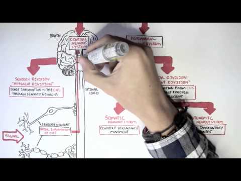
0:00 On Monday, how's it going on biology and medicine videos? 0:03 Please make sure to subscribe, join the forum and group for the latest videos, 0:06 please visit 0:06 Facebook and Monday's video. 0:08 Please like and here you can also ask questions, answer questions and post some 0:10 interesting 0:11 things including our works. 0:13 You can also change the quality settings to the highest one for better graphics 0:16 . 0:16 In this video, we're going to look at the cerebellum. 0:18 Now the cerebellum, where is it and what is its? 0:21 What is its function? 0:23 Here we have a sagittal view of our brain. 0:27 Now here we have the cerebrum. 0:29 Cerebellums sometimes get confused between the cerebrum and the cerebellum. 0:32 The cerebellum is located below the cerebrum, below the occipital lobe. 0:36 The cerebellum is also known as the little brain and is located in the dorsal 0:41 aspect of 0:41 the pons, behind the pons. 0:43 Roughly in between the cerebellum and the pons, we have what's called the 0:46 fourth ventricle. 0:48 The ventricle within the brain is important for the brain because it contains 0:52 cerebrospinal 0:53 fluid. 0:54 The cerebrospinal fluid or CSF, its main role in the brain is to provide 1:00 nutrition for the 1:01 different structures in the brain. 1:04 If here is the fourth ventricle, an important interesting to note is the third 1:08 ventricle is 1:08 situated here near the thalamus. 1:11 Now, the cerebellum, its main role in our body is to coordinate skeletal muscle 1:17 contraction 1:18 so that its precise, so that we don't fall down, so its basically helps us 1:23 coordinate 1:24 our movements. 1:25 So let's have a closer look at the cerebellum and have a closer look at the 1:29 anatomy. 1:30 So here we have the brain stem, what part of the brain stem? 1:33 Here we have the pons and here we have the cerebellum, dorsal of the pons. 1:36 The cerebellum consists of anterior lobes and posterior lobes. 1:42 It also consists of this tree-like structure which is composed of white matter 1:47 known as 1:48 the arbravite, A as in plural. 1:53 So now a very important thing to note is that the cerebellum connects to the p 1:59 ons, the midbrain 2:00 and the medulla oblamgara through the cerebular pendicles. 2:07 I hope I pronounced that right, cerebular pendicles. 2:09 And there are three cerebellar pendicles which connects the cerebellum 2:16 essentially to the 2:17 brain stem. 2:19 And these three are the superior pendicle, the middle pendicle and the inferior 2:25 pendicle. 2:25 So they connect again the cerebellum to the brain stem. 2:30 And they are important because they also send in and send out signals from the 2:36 cerebellum. 2:38 Another final thing to note is that cerebellum consists of two hemispheres. 2:43 The cerebrum, the main part of the brain you can say, also consists of two hem 2:46 ispheres. 2:47 And so from this we can deduce that the cerebellum also has two hemispheres. 2:53 But through this diagram we can't really tell. 2:54 So let's look at it from a different diagram from the bottom view, let's just 2:59 say. 3:00 So here we have another image diagram of the cerebellum. 3:04 Cerebellum here as you can see consists of two hemispheres. 3:09 And here we have the brain stem coming down. 3:13 What separates the two hemispheres or joins them together is an area known as 3:17 the vermis. 3:18 And here we have the anterior lobe and the posterior lobe of the cerebellum. 3:23 And this red line I'm drawing as you can see shows that the cerebellum consists 3:27 of two hemispheres. 3:28 Now let's stop there and go back to the cerebular pentacles. 3:35 Those three pentacles that are important in connecting the cerebellum to the 3:40 brain stem. 3:41 And they're important because it allows signals to be sent in and out of the 3:46 cerebellum. 3:47 So now here is a diagram of only the brain stem with a thalamus on the top. 3:53 Now this brain stem consists of the midbrain here, the pons and the medulla ob 3:58 longata. 3:59 And we're only concentrating on this area here which is the cerebellar pent 4:03 acles, part 4:04 of it, which connects the cerebellum to the brain stem. 4:08 Of course in this diagram I did not draw the cerebellum because we're only 4:12 concentrating 4:13 on these fibers. 4:14 I remember that we have three cerebellar pentacles or peduncles, some was 4:23 probably cold, starting 4:26 with the superior peduncle which connects the cerebellum to the midbrain and it 4:29 also provides 4:30 feedback signals to the cerebrum. 4:32 So the superior peduncle provides signals from the cerebellum to the cerebrum. 4:38 Next we have the middle peduncle which conveys impulses of voluntary movements 4:42 from the cerebrum 4:44 through the pons and to the cerebellum. 4:47 So the middle peduncle's role essentially is in giving signals from the cerebr 4:53 um, the 4:53 main part of the brain, to the cerebellum. 4:55 So it's the opposite of the superior peduncle. 4:58 The superior peduncle conveys impulses from the cerebellum to the cerebrum. 5:02 The middle peduncle conveys impulses from the cerebrum to the cerebellum, so 5:08 opposite. 5:09 And finally we have the inferior peduncle which connects the cerebellum to the 5:13 medulla 5:13 oblongara and the spinal cord. 5:16 And it contains both incoming vestibular and proprioceptive fibers and outgoing 5:21 motor fibers. 5:23 What the inferior peduncle essentially does is that it receives information 5:27 from our limbs 5:27 you can say, from our joints, from our muscles and interprets this. 5:32 And then it also sends out signals for coordinated movements together with the 5:37 cerebrum. 5:38 I hope that middle makes sense. 5:42 You have to understand it's not the cerebellum that sends out the motor signals 5:47 , it's the 5:48 cerebrum which actually sends out the motor signals, but it's the cerebellum 5:52 which assists 5:53 in coordination and interpreting the best possible way to send out the motor 5:59 signal being 6:00 sent out by the cerebrum if that makes sense. 6:03 Okay, let's just look at an overall picture first, so we can get a better 6:07 understanding 6:08 of the role the cerebellum has in the human body because it has a very big role 6:13 . 6:14 So if here is our cerebral cortex, the one that's actually going to send the 6:18 motor signals, 6:19 it will send the signal to the brain stem and the spinal cord or the spinal 6:23 cord. 6:23 And from the brain stem it will pass onto the spinal cord and then it will go 6:27 to the limbs 6:28 or different parts of our body for a movement. 6:32 I wrote here coordinated movements because the final product, the final output 6:37 is a coordinated 6:38 movement, but a coordinated movement cannot happen without the assistance of 6:43 the cerebellum. 6:45 So before the cerebral cortex can actually send out a coordinated movements, 6:51 the spinal 6:52 cord will send signals to the brain stem or to the cerebellum straight away 6:57 which will 6:58 tell the cerebellum the position of the muscle and the joints of different 7:03 limbs. 7:04 You can, for example, so with this information, the cerebellum can create a 7:11 blueprint of the 7:12 best possible way that the cerebral cortex can initiate a very good movement. 7:19 So cerebellum will calculate and interpret the signals coming from the muscle 7:22 and from 7:23 the limbs and send a blueprint to the cerebral cortex. 7:26 It will send it first to the thalamus and from the thalamus the blueprint will 7:30 go to the 7:30 cerebral cortex. 7:32 And with this blueprint, the cerebral cortex can then send out a coordinated 7:36 movement or 7:37 send out signals to the brain stem and the spinal cord to initiate a 7:41 coordinated movement. 7:43 The cerebellum itself will also send a signal to the brain stem to the spinal 7:48 cord to help 7:48 in this coordination process. 7:51 So it's fundamental to know that a coordinated movement requires a cerebellum. 7:59 I hope that makes a bit of sense. 8:01 Now let's look at a diagram picture of this overall process you can say. 8:06 So here we have a central view of the brain again. 8:09 So here we have the thalamus and it's a relay and it contains relay nucleus 8:13 from the motor 8:13 cortex to the brain stem. 8:15 We have the motor cortex here in this part of the cerebral cortex and we have 8:20 the somatosensory 8:21 cortex. 8:22 The pon situated here and the medulla obangara. 8:26 Another important nuclei to know within the medulla obangara is the accessory c 8:30 uneate nucleus. 8:33 And we'll soon see it's its role. 8:37 Now with this diagram let's see an overall picture of how the motor cortex or 8:43 how we will 8:44 initiate a motor movement such as moving our hands or moving our legs when we 8:51 walk. 8:52 First of all notice the middle peduncle's role in that it conveys impulses of 8:56 voluntary 8:57 movements from the cerebrum through the pons into the cerebellum. 9:00 So notice that first and we'll use that in our first explanation. 9:04 So what happens first is that the motor cortex will send signals, the relay 9:10 nuclei in the 9:11 thalamus to the cerebellum to notify the cerebellum of its intent to initiate 9:20 movement, a movement 9:22 in the biceps for example and it sends these signals through the middle ped 9:29 uncle and at 9:30 the same time the cerebellum actually receives signals from many many many 9:36 different limbs 9:37 continuously for example it receives signals from proprio receptors in the b 9:43 iceps and in 9:44 the joints of the biceps to notify the cerebellum of its position. 9:50 So these signals from the proprio receptors will travel via the accessory cune 9:55 ate nucleus 9:56 into the cerebellum and it enters a cerebellum through the inferior peduncle. 10:04 And so with this information from the motor cortex and from the proprio 10:08 receptors the 10:09 cerebellum can then calculate the best option to coordinate muscle contraction 10:16 and it also 10:16 of course involves balance and the force required. 10:21 And then after calculating the best option it will the cerebellum will send 10:25 this blueprint 10:26 to the motor cortex it will first send it out of the cerebellum through the 10:30 superior peduncle 10:31 and then through the thalamus into the motor cortex and then with this the 10:36 motor cortex 10:36 can then send out a signal down the palms down the brain stem to the spinal 10:43 cord to 10:44 send out a coordinated movement to the biceps in this case to move the arms. 10:51 I hope that all makes sense and I hope you can appreciate the role the cerebell 10:56 um has 10:57 in our body in that it provides balance of coordination of our voluntary 11:03 movements. 11:05 And by the way this is just an oval picture it definitely goes into more 11:09 difficulty if 11:10 you look more deeper into this nerves and fibers and stuff. 11:15 Hope you enjoyed it though thanks for watching.